 ムービー
ムービー コントローラー
 構造ビューア
 EMN検索について


-検索条件
-検索結果
検索 (著者・登録者: nicholas & ci)の結果177件中、1から50件目までを表示しています
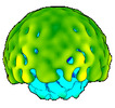
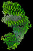
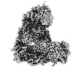
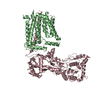
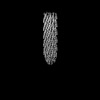
EMDB-50358: 
In vitro-induced genome-releasing intermediate of Rhodobacter microvirus Ebor computed with C5 symmetry
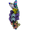
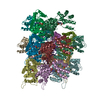
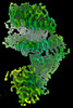
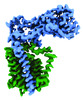
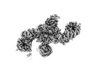
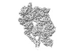
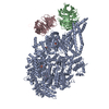
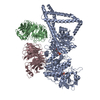
EMDB-19075:
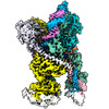
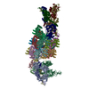
Conformational Landscape of the Type V-K CRISPR-associated TransposonIntegration Assembly CAST V-K composite map
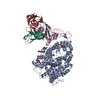
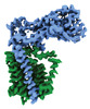
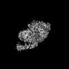
EMDB-19282:
Conformational Landscape of the Type V-K CRISPR-associated TransposonIntegration Assembly CAST V-K Cas12k domain local-refinement map
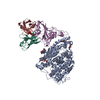
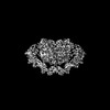
EMDB-19283:
Conformational Landscape of the Type V-K CRISPR-associated TransposonIntegration Assembly CAST V-K TnsC domain local-refinement map
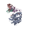
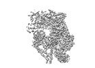
EMDB-19284:
Conformational Landscape of the Type V-K CRISPR-associated TransposonIntegration Assembly CAST V-K TnsB domain local-refinement map
EMDB-19286:
consensus map of the V-K CRISPR-associated Transposon Integration Assembly
PDB-8rdu:
Conformational Landscape of the Type V-K CRISPR-associated TransposonIntegration Assembly CAST V-K composite map
PDB-8rkt:
Conformational Landscape of the Type V-K CRISPR-associated TransposonIntegration Assembly CAST V-K Cas12k domain local-refinement map
PDB-8rku:
Conformational Landscape of the Type V-K CRISPR-associated TransposonIntegration Assembly CAST V-K TnsC domain local-refinement map
PDB-8rkv:
Conformational Landscape of the Type V-K CRISPR-associated TransposonIntegration Assembly CAST V-K TnsB domain local-refinement map
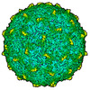
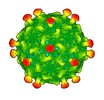
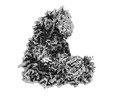
EMDB-50356:
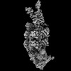
Empty capsid of Rhodobacter microvirus Ebor computed with I4 symmetry
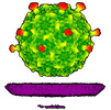
EMDB-50357:
Native capsid of Rhodobacter microvirus Ebor computed with I4 symmetry
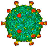
EMDB-50359:
Rhodobacter microvirus Ebor attached to B10 host cell reconstructed by single particle analysis with applied C5 symmetry
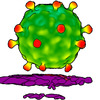
EMDB-50360:
Rhodobacter microvirus Ebor attached to the outer membrane vesicle
EMDB-50361:
Rhodobacter microvirus Ebor attached to the host cell reconstructed by subtomogram averaging

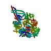
EMDB-15697:
Cryo-EM structure of shCas12k-sgRNA-dsDNA ternary complex (type V-K CRISPR-associated transposon)

PDB-8axa:
Cryo-EM structure of shCas12k-sgRNA-dsDNA ternary complex (type V-K CRISPR-associated transposon)

EMDB-29530:
SARS-CoV-2 XBB.1 spike RBD bound to the human ACE2 ectodomain and the S309 neutralizing antibody Fab fragment
EMDB-29531:
SARS-CoV-2 BQ.1.1 spike RBD bound to the human ACE2 ectodomain and the S309 neutralizing antibody Fab fragment
EMDB-40240:
SARS-CoV-2 BN.1 spike RBD bound to the human ACE2 ectodomain and the S309 neutralizing antibody Fab fragment
PDB-8fxb:
SARS-CoV-2 XBB.1 spike RBD bound to the human ACE2 ectodomain and the S309 neutralizing antibody Fab fragment
PDB-8fxc:
SARS-CoV-2 BQ.1.1 spike RBD bound to the human ACE2 ectodomain and the S309 neutralizing antibody Fab fragment
PDB-8s9g:
SARS-CoV-2 BN.1 spike RBD bound to the human ACE2 ectodomain and the S309 neutralizing antibody Fab fragment
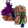
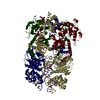
EMDB-41140:
APC/C-CDH1-UBE2C-Ubiquitin-CyclinB-NTD
EMDB-41142:
APC/C-CDH1-UBE2C-UBE2S-Ubiquitin-CyclinB

PDB-8tar:
APC/C-CDH1-UBE2C-Ubiquitin-CyclinB-NTD

PDB-8tau:
APC/C-CDH1-UBE2C-UBE2S-Ubiquitin-CyclinB
EMDB-41299:
Structural basis of peptidoglycan synthesis by E. coli RodA-PBP2 complex

EMDB-41303:
Transmembrane map
EMDB-41304:
Periplasmic map
PDB-8tj3:
Structural basis of peptidoglycan synthesis by E. coli RodA-PBP2 complex
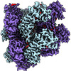
EMDB-16087:
Structure of the human UBR5 Dimer.
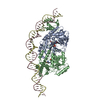
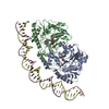
EMDB-14778:
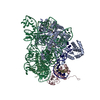
Cryo-EM structure of holo-PdxR from Bacillus clausii bound to its target DNA in the half-closed conformation
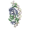
EMDB-14801:
Cryo-EM structure of holo-PdxR from Bacillus clausii bound to its target DNA in the closed conformation, C2 symmetry
EMDB-14852:
Cryo-EM structure of holo-PdxR from Bacillus clausii bound to its target DNA in the closed conformation, C1 symmetry

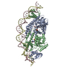
EMDB-14960:
Cryo-EM structure of holo-PdxR from Bacillus clausii bound to its target DNA in the open conformation
PDB-7zla:
Cryo-EM structure of holo-PdxR from Bacillus clausii bound to its target DNA in the half-closed conformation
PDB-7zn5:
Cryo-EM structure of holo-PdxR from Bacillus clausii bound to its target DNA in the closed conformation, C2 symmetry.
PDB-7zpa:
Cryo-EM structure of holo-PdxR from Bacillus clausii bound to its target DNA in the closed conformation, C1 symmetry
PDB-7zth:
Cryo-EM structure of holo-PdxR from Bacillus clausii bound to its target DNA in the open conformation

EMDB-28999:
Engineered human dynein motor domain in microtubule-bound state

EMDB-29003:
Engineered human dynein motor domain in the microtubule-unbound state in the buffer containing ATP-Vi
EMDB-29012:
Engineered human dynein motor domain in the microtubule-unbound state with LIS1 complex in the buffer containing ATP-Vi
EMDB-29014:
Engineered human dynein motor domain in the microtubule-unbound state with LIS1 complex in the buffer containing ATP-Vi (local refined on AAA3-AAA5 and LIS1)

PDB-8fcy:
Engineered human dynein motor domain in microtubule-bound state
PDB-8fd6:
Engineered human dynein motor domain in the microtubule-unbound state in the buffer containing ATP-Vi
PDB-8fdt:
Engineered human dynein motor domain in the microtubule-unbound state with LIS1 complex in the buffer containing ATP-Vi
PDB-8fdu:
Engineered human dynein motor domain in the microtubule-unbound state with LIS1 complex in the buffer containing ATP-Vi (local refined on AAA3-AAA5 and LIS1)

EMDB-15831:
CryoEM structure of the pointy tip (proteins pIII/pVI/pVIII) from the f1 filamentous bacteriophage
EMDB-15832:
CryoEM structure of the round tip (proteins pVII/pVIII/pIX) from the f1 filamentous bacteriophage
ページ:
 wwPDBはEMDBデータモデルのバージョン3へ移行します
