National Institutes of Health/National Institute of General Medical Sciences (NIH/NIGMS)
R35 GM132120
United States
Citation
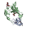
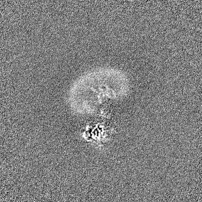
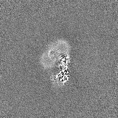
Journal: Nat Commun / Year: 2023 Title: Structural basis of peptidoglycan synthesis by E. coli RodA-PBP2 complex. Authors: Rie Nygaard / Chris L B Graham / Meagan Belcher Dufrisne / Jonathan D Colburn / Joseph Pepe / Molly A Hydorn / Silvia Corradi / Chelsea M Brown / Khuram U Ashraf / Owen N Vickery / Nicholas ...Authors: Rie Nygaard / Chris L B Graham / Meagan Belcher Dufrisne / Jonathan D Colburn / Joseph Pepe / Molly A Hydorn / Silvia Corradi / Chelsea M Brown / Khuram U Ashraf / Owen N Vickery / Nicholas S Briggs / John J Deering / Brian Kloss / Bruno Botta / Oliver B Clarke / Linda Columbus / Jonathan Dworkin / Phillip J Stansfeld / David I Roper / Filippo Mancia / Abstract: Peptidoglycan (PG) is an essential structural component of the bacterial cell wall that is synthetized during cell division and elongation. PG forms an extracellular polymer crucial for cellular ...Peptidoglycan (PG) is an essential structural component of the bacterial cell wall that is synthetized during cell division and elongation. PG forms an extracellular polymer crucial for cellular viability, the synthesis of which is the target of many antibiotics. PG assembly requires a glycosyltransferase (GT) to generate a glycan polymer using a Lipid II substrate, which is then crosslinked to the existing PG via a transpeptidase (TP) reaction. A Shape, Elongation, Division and Sporulation (SEDS) GT enzyme and a Class B Penicillin Binding Protein (PBP) form the core of the multi-protein complex required for PG assembly. Here we used single particle cryo-electron microscopy to determine the structure of a cell elongation-specific E. coli RodA-PBP2 complex. We combine this information with biochemical, genetic, spectroscopic, and computational analyses to identify the Lipid II binding sites and propose a mechanism for Lipid II polymerization. Our data suggest a hypothesis for the movement of the glycan strand from the Lipid II polymerization site of RodA towards the TP site of PBP2, functionally linking these two central enzymatic activities required for cell wall peptidoglycan biosynthesis.
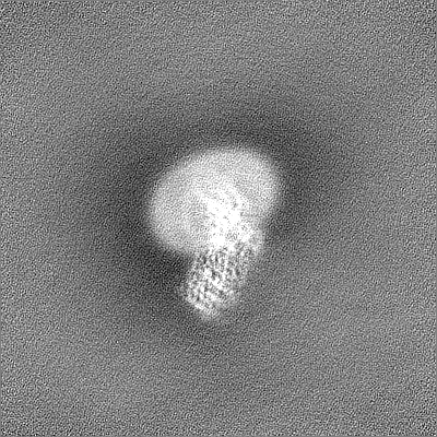
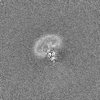
Name: RodA-PBP2 / type: complex / ID: 1 / Parent: 0 / Macromolecule list: #1-#2 / Details: Nanodisc were formed using MSP1E3D1 and POPG lipid
Source (natural)
Organism: Escherichia coli (E. coli)
Molecular weight
Theoretical: 111.803 KDa
-
Experimental details
-
Structure determination
Method
cryo EM
Processing
single particle reconstruction
Aggregation state
particle
-
Sample preparation
Concentration
0.66 mg/mL
Buffer
pH: 7 Component:
Concentration
Name
Formula
20.0 mM
HEPES
150.0 mM
Sodium Chloride
NaCl
1.0 mM
TCEP
tris(2-carboxyethyl)phosphine
Vitrification
Cryogen name: ETHANE / Chamber humidity: 95 % / Chamber temperature: 277 K / Instrument: FEI VITROBOT MARK IV
-
Electron microscopy
Microscope
FEI TITAN
Image recording
Film or detector model: GATAN K3 BIOQUANTUM (6k x 4k) / Number grids imaged: 1 / Number real images: 11120 / Average exposure time: 2.5 sec. / Average electron dose: 58.5 e/Å2
Electron beam
Acceleration voltage: 300 kV / Electron source: FIELD EMISSION GUN
In the structure databanks used in Yorodumi, some data are registered as the other names, "COVID-19 virus" and "2019-nCoV". Here are the details of the virus and the list of structure data.
Jan 31, 2019. EMDB accession codes are about to change! (news from PDBe EMDB page)
EMDB accession codes are about to change! (news from PDBe EMDB page)
The allocation of 4 digits for EMDB accession codes will soon come to an end. Whilst these codes will remain in use, new EMDB accession codes will include an additional digit and will expand incrementally as the available range of codes is exhausted. The current 4-digit format prefixed with “EMD-” (i.e. EMD-XXXX) will advance to a 5-digit format (i.e. EMD-XXXXX), and so on. It is currently estimated that the 4-digit codes will be depleted around Spring 2019, at which point the 5-digit format will come into force.
The EM Navigator/Yorodumi systems omit the EMD- prefix.
Related info.:Q: What is EMD? / ID/Accession-code notation in Yorodumi/EM Navigator
Yorodumi is a browser for structure data from EMDB, PDB, SASBDB, etc.
This page is also the successor to EM Navigator detail page, and also detail information page/front-end page for Omokage search.
The word "yorodu" (or yorozu) is an old Japanese word meaning "ten thousand". "mi" (miru) is to see.
Related info.:EMDB / PDB / SASBDB / Comparison of 3 databanks / Yorodumi Search / Aug 31, 2016. New EM Navigator & Yorodumi / Yorodumi Papers / Jmol/JSmol / Function and homology information / Changes in new EM Navigator and Yorodumi
 Movie
Movie Controller


 Open data
 Basic information
 Map data
 Sample
 Keywords
 Authors
 United States, 1 items
  Citation

 Structure visualization
 Downloads & links
 EMDB map data format
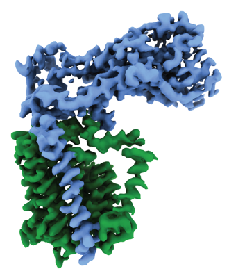 emd_41304.png
 http://ftp.pdbj.org/pub/emdb/structures/EMD-41304


 Z (Sec.)
 Y (Row.)
 X (Col.)


 Sample components
 Processing
 Electron microscopy
 FIELD EMISSION GUN