Movie
Movie Controller

[English] 日本語
 Yorodumi
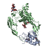
- PDB-8tj3: Structural basis of peptidoglycan synthesis by E. coli RodA-PBP2 ... -
+ Open data

- Basic information
| Entry | Database: PDB / ID: 8tj3 | ||||||
|---|---|---|---|---|---|---|---|
| Title | Structural basis of peptidoglycan synthesis by E. coli RodA-PBP2 complex | ||||||
 Components Components |
| ||||||
 Keywords Keywords | MEMBRANE PROTEIN / Peptidoglycan / glycosyltransferase / enzyme | ||||||
| Function / homology |  Function and homology information Function and homology informationlipid-linked peptidoglycan transporter activity / peptidoglycan glycosyltransferase / peptidoglycan L,D-transpeptidase activity / peptidoglycan glycosyltransferase activity / serine-type D-Ala-D-Ala carboxypeptidase / serine-type D-Ala-D-Ala carboxypeptidase activity / cell division site / glycosyltransferase activity / penicillin binding / peptidoglycan biosynthetic process ...lipid-linked peptidoglycan transporter activity / peptidoglycan glycosyltransferase / peptidoglycan L,D-transpeptidase activity / peptidoglycan glycosyltransferase activity / serine-type D-Ala-D-Ala carboxypeptidase / serine-type D-Ala-D-Ala carboxypeptidase activity / cell division site / glycosyltransferase activity / penicillin binding / peptidoglycan biosynthetic process / cell wall organization / regulation of cell shape / cell division / proteolysis / plasma membrane Similarity search - Function | ||||||
| Biological species |  | ||||||
| Method | ELECTRON MICROSCOPY / single particle reconstruction / cryo EM / Resolution: 3.2 Å | ||||||
 Authors Authors | Nygaard, R. / Mancia, F. | ||||||
| Funding support |  United States, 1items United States, 1items
| ||||||
 Citation Citation | Journal: Nat Commun / Year: 2023 Title: Structural basis of peptidoglycan synthesis by E. coli RodA-PBP2 complex. Authors: Rie Nygaard / Chris L B Graham / Meagan Belcher Dufrisne / Jonathan D Colburn / Joseph Pepe / Molly A Hydorn / Silvia Corradi / Chelsea M Brown / Khuram U Ashraf / Owen N Vickery / Nicholas ...Authors: Rie Nygaard / Chris L B Graham / Meagan Belcher Dufrisne / Jonathan D Colburn / Joseph Pepe / Molly A Hydorn / Silvia Corradi / Chelsea M Brown / Khuram U Ashraf / Owen N Vickery / Nicholas S Briggs / John J Deering / Brian Kloss / Bruno Botta / Oliver B Clarke / Linda Columbus / Jonathan Dworkin / Phillip J Stansfeld / David I Roper / Filippo Mancia /   Abstract: Peptidoglycan (PG) is an essential structural component of the bacterial cell wall that is synthetized during cell division and elongation. PG forms an extracellular polymer crucial for cellular ...Peptidoglycan (PG) is an essential structural component of the bacterial cell wall that is synthetized during cell division and elongation. PG forms an extracellular polymer crucial for cellular viability, the synthesis of which is the target of many antibiotics. PG assembly requires a glycosyltransferase (GT) to generate a glycan polymer using a Lipid II substrate, which is then crosslinked to the existing PG via a transpeptidase (TP) reaction. A Shape, Elongation, Division and Sporulation (SEDS) GT enzyme and a Class B Penicillin Binding Protein (PBP) form the core of the multi-protein complex required for PG assembly. Here we used single particle cryo-electron microscopy to determine the structure of a cell elongation-specific E. coli RodA-PBP2 complex. We combine this information with biochemical, genetic, spectroscopic, and computational analyses to identify the Lipid II binding sites and propose a mechanism for Lipid II polymerization. Our data suggest a hypothesis for the movement of the glycan strand from the Lipid II polymerization site of RodA towards the TP site of PBP2, functionally linking these two central enzymatic activities required for cell wall peptidoglycan biosynthesis. | ||||||
| History |
|
- Structure visualization
| Structure viewer | Molecule: MolmilJmol/JSmol |
|---|
- Downloads & links
-Download
| PDBx/mmCIF format | 8tj3.cif.gz | 283.6 KB | Display | PDBx/mmCIF format |
|---|---|---|---|---|
| PDB format | pdb8tj3.ent.gz | 226.8 KB | Display | PDB format |
| PDBx/mmJSON format | 8tj3.json.gz | Tree view | PDBx/mmJSON format | |
| Others |  Other downloads Other downloads |
-Validation report
| Arichive directory | https://data.pdbj.org/pub/pdb/validation_reports/tj/8tj3ftp://data.pdbj.org/pub/pdb/validation_reports/tj/8tj3 | HTTPS FTP |
|---|
-Related structure data
| Related structure data |  41299MC M: map data used to model this data C: citing same article ( |
|---|---|
| Similar structure data |


-Links
 PDBj
PDBj
- Assembly
| Deposited unit | 
|
|---|---|
| 1 |
|
-Components
| #1: Protein | Mass: 40508.766 Da / Num. of mol.: 1 Source method: isolated from a genetically manipulated source Source: (gene. exp.) |
|---|---|
| #2: Protein | Mass: 70943.414 Da / Num. of mol.: 1 Source method: isolated from a genetically manipulated source Source: (gene. exp.) |
-Experimental details
-Experiment
| Experiment | Method: ELECTRON MICROSCOPY |
|---|---|
| EM experiment | Aggregation state: PARTICLE / 3D reconstruction method: single particle reconstruction |
- Sample preparation
| Component | Name: RodA-PBP2 / Type: COMPLEX / Details: Nanodisc were formed using MSP1E3D1 and POPG lipid / Entity ID: all / Source: RECOMBINANT | ||||||||||||||||||||
|---|---|---|---|---|---|---|---|---|---|---|---|---|---|---|---|---|---|---|---|---|---|
| Molecular weight | Value: 0.111803 MDa / Experimental value: NO | ||||||||||||||||||||
| Source (natural) | Organism: | ||||||||||||||||||||
| Source (recombinant) | Organism: | ||||||||||||||||||||
| Buffer solution | pH: 7 | ||||||||||||||||||||
| Buffer component |
| ||||||||||||||||||||
| Specimen | Conc.: 0.66 mg/ml / Embedding applied: NO / Shadowing applied: NO / Staining applied: NO / Vitrification applied: YES | ||||||||||||||||||||
| Vitrification | Instrument: FEI VITROBOT MARK IV / Cryogen name: ETHANE / Humidity: 95 % / Chamber temperature: 277 K |
- Electron microscopy imaging
| Microscopy | Model: FEI TITAN |
|---|---|
| Electron gun | Electron source:  FIELD EMISSION GUN / Accelerating voltage: 300 kV / Illumination mode: FLOOD BEAM FIELD EMISSION GUN / Accelerating voltage: 300 kV / Illumination mode: FLOOD BEAM |
| Electron lens | Mode: BRIGHT FIELD / Nominal defocus max: 2500 nm / Nominal defocus min: 1000 nm |
| Image recording | Average exposure time: 2.5 sec. / Electron dose: 58.5 e/Å2 / Film or detector model: GATAN K3 BIOQUANTUM (6k x 4k) / Num. of grids imaged: 1 / Num. of real images: 11120 |
- Processing
| EM software |
| ||||||||||||||||||||||||||||||
|---|---|---|---|---|---|---|---|---|---|---|---|---|---|---|---|---|---|---|---|---|---|---|---|---|---|---|---|---|---|---|---|
| CTF correction | Details: Patch CTF / Type: PHASE FLIPPING AND AMPLITUDE CORRECTION | ||||||||||||||||||||||||||||||
| Particle selection | Num. of particles selected: 4415933 | ||||||||||||||||||||||||||||||
| 3D reconstruction | Resolution: 3.2 Å / Resolution method: FSC 0.143 CUT-OFF / Num. of particles: 399000 / Symmetry type: POINT | ||||||||||||||||||||||||||||||
| Atomic model building | PDB-ID: 4F0A Accession code: 4F0A / Source name: PDB / Type: experimental model | ||||||||||||||||||||||||||||||
| Refine LS restraints |
|