 Movie
Movie Controller
 Structure viewers
 About EMN search


-Search query
-Search result
Showing 1 - 50 of 95 items for (author: matthies & m)

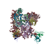
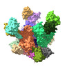
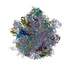
EMDB-29783: 
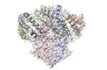
Cryo-EM structure of T/F100 SOSIP.664 HIV-1 Env trimer with LMHS mutations in complex with 8ANC195 and 10-1074

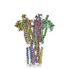
EMDB-41613:
Cryo-EM structure of BG505 SOSIP.664 HIV-1 Env trimer in complex with temsavir, 8ANC195, and 10-1074
PDB-8g6u:
Cryo-EM structure of T/F100 SOSIP.664 HIV-1 Env trimer with LMHS mutations in complex with 8ANC195 and 10-1074

PDB-8ttw:
Cryo-EM structure of BG505 SOSIP.664 HIV-1 Env trimer in complex with temsavir, 8ANC195, and 10-1074

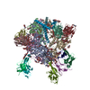
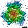
EMDB-41624:
Cryo-EM structure of the human MRS2 magnesium channel under Mg2+ condition

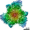
EMDB-41628:
Cryo-EM structure of the human MRS2 magnesium channel under Mg2+-free condition

EMDB-41629:
Cryo-EM structure of the human MRS2 magnesium channel under Mg2+ condition (C1 map)
EMDB-41630:
Cryo-EM structure of the human MRS2 magnesium channel under Mg2+-free condition (C1 map)
PDB-8tul:
Cryo-EM structure of the human MRS2 magnesium channel under Mg2+ condition
PDB-8tup:
Cryo-EM structure of the human MRS2 magnesium channel under Mg2+-free condition
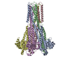
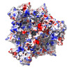
EMDB-27596:
Cryo-EM structure of T/F100 SOSIP.664 HIV-1 Env trimer in complex with 8ANC195 and 10-1074
PDB-8dok:
Cryo-EM structure of T/F100 SOSIP.664 HIV-1 Env trimer in complex with 8ANC195 and 10-1074
EMDB-27103:
Cryo-EM structure of T/F100 SOSIP.664 HIV-1 Env trimer with LMHS mutations in complex with Temsavir, 8ANC195, and 10-1074

PDB-8czz:
Cryo-EM structure of T/F100 SOSIP.664 HIV-1 Env trimer with LMHS mutations in complex with Temsavir, 8ANC195, and 10-1074

EMDB-27148:
Zebrafish MFSD2A isoform B in inward open ligand bound conformation
EMDB-27149:
Zebrafish MFSD2A isoform B in inward open ligand-free conformation

EMDB-27150:
Zebrafish MFSD2A isoform B in inward open ligand 1A conformation

EMDB-27151:
Zebrafish MFSD2A isoform B in inward open ligand 1B conformation
EMDB-27152:
Zebrafish MFSD2A isoform B in inward open ligand 2B conformation
EMDB-27153:
Zebrafish MFSD2A isoform B in inward open ligand 3C conformation
PDB-8d2s:
Zebrafish MFSD2A isoform B in inward open ligand bound conformation
PDB-8d2t:
Zebrafish MFSD2A isoform B in inward open ligand-free conformation

PDB-8d2u:
Zebrafish MFSD2A isoform B in inward open ligand 1A conformation

PDB-8d2v:
Zebrafish MFSD2A isoform B in inward open ligand 1B conformation

PDB-8d2w:
Zebrafish MFSD2A isoform B in inward open ligand 2B conformation

PDB-8d2x:
Zebrafish MFSD2A isoform B in inward open ligand 3C conformation

EMDB-28538:
Structure of SARS-CoV-2 Orf3a in late endosome/lysosome-like membrane environment, MSP1D1 nanodisc

EMDB-28544:
Structure of SARS-CoV-1 Orf3a in late endosome/lysosome-like environment, MSP1D1 nanodisc

EMDB-28545:
Structure of SARS-CoV-2 Orf3a in plasma membrane-like environment, MSP1D1 nanodisc
EMDB-28546:
Structure of SARS-CoV-2 Orf3a in late endosome/lysosome-like environment, Saposin A nanodisc

PDB-8eqj:
Structure of SARS-CoV-2 Orf3a in late endosome/lysosome-like membrane environment, MSP1D1 nanodisc

PDB-8eqs:
Structure of SARS-CoV-1 Orf3a in late endosome/lysosome-like environment, MSP1D1 nanodisc

PDB-8eqt:
Structure of SARS-CoV-2 Orf3a in plasma membrane-like environment, MSP1D1 nanodisc

PDB-8equ:
Structure of SARS-CoV-2 Orf3a in late endosome/lysosome-like environment, Saposin A nanodisc
EMDB-25132:
Androgen receptor bound to DNA - Entrenched state

EMDB-25133:
Androgen receptor bound to DNA - Splayed state
EMDB-25134:
Androgen receptor bound to DNA - Divorced state
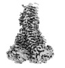
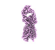
EMDB-13089:
major seeded in vitro fibril morphology from murine SAA1.1 protein

PDB-7ovt:
major seeded in vitro fibril morphology from murine SAA1.1 protein
PDB-7b5k:
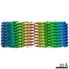
E. coli 70S containing suppressor tRNA in the A-site stabilized by a Negamycin analogue and P-site tRNA-nascent chain.

EMDB-22022:
hEAAT3-IFS-in10mMGlu

EMDB-22023:
hEAAT3-IFS-in250mM-KCl
EMDB-22024:
hEAAT3-Asymmetric-2o1i-in20mM-Asp
EMDB-22011:
hEAAT3-IFS-Na

EMDB-22014:
hEAAT3-OFS-Asp

EMDB-22020:
hEAAT3-Asymmetric-1o2i

EMDB-22021:
hEAAT3-IFS-Apo

PDB-6x2l:
hEAAT3-IFS-Na

PDB-6x2z:
hEAAT3-OFS-Asp

PDB-6x3e:
hEAAT3-Asymmetric-1o2i
Pages:
 wwPDB to switch to version 3 of the EMDB data model
