 Movie
Movie Controller

[English] 日本語
 Yorodumi
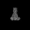
- PDB-8tup: Cryo-EM structure of the human MRS2 magnesium channel under Mg2+-... -
+ Open data

- Basic information
| Entry | Database: PDB / ID: 8tup | ||||||
|---|---|---|---|---|---|---|---|
| Title | Cryo-EM structure of the human MRS2 magnesium channel under Mg2+-free condition | ||||||
 Components Components | Magnesium transporter MRS2 homolog, mitochondrial | ||||||
 Keywords Keywords | METAL TRANSPORT / Magnesium / Ion channel / Membrane protein / Ion Translocation / Divalent Ion / Pentamer | ||||||
| Function / homology |  Function and homology information Function and homology informationmitochondrial magnesium ion transmembrane transport / Miscellaneous transport and binding events / magnesium ion transmembrane transporter activity / lactate metabolic process / transmembrane transport / mitochondrial inner membrane / mitochondrion / metal ion binding Similarity search - Function | ||||||
| Biological species |  Homo sapiens (human) Homo sapiens (human) | ||||||
| Method | ELECTRON MICROSCOPY / single particle reconstruction / cryo EM / Resolution: 3.3 Å | ||||||
 Authors Authors | Lai, L.T.F. / Balaraman, J. / Zhou, F. / Matthies, D. | ||||||
| Funding support |  United States, 1items United States, 1items
| ||||||
 Citation Citation | Journal: Nat Commun / Year: 2023 Title: Cryo-EM structures of human magnesium channel MRS2 reveal gating and regulatory mechanisms. Authors: Louis Tung Faat Lai / Jayashree Balaraman / Fei Zhou / Doreen Matthies / Abstract: Magnesium ions (Mg) play an essential role in cellular physiology. In mitochondria, protein and ATP synthesis and various metabolic pathways are directly regulated by Mg. MRS2, a magnesium channel ...Magnesium ions (Mg) play an essential role in cellular physiology. In mitochondria, protein and ATP synthesis and various metabolic pathways are directly regulated by Mg. MRS2, a magnesium channel located in the inner mitochondrial membrane, mediates the influx of Mg into the mitochondrial matrix and regulates Mg homeostasis. Knockdown of MRS2 in human cells leads to reduced uptake of Mg into mitochondria and disruption of the mitochondrial metabolism. Despite the importance of MRS2, the Mg translocation and regulation mechanisms of MRS2 are still unclear. Here, using cryo-EM we report the structures of human MRS2 in the presence and absence of Mg at 2.8 Å and 3.3 Å, respectively. From the homo-pentameric structures, we identify R332 and M336 as major gating residues, which are then tested using mutagenesis and two cellular divalent ion uptake assays. A network of hydrogen bonds is found connecting the gating residue R332 to the soluble domain, potentially regulating the gate. Two Mg-binding sites are identified in the MRS2 soluble domain, distinct from the two sites previously reported in CorA, a homolog of MRS2 in prokaryotes. Altogether, this study provides the molecular basis for understanding the Mg translocation and regulatory mechanisms of MRS2. | ||||||
| History |
|
- Structure visualization
| Structure viewer | Molecule: MolmilJmol/JSmol |
|---|
- Downloads & links
-Download
| PDBx/mmCIF format | 8tup.cif.gz | 323.4 KB | Display | PDBx/mmCIF format |
|---|---|---|---|---|
| PDB format | pdb8tup.ent.gz | 259 KB | Display | PDB format |
| PDBx/mmJSON format | 8tup.json.gz | Tree view | PDBx/mmJSON format | |
| Others |  Other downloads Other downloads |
-Validation report
| Arichive directory | https://data.pdbj.org/pub/pdb/validation_reports/tu/8tupftp://data.pdbj.org/pub/pdb/validation_reports/tu/8tup | HTTPS FTP |
|---|
-Related structure data
| Related structure data | 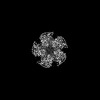 41628MC 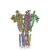 8tulC C: citing same article ( M: map data used to model this data |
|---|---|
| Similar structure data |


-Links
 PDBj
PDBj
- Assembly
| Deposited unit | 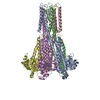
|
|---|---|
| 1 |
|
-Components
| #1: Protein | Mass: 51373.516 Da / Num. of mol.: 5 Source method: isolated from a genetically manipulated source Source: (gene. exp.) Homo sapiens (human) / Gene: MRS2 / Cell line (production host): Expi293F / Production host: Homo sapiens (human) / References: UniProt: Q9HD23#2: Chemical |   Mass: 24.305 Da / Num. of mol.: 3 / Source method: obtained synthetically / Formula: Mg / Feature type: SUBJECT OF INVESTIGATION Mass: 24.305 Da / Num. of mol.: 3 / Source method: obtained synthetically / Formula: Mg / Feature type: SUBJECT OF INVESTIGATION#3: Water | ChemComp-HOH / |  Mass: 18.015 Da / Num. of mol.: 66 / Source method: isolated from a natural source / Formula: H2O Mass: 18.015 Da / Num. of mol.: 66 / Source method: isolated from a natural source / Formula: H2OHas ligand of interest | Y | |
|---|
-Experimental details
-Experiment
| Experiment | Method: ELECTRON MICROSCOPY |
|---|---|
| EM experiment | Aggregation state: PARTICLE / 3D reconstruction method: single particle reconstruction |
- Sample preparation
| Component | Name: Cryo-EM structure of the pentameric human MRS2 magnesium channel under Mg2+-free condition at an average resolution of 3.3 A, filtered to local resolution, C5 Type: COMPLEX / Entity ID: #1 / Source: RECOMBINANT | ||||||||||||||||||||
|---|---|---|---|---|---|---|---|---|---|---|---|---|---|---|---|---|---|---|---|---|---|
| Molecular weight | Value: 0.219 MDa / Experimental value: NO | ||||||||||||||||||||
| Source (natural) | Organism: Homo sapiens (human) | ||||||||||||||||||||
| Source (recombinant) | Organism: Homo sapiens (human) / Cell: Expi293F / Plasmid: pCMV | ||||||||||||||||||||
| Buffer solution | pH: 7.3 Details: 20 mM HEPES, 150 mM NaCl, 1mM EDTA, 0.003% LMNG, pH 7.3 | ||||||||||||||||||||
| Buffer component |
| ||||||||||||||||||||
| Specimen | Conc.: 0.5 mg/ml / Embedding applied: NO / Shadowing applied: NO / Staining applied: NO / Vitrification applied: YES / Details: This sample was monodisperse | ||||||||||||||||||||
| Specimen support | Grid material: COPPER / Grid mesh size: 400 divisions/in. / Grid type: Quantifoil R1.2/1.3 | ||||||||||||||||||||
| Vitrification | Instrument: LEICA EM GP / Cryogen name: ETHANE / Humidity: 95 % / Chamber temperature: 277 K Details: 400-mesh R1.2/1.3 Cu grids (Quantifoil) were made hydrophilic by glow discharging for 60 seconds with a current of 15 mA in a PELCO easiGlow system. The cryo grids were produced using a ...Details: 400-mesh R1.2/1.3 Cu grids (Quantifoil) were made hydrophilic by glow discharging for 60 seconds with a current of 15 mA in a PELCO easiGlow system. The cryo grids were produced using a Leica EM GP2 (Leica). The chamber was kept at 4 C and set to 95% humidity. 3 microliter sample at 0.5 mg/ml was applied to a glow-discharged holey grid, blotted for 6 s, and plunge frozen into liquid ethane and stored in liquid nitrogen. |
- Electron microscopy imaging
| Experimental equipment |  Model: Titan Krios / Image courtesy: FEI Company |
|---|---|
| Microscopy | Model: FEI TITAN KRIOS |
| Electron gun | Electron source:  FIELD EMISSION GUN / Accelerating voltage: 300 kV / Illumination mode: FLOOD BEAM FIELD EMISSION GUN / Accelerating voltage: 300 kV / Illumination mode: FLOOD BEAM |
| Electron lens | Mode: BRIGHT FIELD / Nominal magnification: 105000 X / Nominal defocus max: 2000 nm / Nominal defocus min: 700 nm / Cs: 2.7 mm / C2 aperture diameter: 100 µm |
| Specimen holder | Cryogen: NITROGEN / Specimen holder model: FEI TITAN KRIOS AUTOGRID HOLDER |
| Image recording | Average exposure time: 3.462 sec. / Electron dose: 50 e/Å2 / Film or detector model: GATAN K3 BIOQUANTUM (6k x 4k) / Num. of grids imaged: 1 / Num. of real images: 3991 Details: Cryo-EM datasets were acquired with SerialEM using a Titan Krios (FEI, now ThermoFisher Scientific) operated at 300 keV and equipped with an energy filter and K3 camera (Gatan Inc.). Movies ...Details: Cryo-EM datasets were acquired with SerialEM using a Titan Krios (FEI, now ThermoFisher Scientific) operated at 300 keV and equipped with an energy filter and K3 camera (Gatan Inc.). Movies of 50 frames with a dose of 1 e-/A2 per frame (50 e-/A2 total dose) were recorded at a nominal magnification of 105,000x, corresponding to a physical pixel size of 0.83 A/px (super-resolution pixel size 0.415 A/px) in CDS mode at a dose rate of 10 e-/px/s and a defocus range of -0.7 to -2.0 um. In total, 9,656 movies were collected. |
| EM imaging optics | Energyfilter name: GIF Bioquantum Details: Cryo-EM datasets were acquired with SerialEM using a Titan Krios (FEI, now ThermoFisher Scientific) operated at 300 keV and equipped with an energy filter and K3 camera (Gatan Inc.). Movies ...Details: Cryo-EM datasets were acquired with SerialEM using a Titan Krios (FEI, now ThermoFisher Scientific) operated at 300 keV and equipped with an energy filter and K3 camera (Gatan Inc.). Movies of 50 frames with a dose of 1 e-/A2 per frame (50 e-/A2 total dose) were recorded at a nominal magnification of 105,000x, corresponding to a physical pixel size of 0.83 A/px (super-resolution pixel size 0.415 A/px) in CDS mode at a dose rate of 10 e-/px/s and a defocus range of -0.7 to -2.0 um. In total, 9,656 movies were collected. Energyfilter slit width: 20 eV |
- Processing
| EM software |
| ||||||||||||||||||||||||||||||||||||||||||||||||||||||||||||
|---|---|---|---|---|---|---|---|---|---|---|---|---|---|---|---|---|---|---|---|---|---|---|---|---|---|---|---|---|---|---|---|---|---|---|---|---|---|---|---|---|---|---|---|---|---|---|---|---|---|---|---|---|---|---|---|---|---|---|---|---|---|
| Image processing | Details: Cryo-EM datasets were acquired with SerialEM using a Titan Krios (FEI, now ThermoFisher Scientific) operated at 300 keV and equipped with an energy filter and K3 camera (Gatan Inc.). Movies ...Details: Cryo-EM datasets were acquired with SerialEM using a Titan Krios (FEI, now ThermoFisher Scientific) operated at 300 keV and equipped with an energy filter and K3 camera (Gatan Inc.). Movies of 50 frames with a dose of 1 e-/A2 per frame (50 e-/A2 total dose) were recorded at a nominal magnification of 105,000x, corresponding to a physical pixel size of 0.83 A/px (super-resolution pixel size 0.415 A/px) in CDS mode at a dose rate of 10 e-/px/s and a defocus range of -0.7 to -2.0 um. In total, 9,656 movies were collected. | ||||||||||||||||||||||||||||||||||||||||||||||||||||||||||||
| CTF correction | Details: Patch CTF / Type: PHASE FLIPPING AND AMPLITUDE CORRECTION | ||||||||||||||||||||||||||||||||||||||||||||||||||||||||||||
| Particle selection | Num. of particles selected: 4802706 Details: Good 2D class averages generated from ~1,000 manually picked particles served as templates for automatic particle picking. | ||||||||||||||||||||||||||||||||||||||||||||||||||||||||||||
| Symmetry | Point symmetry: C5 (5 fold cyclic) | ||||||||||||||||||||||||||||||||||||||||||||||||||||||||||||
| 3D reconstruction | Resolution: 3.3 Å / Resolution method: FSC 0.143 CUT-OFF / Num. of particles: 1744117 / Algorithm: BACK PROJECTION Details: Final non-uniform refinement by using 1,744,117 particles yielded maps at 3.3 A (with C5 symmetry applied) and 3.6 A (with C1 symmetry applied) according to gold-standard FSC = 0.143 criterion. Num. of class averages: 1 / Symmetry type: POINT | ||||||||||||||||||||||||||||||||||||||||||||||||||||||||||||
| Atomic model building |
| ||||||||||||||||||||||||||||||||||||||||||||||||||||||||||||
| Atomic model building |
| ||||||||||||||||||||||||||||||||||||||||||||||||||||||||||||
| Refine LS restraints |
|
