 ムービー
ムービー コントローラー
 構造ビューア
 EMN検索について


-検索条件
-検索結果
検索 (著者・登録者: koen & g)の結果91件中、1から50件目までを表示しています

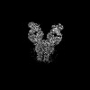
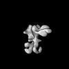
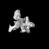
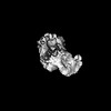
EMDB-53563: 
Non-uniform refine map MiDAC complex

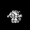
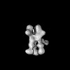
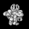
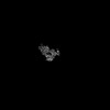
EMDB-53564:
Focussed map (top) MiDAC complex

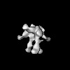
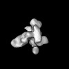
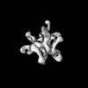
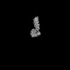
EMDB-53565:
Focussed map (bottom) MiDAC complex

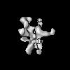
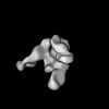
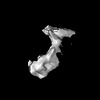
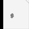
EMDB-53566:
Focussed map (middle) MiDAC complex

EMDB-53847:
Cryo-EM structure of human ATP citrate lyase in complex with inhibitor EVT0185-CoA

PDB-9r90:
Cryo-EM structure of human ATP citrate lyase in complex with inhibitor EVT0185-CoA

EMDB-53567:
An auto inhibitory loop in the MiDAC histone deacetylase complex

PDB-9r4i:
An auto inhibitory loop in the MiDAC histone deacetylase complex

EMDB-51786:
Cryo-EM Structure of human OAS2 Dimer

PDB-9h1z:
Cryo-EM Structure of human OAS2 Dimer
EMDB-43745:
SARS-CoV-2 M protein dimer in complex with JNJ-9676 and Fab-B

PDB-8w2e:
SARS-CoV-2 M protein dimer in complex with JNJ-9676 and Fab-B
EMDB-41869:
BG505.664 SOSIP in complex with polyclonal antibodies from NHP 8131 (gp120 glycan hole, gp41 glycan hole/fusion peptide and trimer base epitopes)
EMDB-41870:
BG505.664 Env SOSIP in complex with polyclonal antibodies from NHP 8131 (gp120-gp120 interface epitope)

EMDB-41871:
BG505.664 Env SOSIP in complex with polyclonal antibodies from NHP 8147 (C3/V5, V1/V2/V3 apex, gp41 glycan hole/fusion peptide and trimer base epitopes)
EMDB-41872:
BG505.664 Env SOSIP in complex with polyclonal antibodies from NHP 8147 (gp120 glycan hole epitope)
EMDB-41972:
GT1.1 SOSIP in complex with wk39 polyclonal antibodies from NHP A12N030 (CD4bs, C3V5 and base epitopes)
EMDB-41973:
GT1.1 SOSIP in complex with wk39 polyclonal antibodies from NHP A12N030 (gp41GH/FP and base epitopes)
EMDB-41974:
GT1.1 SOSIP in complex with wk39 polyclonal antibodies from NHP DC8G (gp41GH/FP and gp120GH epitopes)
EMDB-41975:
GT1.1 SOSIP in complex with wk39 polyclonal antibodies from NHP DC8G (gp120GH and base epitopes)
EMDB-41976:
GT1.1 SOSIP in complex with wk39 polyclonal antibodies from NHP DC8G (V1V2V3 and gp120GH epitopes)
EMDB-41977:
GT1.1 SOSIP-CC2 in complex with wk80 polyclonal antibodies from NHP 8229 (V1V2V3, C3V5, CD4bs, gp41GH/FP and base epitopes)
EMDB-41978:
GT1.1 SOSIP-CC2 in complex with wk80 polyclonal antibodies from NHP 8239 (gp120GH, gp41GH/FP, CD4bs, and base epitopes)

EMDB-50815:
Rea1 delta AAA2H2alpha ATPgS structure

EMDB-50816:
Rea1 D2915A-R2976A-D3042A mutant ATP conformation I
EMDB-50817:
Rea1 D2915A-R2976A-D3042A ATP conformation II
EMDB-50818:
Rea1 D2915A-R2976A-D3042A mutant ATP conformation III

EMDB-18702:
Endosomal membrane tethering complex CORVET, Vps8-Vps11 local refinement map

EMDB-18703:
Endosomal membrane tethering complex CORVET, Vps8 beta propeller local refinement map
EMDB-18704:
Endosomal membrane tethering complex CORVET, SNARE binding module local refinement map

EMDB-18705:
Endosomal membrane tethering complex CORVET, core local refinement map

EMDB-18706:
Endosomal membrane tethering complex CORVET, Vps18 beta propeller local refinement map

EMDB-18707:
Endosomal membrane tethering complex CORVET, consensus map

EMDB-18708:
Endosomal membrane tethering complex CORVET, Vps11deltaN mutant
EMDB-18560:
SARS-CoV-2 S protein bound to human neutralising antibody UZGENT_G5
EMDB-18571:
SARS-CoV-2 S protein bound to neutralising antibody UZGENT_A3

PDB-8qpr:
SARS-CoV-2 S protein bound to human neutralising antibody UZGENT_G5

PDB-8qq0:
SARS-CoV-2 S protein bound to neutralising antibody UZGENT_A3

EMDB-15677:
Cryo-EM structure for mouse leptin in complex with the mouse LEP-R ectodomain (1:2 mLEP:mLEPR model)

EMDB-15678:
Mouse leptin:LEP-R complex cryoEM structure (3:3 model)

EMDB-15679:
Cryo-EM structure for a 3:3 complex between mouse leptin and the mouse LEP-R ectodomain (local refinement)

EMDB-15680:
Human leptin in complex with the human LEP-R ectodomain fused to a C-terminal trimeric isoleucine GCN4 zipper (2:2 model)

EMDB-15681:
Human leptin in complex with the human LEP-R ectodomain fused to a C-terminal trimeric isoleucine GCN4 zipper (closed 3:3 model)

EMDB-15683:
Human leptin in complex with the human LEP-R ectodomain fused to a C-terminal trimeric isoleucine GCN4 zipper (open 3:3 model).

EMDB-15899:
Cryo-EM structure for the mouse LEPR-CRH2:Leptin:LEPR-Ig complex following symmetry expansion in combination with local refinement

PDB-8avb:
Cryo-EM structure for mouse leptin in complex with the mouse LEP-R ectodomain (1:2 mLEP:mLEPR model).

PDB-8avc:
Mouse leptin:LEP-R complex cryoEM structure (3:3 model)

PDB-8avd:
Cryo-EM structure for a 3:3 complex between mouse leptin and the mouse LEP-R ectodomain (local refinement)

PDB-8ave:
Human leptin in complex with the human LEP-R ectodomain fused to a C-terminal trimeric isoleucine GCN4 zipper (2:2 model)
PDB-8avf:
Human leptin in complex with the human LEP-R ectodomain fused to a C-terminal trimeric isoleucine GCN4 zipper (closed 3:3 model)
ページ:
 wwPDBはEMDBデータモデルのバージョン3へ移行します
