 Movie
Movie Controller
 Structure viewers
 About EMN search


-Search query
-Search result
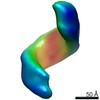
Showing 1 - 50 of 66 items for (author: klumpe & s)
EMDB-19643: 
Tomographic reconstruction of the follicle cell nuclear periphery from high pressure frozen, freeze substituted and resin embedded D. melanogaster egg chamber
EMDB-19644:
Tomographic reconstruction of the follicle cell nuclear periphery from high pressure frozen, freeze substituted and resin embedded D. melanogaster egg chamber
EMDB-19645:
Tomographic reconstruction of the follicle cell nuclear periphery from high pressure frozen, freeze substituted and resin embedded D. melanogaster egg chamber
EMDB-19646:
Tomographic reconstruction of the follicle cell nuclear periphery from high pressure frozen, freeze substituted and resin embedded D. melanogaster egg chamber
EMDB-19647:
Tomographic reconstruction of nuclear copia VLP clusters in D. melanogaster ovarian somatic cells
EMDB-19648:
Tomogram of the nuclear periphery of a follicle cell from lift-out experiments on D. melanogaster egg chambers
EMDB-19649:
In situ copia VLP cluster subtomogram averaging, C1-C1 contact map
EMDB-19650:
In situ copia VLP cluster subtomogram averaging, C1-C5 contact map
EMDB-19651:
In situ copia VLP cluster subtomogram averaging, C5-C5 contact map
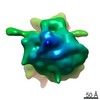
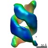
EMDB-19708:
The structure of the copia retrotransposon icosahedral capsid (T=9)
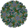
PDB-8s41:
The structure of the copia retrotransposon icosahedral capsid (T=9)

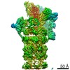
EMDB-17241:
C. elegans L1 80S ribosome

EMDB-17242:
C. elegans L1 larva 80S ribosome class 1

EMDB-17243:
C. elegans L1 larva 80S ribosome class 2

EMDB-17244:
C. elegans L1 larva 80S ribosome class 3

EMDB-17245:
C. elegans L1 larva 80S ribosome class 4

EMDB-17246:
C. elegans L1 larva body wall muscle tomogram

EMDB-17247:
C. elegans L1 larva ventral pharyngeal periphery tomogram

EMDB-17248:
Tomogram of the nucleus of a C. elegans L1 larva

EMDB-18186:
C. elegans L1 larva

EMDB-18187:
Focused refinment of 11-protofilament microtubule from C. elegans
EMDB-16537:
Optimizing Cryo-FIB Lamellas for sub-5 Angstrom in situ Structural Biology : Subtomogram average of the Large Subunit of S.Cerevisiae 80S Ribosome
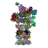
EMDB-14083:
26S proteasome WT-Ubp6-UbVS complex in the si state (ATPases, Rpn1, Ubp6, and UbVS)
PDB-7qo4:
26S proteasome WT-Ubp6-UbVS complex in the si state (ATPases, Rpn1, Ubp6, and UbVS)
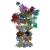
EMDB-14082:
Structure of the 26S proteasome-Ubp6 complex in the si state (Core Particle and Lid)

PDB-7qo3:
Structure of the 26S proteasome-Ubp6 complex in the si state (Core Particle and Lid)
EMDB-14084:
26S proteasome Rpt1-RK -Ubp6-UbVS complex in the si state
EMDB-14085:
26S proteasome Rpt1-RK -Ubp6-UbVS complex in the s2 state
PDB-7qo5:
26S proteasome Rpt1-RK -Ubp6-UbVS complex in the si state
PDB-7qo6:
26S proteasome Rpt1-RK -Ubp6-UbVS complex in the s2 state

EMDB-13878:
Cryo-electron tomogram from a cryo-FIB lift-out lamella of Drosophila melanogaster egg chambers

EMDB-13832:
Subtomogram average of 80S ribosomes from a cryo-FIB-lamella of Sum159 human cell line
EMDB-13833:
Cryo-electron tomograms from cryo-FIB-lamellae of Sum159 human cell line
EMDB-13834:
Subtomogram average of 80S ribosomes from a cryo-FIB-lamella of Sum159 human cell line prepared after cryo-FIB-SEM volume imaging

EMDB-13835:
Subtomogram average of 80S ribosomes a cryo-FIB-lamella of Sum159 human cell line prepared after cryo-FIB-SEM volume imaging
EMDB-13836:
Cryo-electron tomogram of a cryo-FIB lamella of a HeLa cell
EMDB-13837:
Cryo-electron tomogram of a cryo-FIB lamella of Emiliania huxleyi cells

EMDB-13838:
Cryo-electron tomogram of a 3D correlated lipid droplet in a cryo-FIB-milled HeLa cell

EMDB-13879:
Subtomogram average of D. melanogaster Ribosomes from tomograms collected on a cryo-lift-out lamella
EMDB-23060:
The stress-sensing domain of activated IRE1a forms helical filaments in narrow ER membrane tubes
EMDB-23058:
The stress-sensing domain of activated IRE1a forms helical filaments in narrow ER membrane tubes

EMDB-12324:
In situ cryo-electron tomogram of the Synechocystis wild-type VIPP1 strain grown in high light

EMDB-12325:
In situ cryo-electron tomogram of the Synechocystis F4E VIPP1 mutant grown in high light

EMDB-12326:
In situ cryo-electron tomogram of the Synechocystis V11E VIPP1 mutant grown in high light

EMDB-12327:
In situ cryo-electron tomogram of a cluster of VIPP1-mCherry structures inside the Chlamydomonas chloroplast

EMDB-12328:
In situ cryo-electron tomogram of a cluster of VIPP1-mCherry structures inside the Chlamydomonas chloroplast

EMDB-12329:
In situ cryo-electron tomogram of a cluster of VIPP1-mCherry structures inside the Chlamydomonas chloroplast

EMDB-12330:
In situ cryo-electron tomogram of a cluster of VIPP1-mCherry structures inside the Chlamydomonas chloroplast

EMDB-12710:
Structural basis for VIPP1 oligomerization and maintenance of thylakoid membrane integrity

EMDB-12711:
Structural basis for VIPP1 oligomerization and maintenance of thylakoid membrane integrity
Pages:
 wwPDB to switch to version 3 of the EMDB data model
