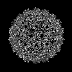
DNA polymerase complex / Hydrolases; Acting on peptide bonds (peptidases); Aspartic endopeptidases / DNA biosynthetic process / DNA integration / aspartic-type endopeptidase activity / nucleic acid binding / proteolysis / zinc ion binding / ATP binding Similarity search - Function
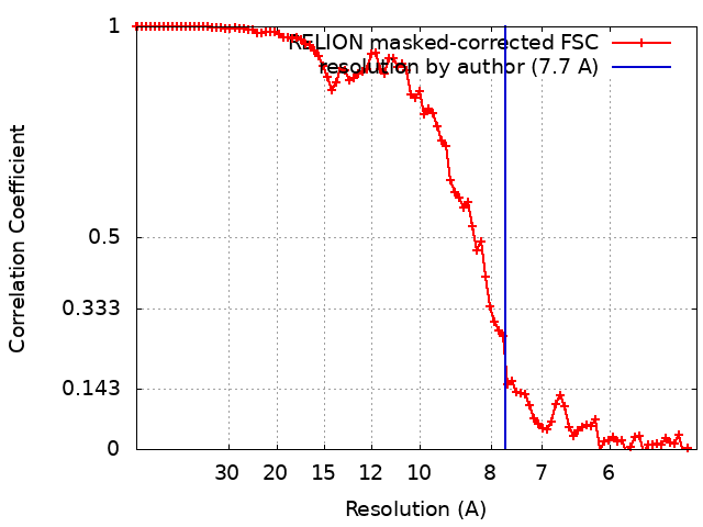
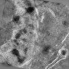
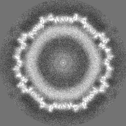
Journal: Cell / Year: 2025 Title: In-cell structure and snapshots of copia retrotransposons in intact tissue by cryo-ET. Authors: Sven Klumpe / Kirsten A Senti / Florian Beck / Jenny Sachweh / Bernhard Hampoelz / Paolo Ronchi / Viola Oorschot / Marlene Brandstetter / Assa Yeroslaviz / John A G Briggs / Julius Brennecke ...Authors: Sven Klumpe / Kirsten A Senti / Florian Beck / Jenny Sachweh / Bernhard Hampoelz / Paolo Ronchi / Viola Oorschot / Marlene Brandstetter / Assa Yeroslaviz / John A G Briggs / Julius Brennecke / Martin Beck / Jürgen M Plitzko / Abstract: Long terminal repeat (LTR) retrotransposons belong to the transposable elements (TEs), autonomously replicating genetic elements that integrate into the host's genome. Among animals, Drosophila ...Long terminal repeat (LTR) retrotransposons belong to the transposable elements (TEs), autonomously replicating genetic elements that integrate into the host's genome. Among animals, Drosophila melanogaster serves as an important model organism for TE research and contains several LTR retrotransposons, including the Ty1-copia family, which is evolutionarily related to retroviruses and forms virus-like particles (VLPs). In this study, we use cryo-focused ion beam (FIB) milling and lift-out approaches to visualize copia VLPs in ovarian cells and intact egg chambers, resolving the in situ copia capsid structure to 7.7 Å resolution by cryoelectron tomography (cryo-ET). Although cytoplasmic copia VLPs vary in size, nuclear VLPs are homogeneous and form densely packed clusters, supporting a model in which nuclear import acts as a size selector. Analyzing flies deficient in the TE-suppressing PIWI-interacting RNA (piRNA) pathway, we observe copia's translocation into the nucleus during spermatogenesis. Our findings provide insights into the replication cycle and cellular structural biology of an active LTR retrotransposon.
In the structure databanks used in Yorodumi, some data are registered as the other names, "COVID-19 virus" and "2019-nCoV". Here are the details of the virus and the list of structure data.
Jan 31, 2019. EMDB accession codes are about to change! (news from PDBe EMDB page)
EMDB accession codes are about to change! (news from PDBe EMDB page)
The allocation of 4 digits for EMDB accession codes will soon come to an end. Whilst these codes will remain in use, new EMDB accession codes will include an additional digit and will expand incrementally as the available range of codes is exhausted. The current 4-digit format prefixed with “EMD-” (i.e. EMD-XXXX) will advance to a 5-digit format (i.e. EMD-XXXXX), and so on. It is currently estimated that the 4-digit codes will be depleted around Spring 2019, at which point the 5-digit format will come into force.
The EM Navigator/Yorodumi systems omit the EMD- prefix.
Related info.:Q: What is EMD? / ID/Accession-code notation in Yorodumi/EM Navigator
Yorodumi is a browser for structure data from EMDB, PDB, SASBDB, etc.
This page is also the successor to EM Navigator detail page, and also detail information page/front-end page for Omokage search.
The word "yorodu" (or yorozu) is an old Japanese word meaning "ten thousand". "mi" (miru) is to see.
Related info.:EMDB / PDB / SASBDB / Comparison of 3 databanks / Yorodumi Search / Aug 31, 2016. New EM Navigator & Yorodumi / Yorodumi Papers / Jmol/JSmol / Function and homology information / Changes in new EM Navigator and Yorodumi
 Movie
Movie Controller

 Yorodumi
 Open data
 Basic information
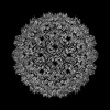
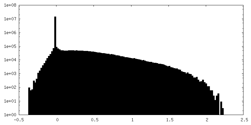
 Map data
 Sample
 Keywords
 Function and homology information
 Authors
 Germany, 1 items
  Citation
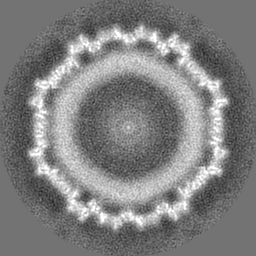
 Structure visualization
 Downloads & links
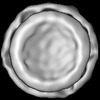
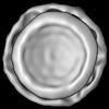
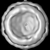
 emd_19708.png
 http://ftp.pdbj.org/pub/emdb/structures/EMD-19708
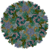

 Z (Sec.)
 Y (Row.)
 X (Col.)


 Sample components
 Processing
 Electron microscopy
 FIELD EMISSION GUN