- EMDB-13834: Subtomogram average of 80S ribosomes from a cryo-FIB-lamella of S... -
+
Open data
ID or keywords:
Loading...
-
Basic information
Entry
Database: EMDB / ID: EMD-13834
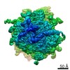
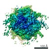
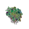
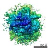
Title
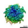
Subtomogram average of 80S ribosomes from a cryo-FIB-lamella of Sum159 human cell line prepared after cryo-FIB-SEM volume imaging
Map data
Subtomogram average of Sum159 ribosomes from 2 tomograms (TS_001.mrc, TS_005.mrc) obtained from a cryo-FIB-milled lamella after cryo-FIB-SEM volume imaging
Sample
Complex: 80S ribosome
Biological species
Homo sapiens (human)
Method
subtomogram averaging / cryo EM / Resolution: 24.0 Å
EIPOD fellowship under Marie Sklodowska-Curie Actions COFUND
664726
European Union
European Research Council (ERC)
760067
European Union
European Union (EU)
871037
European Union
Citation
Journal: Elife / Year: 2021 Title: A modular platform for automated cryo-FIB workflows. Authors: Sven Klumpe / Herman Kh Fung / Sara K Goetz / Ievgeniia Zagoriy / Bernhard Hampoelz / Xiaojie Zhang / Philipp S Erdmann / Janina Baumbach / Christoph W Müller / Martin Beck / Jürgen M ...Authors: Sven Klumpe / Herman Kh Fung / Sara K Goetz / Ievgeniia Zagoriy / Bernhard Hampoelz / Xiaojie Zhang / Philipp S Erdmann / Janina Baumbach / Christoph W Müller / Martin Beck / Jürgen M Plitzko / Julia Mahamid / Abstract: Lamella micromachining by focused ion beam milling at cryogenic temperature (cryo-FIB) has matured into a preparation method widely used for cellular cryo-electron tomography. Due to the limited ...Lamella micromachining by focused ion beam milling at cryogenic temperature (cryo-FIB) has matured into a preparation method widely used for cellular cryo-electron tomography. Due to the limited ablation rates of low Ga ion beam currents required to maintain the structural integrity of vitreous specimens, common preparation protocols are time-consuming and labor intensive. The improved stability of new-generation cryo-FIB instruments now enables automated operations. Here, we present an open-source software tool, SerialFIB, for creating automated and customizable cryo-FIB preparation protocols. The software encompasses a graphical user interface for easy execution of routine lamellae preparations, a scripting module compatible with available Python packages, and interfaces with three-dimensional correlative light and electron microscopy (CLEM) tools. SerialFIB enables the streamlining of advanced cryo-FIB protocols such as multi-modal imaging, CLEM-guided lamella preparation and in situ lamella lift-out procedures. Our software therefore provides a foundation for further development of advanced cryogenic imaging and sample preparation protocols.
History
Deposition
Nov 3, 2021
-
Header (metadata) release
Jan 12, 2022
-
Map release
Jan 12, 2022
-
Update
Jan 12, 2022
-
Current status
Jan 12, 2022
Processing site: PDBe / Status: Released
-
Structure visualization
Movie
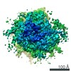
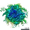
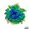
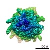
Surface view with section colored by density value
Download / File: emd_13834.map.gz / Format: CCP4 / Size: 10.5 MB / Type: IMAGE STORED AS FLOATING POINT NUMBER (4 BYTES)
Annotation
Subtomogram average of Sum159 ribosomes from 2 tomograms (TS_001.mrc, TS_005.mrc) obtained from a cryo-FIB-milled lamella after cryo-FIB-SEM volume imaging
In the structure databanks used in Yorodumi, some data are registered as the other names, "COVID-19 virus" and "2019-nCoV". Here are the details of the virus and the list of structure data.
Jan 31, 2019. EMDB accession codes are about to change! (news from PDBe EMDB page)
EMDB accession codes are about to change! (news from PDBe EMDB page)
The allocation of 4 digits for EMDB accession codes will soon come to an end. Whilst these codes will remain in use, new EMDB accession codes will include an additional digit and will expand incrementally as the available range of codes is exhausted. The current 4-digit format prefixed with “EMD-” (i.e. EMD-XXXX) will advance to a 5-digit format (i.e. EMD-XXXXX), and so on. It is currently estimated that the 4-digit codes will be depleted around Spring 2019, at which point the 5-digit format will come into force.
The EM Navigator/Yorodumi systems omit the EMD- prefix.
Related info.:Q: What is EMD? / ID/Accession-code notation in Yorodumi/EM Navigator
Yorodumi is a browser for structure data from EMDB, PDB, SASBDB, etc.
This page is also the successor to EM Navigator detail page, and also detail information page/front-end page for Omokage search.
The word "yorodu" (or yorozu) is an old Japanese word meaning "ten thousand". "mi" (miru) is to see.
Related info.:EMDB / PDB / SASBDB / Comparison of 3 databanks / Yorodumi Search / Aug 31, 2016. New EM Navigator & Yorodumi / Yorodumi Papers / Jmol/JSmol / Function and homology information / Changes in new EM Navigator and Yorodumi
 Movie
Movie Controller

 Yorodumi
 Open data
 Basic information
 Map data
 Sample
 Homo sapiens (human)
 Authors
 Citation
 Structure visualization
 Movie viewer
 Downloads & links
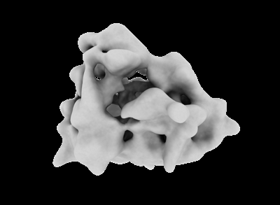 emd_13834.png
 http://ftp.pdbj.org/pub/emdb/structures/EMD-13834


 Z (Sec.)
 Y (Row.)
 X (Col.)


 Sample components
 Processing
 Electron microscopy
 FIELD EMISSION GUN

