 Movie
Movie Controller
 Structure viewers
 About EMN search


-Search query
-Search result
Showing 1 - 50 of 56 items for (author: kim & cy)
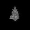
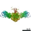
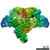
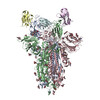
EMDB-42524: 
Cryo-EM Structure of Full-Length Spike Protein of Omicron XBB.1.5
Method: single particle / : Huynh KW, Chang JS, Fennell KF, Che Y, Wu H
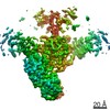
PDB-8usz:
Cryo-EM Structure of Full-Length Spike Protein of Omicron XBB.1.5
Method: single particle / : Huynh KW, Chang JS, Fennell KF, Che Y, Wu H
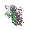
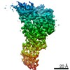
EMDB-28138:
3D reconstruction of the apical complex of Plasmodium falciparum (3D7) free merozoite
Method: electron tomography / : Segev-Zarko L, Sun SY, Kim CY
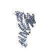
EMDB-28141:
3D reconstruction of the apical complex of Plasmodium falciparum (3D7) free merozoite
Method: electron tomography / : Segev-Zarko L, Sun SY, Kim CY

EMDB-28142:
3D Reconstruction of Plasmodium falciparum (3D7) free merozoite
Method: electron tomography / : Segev-Zarko L, Sun SY, Kim CY

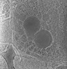
EMDB-28125:
Plasmodium falciparum merozoites apical 2-ring units
Method: subtomogram averaging / : Sun SY, Pintilie GD

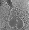
EMDB-28126:
Toxoplasma apical rings
Method: subtomogram averaging / : Sun SY, Pintilie GD
EMDB-28139:
Toxoplasma gondii apical complex (ionophore stimulated)
Method: electron tomography / : Segev-Zarko L, Sun SY, Kim CY, Egan ES, Chiu W, Boothroyd JC
EMDB-28140:
Toxoplasma gondii apical complex (non-stimulated)
Method: electron tomography / : Segev-Zarko L, Sun SY, Chiu W, Boothroyd JC

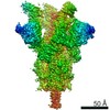
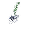
EMDB-27438:
Cryo-EM structure of SARS-CoV-2 Beta (B.1.351) spike protein in complex with VH domain F6
Method: single particle / : Zhu X, Saville JW, Mannar D, Berezuk AM, Subramaniam S

EMDB-27439:
Cryo-EM structure of SARS-CoV-2 Beta (B.1.351) spike protein in complex with VH domain F6 (focused refinement of RBD and VH F6)
Method: single particle / : Zhu X, Saville JW, Mannar D, Berezuk AM, Subramaniam S

PDB-8di5:
Cryo-EM structure of SARS-CoV-2 Beta (B.1.351) spike protein in complex with VH domain F6 (focused refinement of RBD and VH F6)
Method: single particle / : Zhu X, Saville JW, Mannar D, Berezuk AM, Subramaniam S
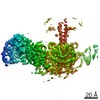
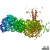
EMDB-24863:
CryoEM map of modular PKS holo-Lsd14 bound to antibody fragment 1B2, stalled at the condensation step, focused refinement of KS-KS'-ACP domains
Method: single particle / : Bagde SR, Kim CY, Fromme JC

EMDB-24862:
CryoEM structure of modular PKS holo-Lsd14 bound to antibody fragment 1B2, stalled at the condensation step, consensus refinement
Method: single particle / : Bagde SR, Kim CY, Fromme JC
EMDB-24864:
CryoEM map of modular PKS holo-Lsd14 bound to antibody fragment 1B2, stalled at the condensation step, focused refinement of LDAT domains
Method: single particle / : Bagde SR, Kim CY, Fromme JC

EMDB-24865:
CryoEM map of modular PKS holo-Lsd14 bound to antibody fragment 1B2, stalled at the condensation step, focused refinement of LD'AT' domains
Method: single particle / : Bagde SR, Kim CY, Fromme JC

EMDB-24866:
CryoEM map of modular PKS holo-Lsd14 bound to antibody fragment 1B2, stalled at the condensation step, focused refinement of KR domain
Method: single particle / : Bagde SR, Kim CY, Fromme JC

EMDB-24867:
CryoEM map of modular PKS holo-Lsd14 bound to antibody fragment 1B2, stalled at the condensation step, focused refinement of DD* and 1B2
Method: single particle / : Bagde SR, Kim CY, Fromme JC

EMDB-24869:
CryoEM map of modular PKS holo-Lsd14 bound to antibody fragment 1B2, consensus refinement
Method: single particle / : Bagde SR, Kim CY, Fromme JC
EMDB-24870:
CryoEM map of modular PKS holo-Lsd14 bound to antibody fragment 1B2, focused refinement of KSAT dimer
Method: single particle / : Bagde SR, Kim CY, Fromme JC
EMDB-24871:
CryoEM map of modular PKS holo-Lsd14 bound to antibody fragment 1B2, focused refinement of LDAT domains
Method: single particle / : Bagde SR, Kim CY, Fromme JC

EMDB-24872:
CryoEM map of modular PKS holo-Lsd14 bound to antibody fragment 1B2, focused refinement of LD'AT' domains
Method: single particle / : Bagde SR, Kim CY, Fromme JC
EMDB-24873:
CryoEM map of modular PKS holo-Lsd14 bound to antibody fragment 1B2, focused refinement of KR domain
Method: single particle / : Bagde SR, Kim CY, Fromme JC
EMDB-24874:
CryoEM map of modular PKS holo-Lsd14 bound to antibody fragment 1B2, focused refinement of DD* and 1B2
Method: single particle / : Bagde SR, Kim CY, Fromme JC
EMDB-24880:
CryoEM map of modular PKS apo-Lsd14 bound to antibody fragment 1B2, consensus refinement
Method: single particle / : Bagde SR, Kim CY, Fromme JC

EMDB-24868:
CryoEM structure of modular PKS holo-Lsd14 stalled at the condensation step and bound to antibody fragment 1B2, composite structure
Method: single particle / : Bagde SR, Kim CY, Fromme JC

EMDB-24875:
CryoEM structure of modular PKS holo-Lsd14 bound to antibody fragment 1B2, composite structure
Method: single particle / : Bagde SR, Kim CY, Fromme JC
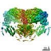
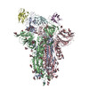
EMDB-11616:
Cryo-EM structure of the SARS-CoV-2 spike protein bound to neutralizing sybodies (Sb23)
Method: single particle / : Hallberg BM, Das H
PDB-7a25:
Cryo-EM structure of the SARS-CoV-2 spike protein bound to neutralizing sybodies (Sb23)
Method: single particle / : Hallberg BM, Das H
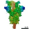
EMDB-11617:
Cryo-EM structure of the SARS-CoV-2 spike protein bound to neutralizing sybodies (Sb23) 2-up conformation
Method: single particle / : Hallberg BM, Das H
PDB-7a29:
Cryo-EM structure of the SARS-CoV-2 spike protein bound to neutralizing sybodies (Sb23) 2-up conformation
Method: single particle / : Hallberg BM, Das H
EMDB-22829:
Human Tom70 in complex with SARS CoV2 Orf9b
Method: single particle / : QCRG Structural Biology Consortium
PDB-7kdt:
Human Tom70 in complex with SARS CoV2 Orf9b
Method: single particle / : QCRG Structural Biology Consortium

EMDB-11002:
CryoEM structure of bovine cytochrome bc1 in complex with a tetrahydroquinolone inhibitor
Method: single particle / : Muench SP, Johnson R, Amporndanai K, Atonyuk S

EMDB-0554:
Cryo-EM Structure of the Lysosomal Folliculin Complex (FLCN-FNIP2-RagA-RagC-Ragulator)
Method: single particle / : Fromm SA, Young LN, Hurley JH

EMDB-0556:
Cryo-EM Map of the active Ragulator-RagA-RagC Complex
Method: single particle / : Yokom AL, Fromm SA, Hurley JH
PDB-5ty4:
MicroED structure of a complex between monomeric TGF-b and its receptor, TbRII, at 2.9 A resolution
Method: electron crystallography / : Weiss SC, de la Cruz MJ, Hattne J, Shi D, Reyes FE, Callero G, Gonen T
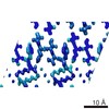
PDB-5k7n:
MicroED structure of tau VQIVYK peptide at 1.1 A resolution
Method: electron crystallography / : de la Cruz MJ, Hattne J, Shi D, Seidler P, Rodriguez J, Reyes FE, Sawaya MR, Cascio D, Eisenberg D, Gonen T
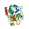
PDB-5k7o:
MicroED structure of lysozyme at 1.8 A resolution
Method: electron crystallography / : de la Cruz MJ, Hattne J, Shi D, Seidler P, Rodriguez J, Reyes FE, Sawaya MR, Cascio D, Eisenberg D, Gonen T
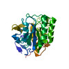
PDB-5k7p:
MicroED structure of xylanase at 2.3 A resolution
Method: electron crystallography / : de la Cruz MJ, Hattne J, Shi D, Seidler P, Rodriguez J, Reyes FE, Sawaya MR, Cascio D, Eisenberg D, Gonen T
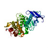
PDB-5k7q:
MicroED structure of thaumatin at 2.5 A resolution
Method: electron crystallography / : de la Cruz MJ, Hattne J, Shi D, Seidler P, Rodriguez J, Reyes FE, Sawaya MR, Cascio D, Eisenberg D, Gonen T
PDB-5k7r:
MicroED structure of trypsin at 1.7 A resolution
Method: electron crystallography / : de la Cruz MJ, Hattne J, Shi D, Seidler P, Rodriguez J, Reyes FE, Sawaya MR, Cascio D, Eisenberg D, Gonen T
PDB-5k7s:
MicroED structure of proteinase K at 1.6 A resolution
Method: electron crystallography / : de la Cruz MJ, Hattne J, Shi D, Seidler P, Rodriguez J, Reyes FE, Sawaya MR, Cascio D, Eisenberg D, Gonen T
PDB-5k7t:
MicroED structure of thermolysin at 2.5 A resolution
Method: electron crystallography / : de la Cruz MJ, Hattne J, Shi D, Seidler P, Rodriguez J, Reyes FE, Sawaya MR, Cascio D, Eisenberg D, Gonen T
EMDB-8216:
MicroED structure of tau VQIVYK peptide at 1.1 A resolution
Method: electron crystallography / : de la Cruz MJ, Hattne J
EMDB-8217:
MicroED structure of lysozyme at 1.8 A resolution
Method: electron crystallography / : de la Cruz MJ, Hattne J
EMDB-8218:
MicroED structure of xylanase at 2.3 A resolution
Method: electron crystallography / : de la Cruz MJ, Hattne J
EMDB-8219:
MicroED structure of thaumatin at 2.5 A resolution
Method: electron crystallography / : de la Cruz MJ, Hattne J

EMDB-8220:
MicroED structure of trypsin at 1.7 A resolution
Method: electron crystallography / : de la Cruz MJ, Hattne J
EMDB-8221:
MicroED structure of proteinase K at 1.6 A resolution
Method: electron crystallography / : de la Cruz MJ, Hattne J
Pages:
 wwPDB to switch to version 3 of the EMDB data model
