 ムービー
ムービー コントローラー
 構造ビューア
 EMN検索について


-検索条件
-検索結果
検索 (著者・登録者: hubert & a)の結果84件中、1から50件目までを表示しています
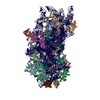
EMDB-18892: 
Lipid droplet-Vacuole contacts in Ldo16 overexpression yeast strain.
EMDB-18893:
Lipid droplet-vacuole and Nucleus-vacuole contacts in WT yeast cell starved for 4 hours
EMDB-18894:
Lipid droplet lipophagy in 4-hour starved WT yeast cell.
EMDB-18895:
Multiple vacuole-lipid droplet-nucleus contacts in 4-hour starved WT yeast cell.
EMDB-18896:
Lipophagy in 5-day starved WT yeast cell.
EMDB-18897:
Lipid droplets in proximity to a vacuole in dLdo strain cell after 5-day starvation.
EMDB-18898:
Vacuolar contents of WT cell after 5-day starvation.

EMDB-18899:
Lipid droplet-nucleus contacts in dLdo yeast strain after 5-day starvation.
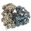
EMDB-17804:
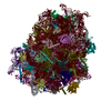
Bat-Hp-CoV Nsp1 and eIF1 bound to the human 40S small ribosomal subunit

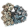
EMDB-17805:
MERS-CoV Nsp1 bound to the human 43S pre-initiation complex
PDB-8ppk:
Bat-Hp-CoV Nsp1 and eIF1 bound to the human 40S small ribosomal subunit

PDB-8ppl:
MERS-CoV Nsp1 bound to the human 43S pre-initiation complex
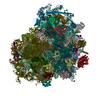
EMDB-33329:
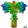
High resolution cry-EM structure of the human 80S ribosome from SNORD127+/+ Kasumi-1 cells
EMDB-33330:
High resolution cry-EM structure of the human 80S ribosome from SNORD127+/- Kasumi-1 cells
PDB-7xnx:
High resolution cry-EM structure of the human 80S ribosome from SNORD127+/+ Kasumi-1 cells
PDB-7xny:
High resolution cry-EM structure of the human 80S ribosome from SNORD127+/- Kasumi-1 cells

EMDB-13404:
Nanodisc reconstituted MsbA in complex with nanobodies, spin-labeled at position A60C

EMDB-13405:
AMP-PNP bound nanodisc reconstituted MsbA with nanobodies, spin-labeled at position A60C

EMDB-13406:
AMP-PNP bound nanodisc reconstituted MsbA with nanobodies, spin-labeled at position T68C

EMDB-13409:
Nanodisc reconstituted MsbA in complex with nanobodies, spin-labeled at position T68C

PDB-7ph2:
Nanodisc reconstituted MsbA in complex with nanobodies, spin-labeled at position A60C

PDB-7ph3:
AMP-PNP bound nanodisc reconstituted MsbA with nanobodies, spin-labeled at position A60C

PDB-7ph4:
AMP-PNP bound nanodisc reconstituted MsbA with nanobodies, spin-labeled at position T68C

PDB-7ph7:
Nanodisc reconstituted MsbA in complex with nanobodies, spin-labeled at position T68C

EMDB-23949:
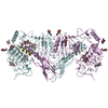
The insulin receptor ectodomain in complex with a venom hybrid insulin analog - "head" region
EMDB-23950:
The insulin receptor ectodomain in complex with four venom hybrid insulins - symmetric conformation
EMDB-23951:
The insulin receptor ectodomain in complex with three venom hybrid insulin molecules - asymmetric conformation
PDB-7mqo:
The insulin receptor ectodomain in complex with a venom hybrid insulin analog - "head" region
PDB-7mqr:
The insulin receptor ectodomain in complex with four venom hybrid insulins - symmetric conformation
PDB-7mqs:
The insulin receptor ectodomain in complex with three venom hybrid insulin molecules - asymmetric conformation

EMDB-25677:
Structure of dimeric phosphorylated Pediculus humanus (Ph) PINK1
EMDB-25678:
Structure of dimeric phosphorylated Pediculus humanus (Ph) PINK1 with kinked alpha-C helix in chain B

EMDB-25679:
Structure of dimeric phosphorylated Pediculus humanus (Ph) PINK1 with extended alpha-C helix in chain B

EMDB-25680:
Structure of dodecameric unphosphorylated Pediculus humanus (Ph) PINK1 D357A mutant

EMDB-25681:
Structure of dimeric unphosphorylated Pediculus humanus (Ph) PINK1 D357A mutant

PDB-7t4k:
Structure of dimeric phosphorylated Pediculus humanus (Ph) PINK1 with kinked alpha-C helix in chain B

PDB-7t4l:
Structure of dimeric phosphorylated Pediculus humanus (Ph) PINK1 with extended alpha-C helix in chain B

PDB-7t4m:
Structure of dodecameric unphosphorylated Pediculus humanus (Ph) PINK1 D357A mutant

PDB-7t4n:
Structure of dimeric unphosphorylated Pediculus humanus (Ph) PINK1 D357A mutant

EMDB-13246:
cryoFIB milling/cryoET of amoeba infected with Legionella pneumophila

EMDB-13247:
cryoFIB milling/cryoET of amoeba infected with Legionella pneumophila

EMDB-13248:
cryoFIB milling/cryoET of amoeba infected with Legionella pneumophila

EMDB-13249:
cryoFIB milling/cryoET of amoeba infected with Legionella pneumophila

EMDB-13187:
Homology model of the full-length AP-3 complex in a compact open conformation

EMDB-13188:
Homology model of the full-length AP-3 complex in an intermediate open conformation

EMDB-13189:
Homology model of the full-length AP-3 complex in a stretched open conformation

PDB-7p3x:
Homology model of the full-length AP-3 complex in a compact open conformation

PDB-7p3y:
Homology model of the full-length AP-3 complex in an intermediate open conformation

PDB-7p3z:
Homology model of the full-length AP-3 complex in a stretched open conformation

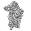
EMDB-12568:
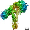
55S mammalian mitochondrial ribosome with mtRRF (pre) and tRNA(P/E)
ページ:
 wwPDBはEMDBデータモデルのバージョン3へ移行します
