 Movie
Movie Controller

[English] 日本語
 Yorodumi
- PDB-7ph3: AMP-PNP bound nanodisc reconstituted MsbA with nanobodies, spin-l... -
+ Open data

- Basic information
| Entry | Database: PDB / ID: 7ph3 | ||||||||||||
|---|---|---|---|---|---|---|---|---|---|---|---|---|---|
| Title | AMP-PNP bound nanodisc reconstituted MsbA with nanobodies, spin-labeled at position A60C | ||||||||||||
 Components Components |
| ||||||||||||
 Keywords Keywords | MEMBRANE PROTEIN / ABC transporter / nanobody / AMP-PNP / Gd-DOTA | ||||||||||||
| Function / homology |  Function and homology information Function and homology informationMsbA transporter complex / lipopolysaccharide floppase activity / lipid translocation / ABC-type lipid A-core oligosaccharide transporter / lipopolysaccharide transport / ATPase-coupled lipid transmembrane transporter activity / ABC-type xenobiotic transporter activity / lipid transport / ATP-binding cassette (ABC) transporter complex / transmembrane transport ...MsbA transporter complex / lipopolysaccharide floppase activity / lipid translocation / ABC-type lipid A-core oligosaccharide transporter / lipopolysaccharide transport / ATPase-coupled lipid transmembrane transporter activity / ABC-type xenobiotic transporter activity / lipid transport / ATP-binding cassette (ABC) transporter complex / transmembrane transport / lipid binding / ATP hydrolysis activity / ATP binding / membrane / identical protein binding / plasma membrane Similarity search - Function | ||||||||||||
| Biological species |   | ||||||||||||
| Method | ELECTRON MICROSCOPY / single particle reconstruction / cryo EM / Resolution: 2.8 Å | ||||||||||||
 Authors Authors | Parey, K. / Januliene, D. / Galazzo, L. / Meier, G. / Vecchis, D. / Striednig, B. / Hilbi, H. / Schaefer, L.V. / Kuprov, I. / Bordignon, E. ...Parey, K. / Januliene, D. / Galazzo, L. / Meier, G. / Vecchis, D. / Striednig, B. / Hilbi, H. / Schaefer, L.V. / Kuprov, I. / Bordignon, E. / Seeger, M.A. / Moeller, A. | ||||||||||||
| Funding support |  Germany, 3items Germany, 3items
| ||||||||||||
 Citation Citation | Journal: Sci Adv / Year: 2022 Title: The ABC transporter MsbA adopts the wide inward-open conformation in cells. Authors: Laura Galazzo / Gianmarco Meier / Dovile Januliene / Kristian Parey / Dario De Vecchis / Bianca Striednig / Hubert Hilbi / Lars V Schäfer / Ilya Kuprov / Arne Moeller / Enrica Bordignon / Markus A Seeger /   Abstract: Membrane proteins are currently investigated after detergent extraction from native cellular membranes and reconstitution into artificial liposomes or nanodiscs, thereby removing them from their ...Membrane proteins are currently investigated after detergent extraction from native cellular membranes and reconstitution into artificial liposomes or nanodiscs, thereby removing them from their physiological environment. However, to truly understand the biophysical properties of membrane proteins in a physiological environment, they must be investigated within living cells. Here, we used a spin-labeled nanobody to interrogate the conformational cycle of the ABC transporter MsbA by double electron-electron resonance. Unexpectedly, the wide inward-open conformation of MsbA, commonly considered a nonphysiological state, was found to be prominently populated in cells. Molecular dynamics simulations revealed that extensive lateral portal opening is essential to provide access of its large natural substrate core lipid A to the binding cavity. Our work paves the way to investigate the conformational landscape of membrane proteins in cells. | ||||||||||||
| History |
|
- Structure visualization
| Structure viewer | Molecule: MolmilJmol/JSmol |
|---|
- Downloads & links
-Download
| PDBx/mmCIF format | 7ph3.cif.gz | 256.3 KB | Display | PDBx/mmCIF format |
|---|---|---|---|---|
| PDB format | pdb7ph3.ent.gz | 203.3 KB | Display | PDB format |
| PDBx/mmJSON format | 7ph3.json.gz | Tree view | PDBx/mmJSON format | |
| Others |  Other downloads Other downloads |
-Validation report
| Arichive directory | https://data.pdbj.org/pub/pdb/validation_reports/ph/7ph3ftp://data.pdbj.org/pub/pdb/validation_reports/ph/7ph3 | HTTPS FTP |
|---|
-Related structure data
| Related structure data |  13405MC  7ndfC  7ph2C  7ph4C  7ph7C M: map data used to model this data C: citing same article ( |
|---|---|
| Similar structure data |


-Links
 PDBj
PDBj


- Assembly
| Deposited unit | 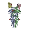
|
|---|---|
| 1 |
|
-Components
-Protein / Antibody / Sugars , 3 types, 6 molecules ABCD
| #1: Protein | Mass: 65760.852 Da / Num. of mol.: 2 Source method: isolated from a genetically manipulated source Source: (gene. exp.) References: UniProt: P60752, ABC-type lipid A-core oligosaccharide transporter #2: Antibody | Mass: 12440.957 Da / Num. of mol.: 2 Source method: isolated from a genetically manipulated source Source: (gene. exp.) #5: Sugar |  Type: D-saccharide / Mass: 510.615 Da / Num. of mol.: 2 / Source method: obtained synthetically / Formula: C24H46O11 / Comment: detergent*YM Type: D-saccharide / Mass: 510.615 Da / Num. of mol.: 2 / Source method: obtained synthetically / Formula: C24H46O11 / Comment: detergent*YM |
|---|
-Non-polymers , 6 types, 14 molecules 


| #3: Chemical |  Mass: 506.196 Da / Num. of mol.: 2 / Source method: obtained synthetically / Formula: C10H17N6O12P3 / Comment: AMP-PNP, energy-carrying molecule analogue*YM Mass: 506.196 Da / Num. of mol.: 2 / Source method: obtained synthetically / Formula: C10H17N6O12P3 / Comment: AMP-PNP, energy-carrying molecule analogue*YM#4: Chemical |  Mass: 24.305 Da / Num. of mol.: 2 / Source method: obtained synthetically / Formula: Mg Mass: 24.305 Da / Num. of mol.: 2 / Source method: obtained synthetically / Formula: Mg#6: Chemical |  Mass: 748.065 Da / Num. of mol.: 2 / Source method: obtained synthetically / Formula: C41H82NO8P / Comment: phospholipid*YM Mass: 748.065 Da / Num. of mol.: 2 / Source method: obtained synthetically / Formula: C41H82NO8P / Comment: phospholipid*YM#7: Chemical |  Mass: 524.524 Da / Num. of mol.: 2 / Source method: obtained synthetically / Formula: C22H32N6O9 / Feature type: SUBJECT OF INVESTIGATION Mass: 524.524 Da / Num. of mol.: 2 / Source method: obtained synthetically / Formula: C22H32N6O9 / Feature type: SUBJECT OF INVESTIGATION#8: Chemical |  Mass: 157.250 Da / Num. of mol.: 2 / Source method: obtained synthetically / Formula: Gd / Feature type: SUBJECT OF INVESTIGATION Mass: 157.250 Da / Num. of mol.: 2 / Source method: obtained synthetically / Formula: Gd / Feature type: SUBJECT OF INVESTIGATION#9: Water | ChemComp-HOH / | Mass: 18.015 Da / Num. of mol.: 4 / Source method: isolated from a natural source / Formula: H2O |
|---|
-Details
| Has ligand of interest | Y |
|---|---|
| Has protein modification | Y |
-Experimental details
-Experiment
| Experiment | Method: ELECTRON MICROSCOPY |
|---|---|
| EM experiment | Aggregation state: PARTICLE / 3D reconstruction method: single particle reconstruction |
- Sample preparation
| Component | Name: E. coli MsbA in complex with nanobody Nb_MsbA#1, labeled with Gd-DOTA at Cys60 Type: COMPLEX / Entity ID: #1-#2 / Source: RECOMBINANT |
|---|---|
| Source (natural) | Organism: |
| Source (recombinant) | Organism: |
| Buffer solution | pH: 7.5 |
| Specimen | Conc.: 2.6 mg/ml / Embedding applied: NO / Shadowing applied: NO / Staining applied: NO / Vitrification applied: YES |
| Vitrification | Cryogen name: ETHANE |
- Electron microscopy imaging
| Experimental equipment |  Model: Titan Krios / Image courtesy: FEI Company |
|---|---|
| Microscopy | Model: FEI TITAN KRIOS |
| Electron gun | Electron source:  FIELD EMISSION GUN / Accelerating voltage: 300 kV / Illumination mode: FLOOD BEAM FIELD EMISSION GUN / Accelerating voltage: 300 kV / Illumination mode: FLOOD BEAM |
| Electron lens | Mode: BRIGHT FIELD / Nominal defocus max: 2500 nm / Nominal defocus min: 800 nm |
| Image recording | Electron dose: 50 e/Å2 / Film or detector model: GATAN K3 BIOQUANTUM (6k x 4k) |
- Processing
| EM software |
| ||||||||||||||||
|---|---|---|---|---|---|---|---|---|---|---|---|---|---|---|---|---|---|
| CTF correction | Type: PHASE FLIPPING AND AMPLITUDE CORRECTION | ||||||||||||||||
| Symmetry | Point symmetry: C2 (2 fold cyclic) | ||||||||||||||||
| 3D reconstruction | Resolution: 2.8 Å / Resolution method: FSC 0.143 CUT-OFF / Num. of particles: 109465 / Symmetry type: POINT | ||||||||||||||||
| Refinement | Highest resolution: 2.8 Å |
