 ムービー
ムービー コントローラー
 構造ビューア
 EMN検索について


-検索条件
-検索結果
検索 (著者・登録者: el & omari & k)の結果全47件を表示しています
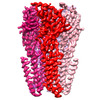
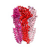
EMDB-16005: 
GABA-A receptor a5 homomer - a5V3 - APO
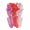
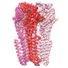
EMDB-16050:
GABA-A receptor a5 homomer - a5V3 - Basmisanil - HR

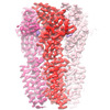
EMDB-16051:
GABA-A receptor a5 homomer - a5V3 - RO154513

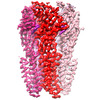
EMDB-16055:
GABA-A receptor a5 homomer - a5V3 - RO5211223
EMDB-16058:
GABA-A receptor a5 homomer - a5V3 - Diazepam

EMDB-16060:
GABA-A receptor a5 homomer - a5V3 - DMCM

EMDB-16063:
GABA-A receptor a5 homomer - a5V3 - L655708
EMDB-16066:
GABA-A receptor a5 homomer - a5V3 - RO7172670
EMDB-16067:
GABA-A receptor a5 homomer - a5V3 - RO7015738
EMDB-16068:
GABA-A receptor a5 homomer - a5V3 - RO4938581

PDB-8bej:
GABA-A receptor a5 homomer - a5V3 - APO

PDB-8bha:
GABA-A receptor a5 homomer - a5V3 - Basmisanil - HR

PDB-8bhb:
GABA-A receptor a5 homomer - a5V3 - RO154513

PDB-8bhi:
GABA-A receptor a5 homomer - a5V3 - RO5211223

PDB-8bhk:
GABA-A receptor a5 homomer - a5V3 - Diazepam

PDB-8bhm:
GABA-A receptor a5 homomer - a5V3 - DMCM

PDB-8bho:
GABA-A receptor a5 homomer - a5V3 - L655708

PDB-8bhq:
GABA-A receptor a5 homomer - a5V3 - RO7172670

PDB-8bhr:
GABA-A receptor a5 homomer - a5V3 - RO7015738

PDB-8bhs:
GABA-A receptor a5 homomer - a5V3 - RO4938581
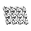
EMDB-13957:
Extended H/L (SLPH/SLPL) complex from C. difficile (CD630 strain) fit into R20291 S-layer negative stain map

PDB-7qgq:
Extended H/L (SLPH/SLPL) complex from C. difficile (CD630 strain) fit into R20291 S-layer negative stain map

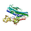
EMDB-13355:
Structure of the Caulobacter crescentus S-layer protein RsaA N-terminal domain bound to LPS and soaked with Holmium

PDB-7peo:
Structure of the Caulobacter crescentus S-layer protein RsaA N-terminal domain bound to LPS and soaked with Holmium

PDB-6rvd:
Revised cryo-EM structure of the human 2:1 Ptch1-Shh complex
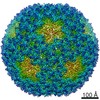
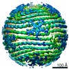
EMDB-10289:
Bacteriophage phi6 dsRNA genome, layer 1, conformation pseudo C2
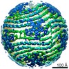
EMDB-10075:
Bacteriophage phi6 dsRNA genome, layer 1, conformation pseudo D3
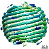
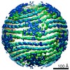
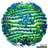
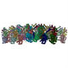
EMDB-0294:
Bacteriophage phi6 dsRNA genome, layer 2
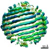
EMDB-0295:
Bacteriophage phi6 dsRNA genome, layer 3
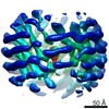
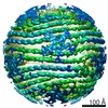
EMDB-0296:
Bacteriophage phi6 dsRNA genome, layers 4 and 5
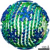
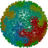
EMDB-0299:
Bacteriophage phi6 nucleocapsid reconstructed with icosahedral symmetry
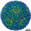
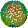
EMDB-0300:
Reconstruction of dsRNA bacteriophage phi6 nucleocapsid with D3 symmetry
EMDB-0302:
Bacteriophage phi6 dsRNA genome, layer 1, conformation pseudo D3'
EMDB-0304:
Bacteriophage phi6 dsRNA genome, layer 1, conformation pseudo D3', sub-conformation 1
EMDB-0305:
Bacteriophage phi6 dsRNA genome, layer 1, conformation pseudo D3', sub-conformation 2
EMDB-0306:
Bacteriophage phi6 dsRNA genome, layer 1, conformation pseudo D3', sub-conformation 3

PDB-6hy0:
Atomic models of P1, P4 C-terminal fragment and P8 fitted in the bacteriophage phi6 nucleocapsid reconstructed with icosahedral symmetry
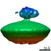
EMDB-9779:
Reconstruction of HRPV6 VP5 spike
PDB-6j7v:
Structure of HRPV6 VP5 fitted in the cryoEM density of the spike
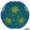
EMDB-3571:
dsRNA bacteriophage phi6 nucleocapsid

EMDB-3572:
Localized reconstruction of bacteriophage phi6 packaging hexamer P4

EMDB-3573:
Localized reconstruction of bacteriophage phi6 vertex
PDB-5muu:
dsRNA bacteriophage phi6 nucleocapsid

PDB-5muv:
Atomic structure fitted into a localized reconstruction of bacteriophage phi6 packaging hexamer P4

PDB-5muw:
Atomic structure of P4 packaging enzyme fitted into a localized reconstruction of bacteriophage phi6 vertex
PDB-5fki:
Pseudorabies virus (PrV) nuclear egress complex proteins fitted as a hexameric lattice into a sub-tomogram average derived from focused- ion beam milled lamellae electron cryo-microscopic data
PDB-4bx4:
Fitting of the bacteriophage Phi8 P1 capsid protein into cryo-EM density
 wwPDBはEMDBデータモデルのバージョン3へ移行します
