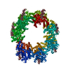
[English] 日本語
Yorodumi
- PDB-7peo: Structure of the Caulobacter crescentus S-layer protein RsaA N-te... -

+
Open data


ID or keywords:

Loading...

-
Basic information

Entry
Database: PDB / ID: 7peo
TitleStructure of the Caulobacter crescentus S-layer protein RsaA N-terminal domain bound to LPS and soaked with Holmium
ComponentsS-layer protein
KeywordsSTRUCTURAL PROTEIN / S-layer protein RsaA bound to LPS and Holmium
Function / homologyRsaA N-terminal domain / S-layer / RTX calcium-binding nonapeptide repeat / RTX calcium-binding nonapeptide repeat (4 copies) / Serralysin-like metalloprotease, C-terminal / calcium ion binding / extracellular region / HOLMIUM ATOM / S-layer protein
Function and homology information
Biological speciesCaulobacter vibrioides (bacteria)
MethodELECTRON MICROSCOPY / single particle reconstruction / cryo EM / Resolution: 4.37 Å
Authorsvon Kugelgen, A. / Bharat, T.A.M.
Funding support United Kingdom, 2items
OrganizationGrant numberCountry
Wellcome Trust202231/Z/16/Z United Kingdom
Leverhulme TrustPhilip Leverhulme Prize United Kingdom
CitationJournal: Structure / Year: 2022
Title: High-resolution mapping of metal ions reveals principles of surface layer assembly in Caulobacter crescentus cells.
Authors: Matthew Herdman / Andriko von Kügelgen / Danguole Kureisaite-Ciziene / Ramona Duman / Kamel El Omari / Elspeth F Garman / Andreas Kjaer / Dimitrios Kolokouris / Jan Löwe / Armin Wagner / ...Authors: Matthew Herdman / Andriko von Kügelgen / Danguole Kureisaite-Ciziene / Ramona Duman / Kamel El Omari / Elspeth F Garman / Andreas Kjaer / Dimitrios Kolokouris / Jan Löwe / Armin Wagner / Phillip J Stansfeld / Tanmay A M Bharat /
Abstract: Surface layers (S-layers) are proteinaceous crystalline coats that constitute the outermost component of most prokaryotic cell envelopes. In this study, we have investigated the role of metal ions in ...Surface layers (S-layers) are proteinaceous crystalline coats that constitute the outermost component of most prokaryotic cell envelopes. In this study, we have investigated the role of metal ions in the formation of the Caulobacter crescentus S-layer using high-resolution structural and cell biology techniques, as well as molecular simulations. Utilizing optical microscopy of fluorescently tagged S-layers, we show that calcium ions facilitate S-layer lattice formation and cell-surface binding. We report all-atom molecular dynamics simulations of the S-layer lattice, revealing the importance of bound metal ions. Finally, using electron cryomicroscopy and long-wavelength X-ray diffraction experiments, we mapped the positions of metal ions in the S-layer at near-atomic resolution, supporting our insights from the cellular and simulations data. Our findings contribute to the understanding of how C. crescentus cells form a regularly arranged S-layer on their surface, with implications on fundamental S-layer biology and the synthetic biology of self-assembling biomaterials.
History
DepositionAug 11, 2021Deposition site: PDBE / Processing site: PDBE
Revision 1.0Dec 1, 2021Provider: repository / Type: Initial release
Revision 1.1Feb 16, 2022Group: Database references / Derived calculations / Category: citation / pdbx_struct_oper_list
Item: _citation.journal_volume / _citation.page_first ..._citation.journal_volume / _citation.page_first / _citation.year / _pdbx_struct_oper_list.name / _pdbx_struct_oper_list.symmetry_operation

-
Structure visualization

Movie
  • Deposited structure unit
  • Imaged by Jmol
  • Download
  • Simplified surface model + fitted atomic model
  • EMDB-13355
  • Imaged by Jmol
  • Download
  • Superimposition on EM map
  • EMDB-13355
  • Imaged by UCSF Chimera
  • Download
Movie viewer
Structure viewerMolecule:
MolmilJmol/JSmol

Downloads & links

-
Assembly

Deposited unit
A: S-layer protein
hetero molecules


Theoretical massNumber of molelcules
Total (without water)28,2305
Polymers25,8201
Non-polymers2,4094
Water0
1
A: S-layer protein
hetero molecules
x 14


Theoretical massNumber of molelcules
Total (without water)395,21470
Polymers361,48514
Non-polymers33,72956
Water0
TypeNameSymmetry operationNumber
identity operation1_555x,y,z1
point symmetry operation13
Noncrystallographic symmetry (NCS)NCS domain:
IDEns-IDDetails
11A
21C
12A
22D
13A
23E
14A
24F
15A
25G
16A
26H
17A
27I
18A
28J
19A
29K
110A
210L
111A
211M
112A
212N
113A
213O
114C
214D
115C
215E
116C
216F
117C
217G
118C
218H
119C
219I
120C
220J
121C
221K
122C
222L
123C
223M
124C
224N
125C
225O
126D
226E
127D
227F
128D
228G
129D
229H
130D
230I
131D
231J
132D
232K
133D
233L
134D
234M
135D
235N
136D
236O
137E
237F
138E
238G
139E
239H
140E
240I
141E
241J
142E
242K
143E
243L
144E
244M
145E
245N
146E
246O
147F
247G
148F
248H
149F
249I
150F
250J
151F
251K
152F
252L
153F
253M
154F
254N
155F
255O
156G
256H
157G
257I
158G
258J
159G
259K
160G
260L
161G
261M
162G
262N
163G
263O
164H
264I
165H
265J
166H
266K
167H
267L
168H
268M
169H
269N
170H
270O
171I
271J
172I
272K
173I
273L
174I
274M
175I
275N
176I
276O
177J
277K
178J
278L
179J
279M
180J
280N
181J
281O
182K
282L
183K
283M
184K
284N
185K
285O
186L
286M
187L
287N
188L
288O
189M
289N
190M
290O
191N
291O

NCS domain segments:
Dom-IDComponent-IDEns-IDRefine codeAuth asym-IDAuth seq-ID
1010A2 - 243
2010C2 - 243
1020A2 - 243
2020D2 - 243
1030A2 - 243
2030E2 - 243
1040A2 - 243
2040F2 - 243
1050A2 - 243
2050G2 - 243
1060A2 - 243
2060H2 - 243
1070A2 - 243
2070I2 - 243
1080A2 - 243
2080J2 - 243
1090A2 - 243
2090K2 - 243
10100A2 - 243
20100L2 - 243
10110A2 - 243
20110M2 - 243
10120A2 - 243
20120N2 - 243
10130A2 - 243
20130O2 - 243
10140C2 - 243
20140D2 - 243
10150C2 - 243
20150E2 - 243
10160C2 - 243
20160F2 - 243
10170C2 - 243
20170G2 - 243
10180C2 - 243
20180H2 - 243
10190C2 - 243
20190I2 - 243
10200C2 - 243
20200J2 - 243
10210C2 - 243
20210K2 - 243
10220C2 - 243
20220L2 - 243
10230C2 - 243
20230M2 - 243
10240C2 - 243
20240N2 - 243
10250C2 - 243
20250O2 - 243
10260D2 - 243
20260E2 - 243
10270D2 - 243
20270F2 - 243
10280D2 - 243
20280G2 - 243
10290D2 - 243
20290H2 - 243
10300D2 - 243
20300I2 - 243
10310D2 - 243
20310J2 - 243
10320D2 - 243
20320K2 - 243
10330D2 - 243
20330L2 - 243
10340D2 - 243
20340M2 - 243
10350D2 - 243
20350N2 - 243
10360D2 - 243
20360O2 - 243
10370E2 - 243
20370F2 - 243
10380E2 - 243
20380G2 - 243
10390E2 - 243
20390H2 - 243
10400E2 - 243
20400I2 - 243
10410E2 - 243
20410J2 - 243
10420E2 - 243
20420K2 - 243
10430E2 - 243
20430L2 - 243
10440E2 - 243
20440M2 - 243
10450E2 - 243
20450N2 - 243
10460E2 - 243
20460O2 - 243
10470F2 - 243
20470G2 - 243
10480F2 - 243
20480H2 - 243
10490F2 - 243
20490I2 - 243
10500F2 - 243
20500J2 - 243
10510F2 - 243
20510K2 - 243
10520F2 - 243
20520L2 - 243
10530F2 - 243
20530M2 - 243
10540F2 - 243
20540N2 - 243
10550F2 - 243
20550O2 - 243
10560G2 - 243
20560H2 - 243
10570G2 - 243
20570I2 - 243
10580G2 - 243
20580J2 - 243
10590G2 - 243
20590K2 - 243
10600G2 - 243
20600L2 - 243
10610G2 - 243
20610M2 - 243
10620G2 - 243
20620N2 - 243
10630G2 - 243
20630O2 - 243
10640H2 - 243
20640I2 - 243
10650H2 - 243
20650J2 - 243
10660H2 - 243
20660K2 - 243
10670H2 - 243
20670L2 - 243
10680H2 - 243
20680M2 - 243
10690H2 - 243
20690N2 - 243
10700H2 - 243
20700O2 - 243
10710I2 - 243
20710J2 - 243
10720I2 - 243
20720K2 - 243
10730I2 - 243
20730L2 - 243
10740I2 - 243
20740M2 - 243
10750I2 - 243
20750N2 - 243
10760I2 - 243
20760O2 - 243
10770J2 - 243
20770K2 - 243
10780J2 - 243
20780L2 - 243
10790J2 - 243
20790M2 - 243
10800J2 - 243
20800N2 - 243
10810J2 - 243
20810O2 - 243
10820K2 - 243
20820L2 - 243
10830K2 - 243
20830M2 - 243
10840K2 - 243
20840N2 - 243
10850K2 - 243
20850O2 - 243
10860L2 - 243
20860M2 - 243
10870L2 - 243
20870N2 - 243
10880L2 - 243
20880O2 - 243
10890M2 - 243
20890N2 - 243
10900M2 - 243
20900O2 - 243
10910N2 - 243
20910O2 - 243

NCS ensembles :
ID
1
2
3
4
5
6
7
8
9
10
11
12
13
14
15
16
17
18
19
20
21
22
23
24
25
26
27
28
29
30
31
32
33
34
35
36
37
38
39
40
41
42
43
44
45
46
47
48
49
50
51
52
53
54
55
56
57
58
59
60
61
62
63
64
65
66
67
68
69
70
71
72
73
74
75
76
77
78
79
80
81
82
83
84
85
86
87
88
89
90
91

-
Components

#1: Protein S-layer protein / / Paracrystalline surface layer protein


Mass: 25820.354 Da / Num. of mol.: 1 / Mutation: TEV site added at the postion 250
Source method: isolated from a genetically manipulated source
Details: LPS O-antigen bound to the protein / Source: (gene. exp.) Caulobacter vibrioides (bacteria) / Strain: YB1001 / Gene: rsaA, CC_1007 / Production host: Caulobacter vibrioides CB15 (bacteria)
Strain (production host): Caulobacter vibrioides (strain ATCC 19089 / CB15)
References: UniProt: P35828
#2: Polysaccharide 4-acetamido-4,6-dideoxy-alpha-D-mannopyranose-(1-3)-4-acetamido-4,6-dideoxy-alpha-D-mannopyranose- ...4-acetamido-4,6-dideoxy-alpha-D-mannopyranose-(1-3)-4-acetamido-4,6-dideoxy-alpha-D-mannopyranose-(1-3)-beta-D-mannopyranose-(1-3)-4-acetamido-4,6-dideoxy-alpha-D-mannopyranose-(1-3)-4-acetamido-4,6-dideoxy-alpha-D-mannopyranose-(1-3)-beta-D-mannopyranose-(1-3)-4-acetamido-4,6-dideoxy-alpha-D-mannopyranose-(1-3)-4-acetamido-4,6-dideoxy-alpha-D-mannopyranose-(1-3)-beta-D-mannopyranose-(1-3)-4-acetamido-4,6-dideoxy-alpha-D-mannopyranose-(1-3)-4-acetamido-4,6-dideoxy-alpha-D-mannopyranose-(1-3)-beta-D-mannopyranose


Type: oligosaccharide / Mass: 2164.121 Da / Num. of mol.: 1
Source method: isolated from a genetically manipulated source
DescriptorTypeProgram
WURCS=2.0/2,12,11/[a1122h-1b_1-5][a1122m-1a_1-5_4*NCC/3=O]/1-2-2-1-2-2-1-2-2-1-2-2/a3-b1_b3-c1_c3-d1_d3-e1_e3-f1_f3-g1_g3-h1_h3-i1_i3-j1_j3-k1_k3-l1WURCSPDB2Glycan 1.1.0
[][D-1-deoxy-Manp]{[(3+1)][a-D-Rhap4NAc]{[(3+1)][a-D-Rhap4NAc]{[(3+1)][b-D-Manp]{[(3+1)][a-D-Rhap4NAc]{[(3+1)][a-D-Rhap4NAc]{[(3+1)][b-D-Manp]{[(3+1)][a-D-Rhap4NAc]{[(3+1)][a-D-Rhap4NAc]{[(3+1)][b-D-Manp]{[(3+1)][a-D-Rhap4NAc]{[(3+1)][a-D-Rhap4NAc]{}}}}}}}}}}}}LINUCSPDB-CARE
#3: Chemical ChemComp-CA / CALCIUM ION


Mass: 40.078 Da / Num. of mol.: 2 / Source method: obtained synthetically / Formula: Ca / Feature type: SUBJECT OF INVESTIGATION
#4: Chemical ChemComp-HO / HOLMIUM ATOM


Mass: 164.930 Da / Num. of mol.: 1 / Source method: obtained synthetically / Formula: Ho / Feature type: SUBJECT OF INVESTIGATION
Has ligand of interestY

-
Experimental details

-
Experiment

ExperimentMethod: ELECTRON MICROSCOPY
EM experimentAggregation state: PARTICLE / 3D reconstruction method: single particle reconstruction

-
Sample preparation

ComponentName: Structure of the Caulobacter crescentus S-layer protein RsaA N-terminal domain bound to LPS and soaked with Holmium
Type: COMPLEX
Details: Structure of the Caulobacter crescentus S-layer protein RsaA N-terminal domain bound to LPS and soaked with Holmium
Entity ID: #1 / Source: MULTIPLE SOURCES
Molecular weightExperimental value: NO
Source (natural)Organism: Caulobacter vibrioides (bacteria) / Strain: YB1001
Buffer solutionpH: 7.5
Details: Buffer solutions were prepared fresh from sterile filtered concentrated stocksolutions. Solutions were filtered through a 0.22 um filter to avoid microbial contamination and degassed using a ...Details: Buffer solutions were prepared fresh from sterile filtered concentrated stocksolutions. Solutions were filtered through a 0.22 um filter to avoid microbial contamination and degassed using a vacuum fold pump. The pH of the HEPES stock solution was adjusted with sodium hydroxide at 4 deg C. 5 mM HoCl3 was added to the specimen 1.5 hours before vitrification.
Buffer component
IDConc.NameFormulaBuffer-ID
125 mMHEPESC8H18N2O4S1
2100 mMsodium chlorideNaClSodium chloride1
31 mMmagnesium chlorideMgCl21
41 mMcalcium chlorideCaCl21
55 mMholmium chlorideHoCl31
SpecimenConc.: 2.25 mg/ml / Embedding applied: NO / Shadowing applied: NO / Staining applied: NO / Vitrification applied: YES
Details: RsaA N-terminal domain with LPS soaked with 5 mM HoCl3 for 1.5 h on ice before vitrification
Specimen supportDetails: 20 seconds, 15 mA / Grid material: COPPER/RHODIUM / Grid mesh size: 200 divisions/in. / Grid type: Quantifoil R2/2
VitrificationInstrument: FEI VITROBOT MARK IV / Cryogen name: ETHANE / Humidity: 100 % / Chamber temperature: 283.15 K
Details: Vitrobot options: Blot time 4 seconds, Blot force -13,1, Wait time 10 seconds, Drain time 0.5 seconds,

-
Electron microscopy imaging

Experimental equipment
Model: Titan Krios / Image courtesy: FEI Company
MicroscopyModel: FEI TITAN KRIOS / Details: EPU software
Electron gunElectron source: FIELD EMISSION GUN / Accelerating voltage: 300 kV / Illumination mode: FLOOD BEAM
Electron lensMode: BRIGHT FIELDBright-field microscopy / Nominal magnification: 130000 X / Calibrated magnification: 130000 X / Nominal defocus max: -4000 nm / Nominal defocus min: -1000 nm / Calibrated defocus min: -1000 nm / Calibrated defocus max: -4000 nm / Cs: 2.7 mm / C2 aperture diameter: 50 µm / Alignment procedure: ZEMLIN TABLEAU
Specimen holderCryogen: NITROGEN / Specimen holder model: FEI TITAN KRIOS AUTOGRID HOLDER / Temperature (max): 70 K / Temperature (min): 70 K
Image recordingAverage exposure time: 8 sec. / Electron dose: 44.8 e/Å2 / Detector mode: COUNTING / Film or detector model: GATAN K2 SUMMIT (4k x 4k) / Num. of grids imaged: 2 / Num. of real images: 2038
Details: Two data collections: First: 0 degree stage tilt with 903 collected movies. Second: 30 degree stage tilt with 1135 collected movies
EM imaging opticsEnergyfilter name: GIF Quantum LS / Energyfilter slit width: 20 eV
Image scansWidth: 3838 / Height: 3710 / Movie frames/image: 20 / Used frames/image: 1-20

-
Processing

SoftwareName: REFMAC / Version: 5.8.0258 / Classification: refinement
EM software
IDNameVersionCategoryDetails
1RELION3particle selectionRELION was used throughout the entire single particle analysis.
2EPUimage acquisition
4CTFFIND4.1.13CTF correctionCTFFIND was used as implemented in Relion 3.0
7UCSF Chimera1.13model fitting
9RELION3initial Euler assignment
10RELION3final Euler assignment
11RELION3classification
12RELION33D reconstruction
19REFMAC5.8.0258model refinement
Image processingDetails: Movies were motion corrected and dose weighted with MotionCor2 (Zheng et al., 2017) implemented in Relion 3.0 (Zivanov et al., 2018). Contrast transfer functions (CTFs) of the resulting ...Details: Movies were motion corrected and dose weighted with MotionCor2 (Zheng et al., 2017) implemented in Relion 3.0 (Zivanov et al., 2018). Contrast transfer functions (CTFs) of the resulting motion corrected micrographs were estimated using CTFFIND4 (Rohou and Grigorieff, 2015).
CTF correctionDetails: RELION refinement with in-built CTF correction. The function is similar to a Wiener filter, so amplitude correction included.
Type: PHASE FLIPPING AND AMPLITUDE CORRECTION
Particle selectionNum. of particles selected: 545533
Details: Initial Particles were extracted in a 2x down-sampled 150 pixel x 150 pixel box and classified using reference-free 2D-classification inside RELION 3.0.
SymmetryPoint symmetry: C1 (asymmetric)
3D reconstructionResolution: 4.37 Å / Resolution method: FSC 0.143 CUT-OFF / Num. of particles: 158430 / Algorithm: FOURIER SPACE
Details: The final map was obtained from 158,430 particles and post-processed using a soft mask focused on the inner fourteen subunits yielding a resolution of 4.37 A according to the gold standard ...Details: The final map was obtained from 158,430 particles and post-processed using a soft mask focused on the inner fourteen subunits yielding a resolution of 4.37 A according to the gold standard Fourier shell correlation criterion of 0.143 (Scheres, 2012) with some anisotropy in Z as judged by directional FSCs (Tan et al., 2017)
Num. of class averages: 1 / Symmetry type: POINT
Atomic model buildingProtocol: RIGID BODY FIT / Space: REAL
Details: The atomic coordinates (PDB ID 6T72) of our previous cryo-EM structure (von Kugelgen et al., 2020) of the RsaANTD oligomer bound to the O-antigen of lipopolysaccharide (LPS) were rigid body ...Details: The atomic coordinates (PDB ID 6T72) of our previous cryo-EM structure (von Kugelgen et al., 2020) of the RsaANTD oligomer bound to the O-antigen of lipopolysaccharide (LPS) were rigid body fitted into the final post-processed map from Relion 3.0 (Zivanov et al., 2018) using UCSF Chimera (Pettersen et al., 2004). The resulting fitted model was subjected to real-space refinement using Refmac5 (Murshudov et al., 2011) inside the CCP-EM suite (Burnely et al., 2017), as described previously (von Kugelgen et al., 2020), using reference restraints of the initial structure (PDB ID 6T72) generated with PROSMART (Nicholls et al. 2012).
Atomic model buildingPDB-ID: 6T72
Pdb chain-ID: A
RefinementResolution: 4.37→162 Å / Cor.coef. Fo:Fc: 0.974 / SU B: 45.344 / SU ML: 0.495
Stereochemistry target values: MAXIMUM LIKELIHOOD WITH PHASES
RfactorNum. reflection% reflection
Rwork0.25486 --
obs0.25486 101719 100 %
Solvent computationSolvent model: PARAMETERS FOR MASK CACLULATION
Displacement parametersBiso mean: 251.647 Å2
Baniso -1Baniso -2Baniso -3
1--5.58 Å21.31 Å2-6.5 Å2
2---6.66 Å2-2.65 Å2
3---12.25 Å2
Refinement stepCycle: 1 / Total: 26278
Refine LS restraints
Refine-IDTypeDev idealDev ideal targetNumber
ELECTRON MICROSCOPYr_bond_refined_d0.0070.01226894
ELECTRON MICROSCOPYr_bond_other_d
ELECTRON MICROSCOPYr_angle_refined_deg1.2281.66636554
ELECTRON MICROSCOPYr_angle_other_deg
ELECTRON MICROSCOPYr_dihedral_angle_1_deg6.47153374
ELECTRON MICROSCOPYr_dihedral_angle_2_deg37.36723.4621092
ELECTRON MICROSCOPYr_dihedral_angle_3_deg16.497153178
ELECTRON MICROSCOPYr_dihedral_angle_4_deg24.9971542
ELECTRON MICROSCOPYr_chiral_restr0.1020.24452
ELECTRON MICROSCOPYr_gen_planes_refined0.0040.0219138
ELECTRON MICROSCOPYr_gen_planes_other
ELECTRON MICROSCOPYr_nbd_refined
ELECTRON MICROSCOPYr_nbd_other
ELECTRON MICROSCOPYr_nbtor_refined
ELECTRON MICROSCOPYr_nbtor_other
ELECTRON MICROSCOPYr_xyhbond_nbd_refined
ELECTRON MICROSCOPYr_xyhbond_nbd_other
ELECTRON MICROSCOPYr_metal_ion_refined
ELECTRON MICROSCOPYr_metal_ion_other
ELECTRON MICROSCOPYr_symmetry_vdw_refined
ELECTRON MICROSCOPYr_symmetry_vdw_other
ELECTRON MICROSCOPYr_symmetry_hbond_refined
ELECTRON MICROSCOPYr_symmetry_hbond_other
ELECTRON MICROSCOPYr_symmetry_metal_ion_refined
ELECTRON MICROSCOPYr_symmetry_metal_ion_other
ELECTRON MICROSCOPYr_mcbond_it17.99324.39513594
ELECTRON MICROSCOPYr_mcbond_other
ELECTRON MICROSCOPYr_mcangle_it29.16636.52616898
ELECTRON MICROSCOPYr_mcangle_other
ELECTRON MICROSCOPYr_scbond_it22.67726.213300
ELECTRON MICROSCOPYr_scbond_other
ELECTRON MICROSCOPYr_scangle_it
ELECTRON MICROSCOPYr_scangle_other
ELECTRON MICROSCOPYr_long_range_B_refined42.75137035
ELECTRON MICROSCOPYr_long_range_B_other
ELECTRON MICROSCOPYr_rigid_bond_restr
ELECTRON MICROSCOPYr_sphericity_free
ELECTRON MICROSCOPYr_sphericity_bonded
Refine LS restraints NCS

Auth asym-ID: A / Refine-ID: ELECTRON MICROSCOPY / Type: interatomic distance / Rms dev position: 0 Å / Weight position: 0.05

Ens-IDDom-IDNumber
1114222
1214222
2114218
2214218
3114212
3214212
4114214
4214214
5114216
5214216
6114216
6214216
7114214
7214214
8114224
8214224
9114216
9214216
10114220
10214220
11114214
11214214
12114210
12214210
13114214
13214214
14114216
14214216
15114216
15214216
16114214
16214214
17114216
17214216
18114218
18214218
19114216
19214216
20114224
20214224
21114218
21214218
22114222
22214222
23114214
23214214
24114212
24214212
25114214
25214214
26114216
26214216
27114218
27214218
28114218
28214218
29114214
29214214
30114214
30214214
31114218
31214218
32114214
32214214
33114218
33214218
34114218
34214218
35114214
35214214
36114214
36214214
37114222
37214222
38114220
38214220
39114218
39214218
40114218
40214218
41114224
41214224
42114220
42214220
43114222
43214222
44114216
44214216
45114212
45214212
46114210
46214210
47114222
47214222
48114220
48214220
49114222
49214222
50114222
50214222
51114222
51214222
52114224
52214224
53114218
53214218
54114214
54214214
55114212
55214212
56114226
56214226
57114218
57214218
58114226
58214226
59114216
59214216
60114222
60214222
61114216
61214216
62114208
62214208
63114206
63214206
64114218
64214218
65114230
65214230
66114220
66214220
67114222
67214222
68114216
68214216
69114210
69214210
70114204
70214204
71114224
71214224
72114224
72214224
73114220
73214220
74114214
74214214
75114210
75214210
76114210
76214210
77114228
77214228
78114228
78214228
79114222
79214222
80114216
80214216
81114212
81214212
82114224
82214224
83114218
83214218
84114214
84214214
85114214
85214214
86114218
86214218
87114216
87214216
88114218
88214218
89114216
89214216
90114212
90214212
91114216
91214216
LS refinement shellResolution: 4.37→4.483 Å / Total num. of bins used: 20
RfactorNum. reflection% reflection
Rfree0 0 -
Rwork0.59 7491 -
obs--100 %

+
About Yorodumi

-
News

-
Feb 9, 2022. New format data for meta-information of EMDB entries

New format data for meta-information of EMDB entries

  • Version 3 of the EMDB header file is now the official format.
  • The previous official version 1.9 will be removed from the archive.

Related info.:EMDB header

External links:wwPDB to switch to version 3 of the EMDB data model

-
Aug 12, 2020. Covid-19 info

Covid-19 info

URL: https://pdbj.org/emnavi/covid19.php

New page: Covid-19 featured information page in EM Navigator.

Related info.:Covid-19 info / Mar 5, 2020. Novel coronavirus structure data

+
Mar 5, 2020. Novel coronavirus structure data

Novel coronavirus structure data

Related info.:Yorodumi Speices / Aug 12, 2020. Covid-19 info

External links:COVID-19 featured content - PDBj / Molecule of the Month (242):Coronavirus Proteases

+
Jan 31, 2019. EMDB accession codes are about to change! (news from PDBe EMDB page)

EMDB accession codes are about to change! (news from PDBe EMDB page)

  • The allocation of 4 digits for EMDB accession codes will soon come to an end. Whilst these codes will remain in use, new EMDB accession codes will include an additional digit and will expand incrementally as the available range of codes is exhausted. The current 4-digit format prefixed with “EMD-” (i.e. EMD-XXXX) will advance to a 5-digit format (i.e. EMD-XXXXX), and so on. It is currently estimated that the 4-digit codes will be depleted around Spring 2019, at which point the 5-digit format will come into force.
  • The EM Navigator/Yorodumi systems omit the EMD- prefix.

Related info.:Q: What is EMD? / ID/Accession-code notation in Yorodumi/EM Navigator

External links:EMDB Accession Codes are Changing Soon! / Contact to PDBj

+
Jul 12, 2017. Major update of PDB

Major update of PDB

  • wwPDB released updated PDB data conforming to the new PDBx/mmCIF dictionary.
  • This is a major update changing the version number from 4 to 5, and with Remediation, in which all the entries are updated.
  • In this update, many items about electron microscopy experimental information are reorganized (e.g. em_software).
  • Now, EM Navigator and Yorodumi are based on the updated data.

External links:wwPDB Remediation / Enriched Model Files Conforming to OneDep Data Standards Now Available in the PDB FTP Archive

-
Yorodumi

Thousand views of thousand structures

  • Yorodumi is a browser for structure data from EMDB, PDB, SASBDB, etc.
  • This page is also the successor to EM Navigator detail page, and also detail information page/front-end page for Omokage search.
  • The word "yorodu" (or yorozu) is an old Japanese word meaning "ten thousand". "mi" (miru) is to see.

Related info.:EMDB / PDB / SASBDB / Comparison of 3 databanks / Yorodumi Search / Aug 31, 2016. New EM Navigator & Yorodumi / Yorodumi Papers / Jmol/JSmol / Function and homology information / Changes in new EM Navigator and Yorodumi

Read more