 Movie
Movie Controller


+ Open data

- Basic information
| Entry | Database: EMDB / ID: EMD-9779 | |||||||||
|---|---|---|---|---|---|---|---|---|---|---|
| Title | Reconstruction of HRPV6 VP5 spike | |||||||||
 Map data Map data | ||||||||||
 Sample Sample |
| |||||||||
 Keywords Keywords | HRPV6 / spike / envelope protein / fusion protein / archaea / haloarchaea / VIRAL PROTEIN | |||||||||
| Function / homology | : / Archaeal pleolipovirus envelope fusion protein / Twin arginine translocation (Tat) signal profile. / Twin-arginine translocation pathway, signal sequence / VP5 Function and homology information Function and homology information | |||||||||
| Biological species |  Halorubrum pleomorphic virus 6 / Halorubrum pleomorphic virus 6 /  Halorubrum (archaea) Halorubrum (archaea) | |||||||||
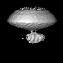
| Method | subtomogram averaging / cryo EM / Resolution: 16.0 Å | |||||||||
 Authors Authors | Li S / Huiskonen JT | |||||||||
| Funding support |  United Kingdom, 1 items United Kingdom, 1 items
| |||||||||
 Citation Citation | Journal: Nat Commun / Year: 2019 Title: The structure of a prokaryotic viral envelope protein expands the landscape of membrane fusion proteins. Authors: Kamel El Omari / Sai Li / Abhay Kotecha / Thomas S Walter / Eduardo A Bignon / Karl Harlos / Pentti Somerharju / Felix De Haas / Daniel K Clare / Mika Molin / Felipe Hurtado / Mengqiu Li / ...Authors: Kamel El Omari / Sai Li / Abhay Kotecha / Thomas S Walter / Eduardo A Bignon / Karl Harlos / Pentti Somerharju / Felix De Haas / Daniel K Clare / Mika Molin / Felipe Hurtado / Mengqiu Li / Jonathan M Grimes / Dennis H Bamford / Nicole D Tischler / Juha T Huiskonen / David I Stuart / Elina Roine /     Abstract: Lipid membrane fusion is an essential function in many biological processes. Detailed mechanisms of membrane fusion and the protein structures involved have been mainly studied in eukaryotic systems, ...Lipid membrane fusion is an essential function in many biological processes. Detailed mechanisms of membrane fusion and the protein structures involved have been mainly studied in eukaryotic systems, whereas very little is known about membrane fusion in prokaryotes. Haloarchaeal pleomorphic viruses (HRPVs) have a membrane envelope decorated with spikes that are presumed to be responsible for host attachment and membrane fusion. Here we determine atomic structures of the ectodomains of the 57-kDa spike protein VP5 from two related HRPVs revealing a previously unreported V-shaped fold. By Volta phase plate cryo-electron tomography we show that VP5 is monomeric on the viral surface, and we establish the orientation of the molecules with respect to the viral membrane. We also show that the viral membrane fuses with the host cytoplasmic membrane in a process mediated by VP5. This sheds light on protein structures involved in prokaryotic membrane fusion. | |||||||||
| History |
|
- Structure visualization
| Movie |
Movie viewer |
|---|---|
| Structure viewer | EM map: SurfViewMolmilJmol/JSmol |
| Supplemental images |

- Downloads & links
-EMDB archive
| Map data | emd_9779.map.gz | 7.3 MB | EMDB map data format | |
|---|---|---|---|---|
| Header (meta data) | emd-9779-v30.xmlemd-9779.xml | 16.3 KB 16.3 KB | Display Display | EMDB header |
| FSC (resolution estimation) | emd_9779_fsc.xml | 5.1 KB | Display | FSC data file |
| Images | 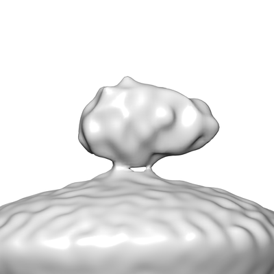 emd_9779.png emd_9779.png | 39.1 KB | ||
| Masks | emd_9779_msk_1.map | 8 MB | Mask map | |
| Filedesc metadata | emd-9779.cif.gz | 6.1 KB | ||
| Others | emd_9779_half_map_1.map.gzemd_9779_half_map_2.map.gz | 7.3 MB 7.3 MB | ||
| Archive directory |  http://ftp.pdbj.org/pub/emdb/structures/EMD-9779ftp://ftp.pdbj.org/pub/emdb/structures/EMD-9779 http://ftp.pdbj.org/pub/emdb/structures/EMD-9779ftp://ftp.pdbj.org/pub/emdb/structures/EMD-9779 | HTTPS FTP |
-Related structure data
| Related structure data | 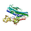 6j7vMC  6qgiC  6qglC M: atomic model generated by this map C: citing same article ( |
|---|---|
| Similar structure data |


-Links
| EMDB pages | EMDB (EBI/PDBe) / EMDataResource |
|---|---|
| Related items in Molecule of the Month |


-Map
| File | Download / File: emd_9779.map.gz / Format: CCP4 / Size: 8 MB / Type: IMAGE STORED AS FLOATING POINT NUMBER (4 BYTES) | ||||||||||||||||||||||||||||||||||||||||||||||||||||||||||||||||||||
|---|---|---|---|---|---|---|---|---|---|---|---|---|---|---|---|---|---|---|---|---|---|---|---|---|---|---|---|---|---|---|---|---|---|---|---|---|---|---|---|---|---|---|---|---|---|---|---|---|---|---|---|---|---|---|---|---|---|---|---|---|---|---|---|---|---|---|---|---|---|
| Projections & slices | Image control
Images are generated by Spider. | ||||||||||||||||||||||||||||||||||||||||||||||||||||||||||||||||||||
| Voxel size | X=Y=Z: 2.24 Å | ||||||||||||||||||||||||||||||||||||||||||||||||||||||||||||||||||||
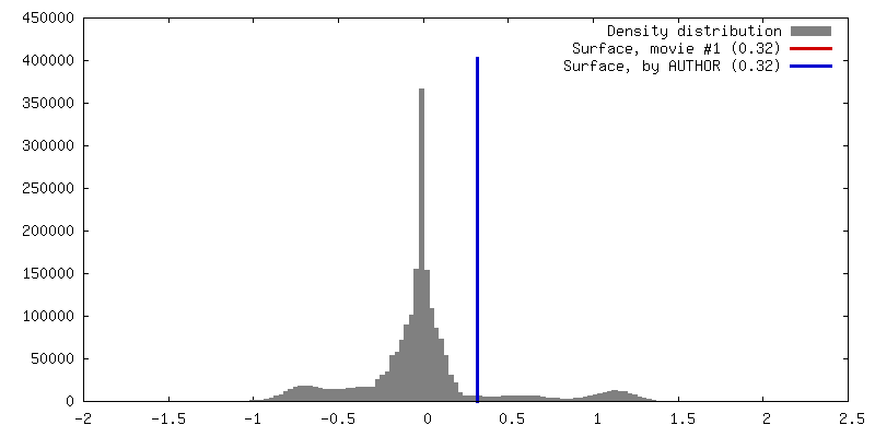
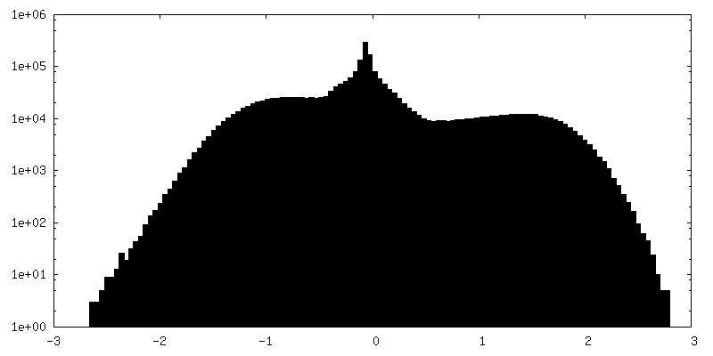
| Density |
| ||||||||||||||||||||||||||||||||||||||||||||||||||||||||||||||||||||
| Symmetry | Space group: 1 | ||||||||||||||||||||||||||||||||||||||||||||||||||||||||||||||||||||
| Details | EMDB XML:
CCP4 map header:
| ||||||||||||||||||||||||||||||||||||||||||||||||||||||||||||||||||||
 Z (Sec.)
 Y (Row.)
 X (Col.)


-Supplemental data
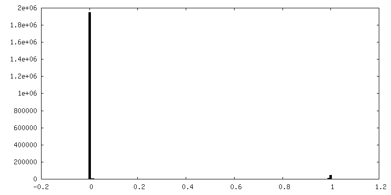
-Mask #1
| File | emd_9779_msk_1.map | ||||||||||||
|---|---|---|---|---|---|---|---|---|---|---|---|---|---|
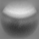
| Projections & Slices |
| ||||||||||||
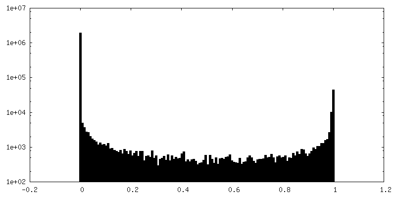
| Density Histograms |


-Half map: #1
| File | emd_9779_half_map_1.map | ||||||||||||
|---|---|---|---|---|---|---|---|---|---|---|---|---|---|
| Projections & Slices |
| ||||||||||||
| Density Histograms |


-Half map: #2
| File | emd_9779_half_map_2.map | ||||||||||||
|---|---|---|---|---|---|---|---|---|---|---|---|---|---|
| Projections & Slices |
| ||||||||||||
| Density Histograms |


- Sample components
-Entire : Halorubrum
| Entire | Name: Halorubrum (archaea) |
|---|---|
| Components |
|
-Supramolecule #1: Halorubrum
| Supramolecule | Name: Halorubrum / type: virus / ID: 1 / Parent: 0 / Macromolecule list: all / NCBI-ID: 56688 / Sci species name: Halorubrum / Virus type: VIRION / Virus isolate: SPECIES / Virus enveloped: Yes / Virus empty: No |
|---|---|
| Host (natural) | Organism: Halorubrum (archaea) |
| Molecular weight | Theoretical: 57 KDa |
| Virus shell | Shell ID: 1 / Name: Envelope |
-Macromolecule #1: VP5
| Macromolecule | Name: VP5 / type: protein_or_peptide / ID: 1 / Number of copies: 1 / Enantiomer: LEVO |
|---|---|
| Source (natural) | Organism: Halorubrum pleomorphic virus 6 |
| Molecular weight | Theoretical: 57.333914 KDa |
| Sequence | String: IAPLVGYAIG AAAISAVGGI GVGWTLREFE VVGSDDPAEG LTPDVLRNQL SDSVVKRKSN NQSTMVDNQN ILDGVEHTAY TEAKIAAIE ELNAGSSESA VLSAANSAID SYETTVRTNF YKSWNETVRE LEAMTQTVIA HADVGLSYIT DFGDPRFGNL A SGTSPNTL ...String: IAPLVGYAIG AAAISAVGGI GVGWTLREFE VVGSDDPAEG LTPDVLRNQL SDSVVKRKSN NQSTMVDNQN ILDGVEHTAY TEAKIAAIE ELNAGSSESA VLSAANSAID SYETTVRTNF YKSWNETVRE LEAMTQTVIA HADVGLSYIT DFGDPRFGNL A SGTSPNTL KDTTVSMPDG TNFTLLTFRH NTGWDSGNAA YSVVEYNPKE VVTSTNSNTY NTVDGTQYMK FSEWNAVETE MD TVFQNVR NGISTWVTNV YGDVQSGAIE ISDLVTPRER ATMMAQEEGM SQAIADLIAL NVPVDAEREA TITIQDTGAT LPG TFALTD SSDGPLSAGQ TYDPSTFSGD VYFTADMSLV EGPWDAINSG VDGGTITITS EPYEGTAIEV TTVESETVSV PAAD WTDNG DGTWSYDASG DLETTITNVD SARFVSTATE TTYDTLQLKG AFTVDKLVNK QSGEEVSSTS FTSSEPQTDS NYITQ DEWD QLEQQNKELI EKYEQSQSGG GLDLGGLDMF GVPGEMVAVG AAAVIGFLML GNN UniProtKB: VP5 |
-Experimental details
-Structure determination
| Method | cryo EM |
|---|---|
 Processing Processing | subtomogram averaging |
| Aggregation state | particle |
-Sample preparation
| Buffer | pH: 7.2 / Component - Concentration: 1.6 M / Component - Formula: NaCl / Component - Name: sodium chloride |
|---|---|
| Vitrification | Cryogen name: ETHANE |
- Electron microscopy
| Microscope | FEI TITAN KRIOS |
|---|---|
| Specialist optics | Phase plate: VOLTA PHASE PLATE / Energy filter - Name: GIF Quantum LS / Energy filter - Slit width: 20 eV |
| Image recording | Film or detector model: GATAN K2 QUANTUM (4k x 4k) / Average exposure time: 1.6 sec. / Average electron dose: 1.6 e/Å2 |
| Electron beam | Acceleration voltage: 300 kV / Electron source:  FIELD EMISSION GUN FIELD EMISSION GUN |
| Electron optics | Illumination mode: FLOOD BEAM / Imaging mode: BRIGHT FIELD |
| Experimental equipment |  Model: Titan Krios / Image courtesy: FEI Company |
-Image processing
| Final reconstruction | Applied symmetry - Point group: C1 (asymmetric) / Resolution.type: BY AUTHOR / Resolution: 16.0 Å / Resolution method: FSC 0.143 CUT-OFF / Software - Name: Dynamo / Number subtomograms used: 6057 |
|---|---|
| Extraction | Number tomograms: 10 / Number images used: 8953 |
| Final angle assignment | Type: OTHER / Software - Name: Dynamo / Details: Sub-volume alignment |
| FSC plot (resolution estimation) |  |
