 Movie
Movie Controller
 Structure viewers
 About EMN search


-Search query
-Search result
Showing 1 - 50 of 87 items for (author: desfosses & a)
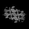
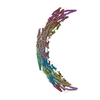
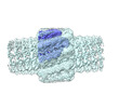
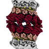
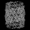
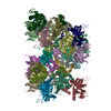
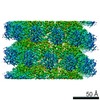
EMDB-18307: 
Native eisosome lattice bound to plasma membrane microdomain
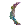
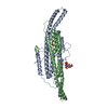
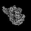
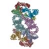
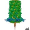
EMDB-18308:
Helical reconstruction of yeast eisosome protein Pil1 bound to membrane composed of lipid mixture -PIP2/+sterol (DOPC, DOPE, DOPS, cholesterol 30:20:20:30)
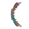
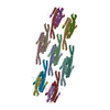
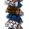
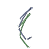
EMDB-18309:
Helical reconstruction of yeast eisosome protein Pil1 bound to membrane composed of lipid mixture +PIP2/-sterol (DOPC, DOPE, DOPS, PI(4,5)P2 50:20:20:10)
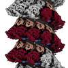
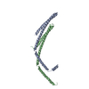
EMDB-18310:
Helical reconstruction of yeast eisosome protein Pil1 bound to membrane composed of lipid mixture +PIP2/+sterol (DOPC, DOPE, DOPS, cholesterol, PI(4,5)P2 35:20:20:15:10)
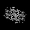
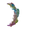
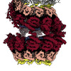
EMDB-18311:
Compact state - Native eisosome lattice bound to plasma membrane microdomain
EMDB-18312:
Stretched state - Native eisosome lattice bound to plasma membrane microdomain
PDB-8qb9:
Helical reconstruction of yeast eisosome protein Pil1 bound to membrane composed of lipid mixture -PIP2/+sterol (DOPC, DOPE, DOPS, cholesterol 30:20:20:30)
PDB-8qbb:
Helical reconstruction of yeast eisosome protein Pil1 bound to membrane composed of lipid mixture +PIP2/-sterol (DOPC, DOPE, DOPS, PI(4,5)P2 50:20:20:10)
PDB-8qbd:
Helical reconstruction of yeast eisosome protein Pil1 bound to membrane composed of lipid mixture +PIP2/+sterol (DOPC, DOPE, DOPS, cholesterol, PI(4,5)P2 35:20:20:15:10)
PDB-8qbe:
Compact state - Pil1 in native eisosome lattice bound to plasma membrane microdomain
PDB-8qbf:
Compact state - Pil1 dimer with lipid headgroups fitted in native eisosome lattice bound to plasma membrane microdomain
PDB-8qbg:
Stretched state - Pil1 in native eisosome lattice bound to plasma membrane microdomain
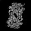
EMDB-17730:
masked refinement giving rise to better defined protruding densities of the potential macrodomain outside the AUD helical assemblies.
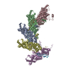
EMDB-18822:
Structure of avian H5N1 influenza A polymerase in complex with human ANP32B.

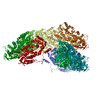
EMDB-17659:
ACAD9-WT in complex with ECSIT-CTER

EMDB-17660:
Cryo-EM structure of human ACAD9-S191A

EMDB-17661:
ACAD9 homodimer WT

PDB-8phe:
ACAD9-WT in complex with ECSIT-CTER

PDB-8phf:
Cryo-EM structure of human ACAD9-S191A
EMDB-17030:
Helical nucleocapsid of the Respiratory Syncytial Virus

EMDB-17031:
Double-ring nucleocapsid of the Respiratory Syncytial Virus
EMDB-17034:
Helical nucleocapsid of the N1-370 mutant of the human Respiratory Syncytial Virus

EMDB-17035:
Subsection of a helical nucleocapsid of the Respiratory Syncytial Virus
EMDB-17036:
Double-headed nucleocapsid of the human Respiratory Syncytial Virus
EMDB-17037:
Ring-capped nucleocapsid of the Respiratory Syncytial Virus

EMDB-17038:
Stacks of nucleocapsid rings of the N1-370 mutant of the human Respiratory Syncytial Virus

PDB-8oou:
Double-ring nucleocapsid of the Respiratory Syncytial Virus
PDB-8op1:
Subsection of a helical nucleocapsid of the Respiratory Syncytial Virus

PDB-8op2:
Stacks of nucleocapsid rings of the N1-370 mutant of the human Respiratory Syncytial Virus
EMDB-13594:
Cryo-EM structure of Saccharomyces cerevisiae TOROID (TORC1 Organized in Inhibited Domains).
EMDB-13595:
Helical reconstruction of TOROID (TORC1 Organized in Inhibited Domains) filaments.
PDB-7pqh:
Cryo-EM structure of Saccharomyces cerevisiae TOROID (TORC1 Organized in Inhibited Domains).

EMDB-14630:
Membrane-bound CHMP2A-CHMP3 filament (430 Angstrom diameter)

EMDB-14631:
Membrane-bound CHMP2A-CHMP3 filament (410 Angstrom diameter)
PDB-7zcg:
CHMP2A-CHMP3 heterodimer (430 Angstrom diameter)
PDB-7zch:
CHMP2A-CHMP3 heterodimer (410 Angstrom diameter)

EMDB-15520:
Cryo-EM structure of human tankyrase 2 SAM-PARP filament (G1032W mutant)
PDB-8aly:
Cryo-EM structure of human tankyrase 2 SAM-PARP filament (G1032W mutant)

EMDB-13261:
Providencia stuartii Arginine decarboxylase (Adc), decamer structure

EMDB-13466:
Providencia stuartii Arginine decarboxylase (Adc), stack structure

PDB-7p9b:
Providencia stuartii Arginine decarboxylase (Adc), decamer structure

PDB-7pk6:
Providencia stuartii Arginine decarboxylase (Adc), stack structure
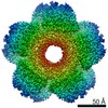
EMDB-10849:
Inducible lysine decarboxylase LdcI decamer, pH 7.0
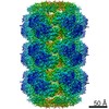
EMDB-10850:
Inducible lysine decarboxylase LdcI stacks, pH 5.7

PDB-6yn5:
Inducible lysine decarboxylase LdcI decamer, pH 7.0

PDB-6yn6:
Inducible lysine decarboxylase LdcI stacks, pH 5.7
EMDB-10160:
In Situ Core-Signalling Unit of E. coli Chemoreceptor Array
EMDB-0142:
Cryo-EM map of in vitro assembled Measles virus N into nucleocapsid-like particles (NCLPs) bound to viral genomic 5-prime RNA hexamers.
PDB-6h5s:
Cryo-EM map of in vitro assembled Measles virus N into nucleocapsid-like particles (NCLPs) bound to viral genomic 5-prime RNA hexamers.
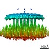
EMDB-4800:
Cryo-EM structure of the anti-feeding prophage (AFP) baseplate in extended state, 3-fold symmetrised
Pages:
 wwPDB to switch to version 3 of the EMDB data model
