Movie
Movie Controller


+ Open data

- Basic information
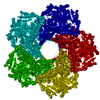
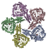
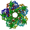
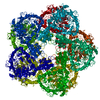
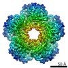
| Entry | Database: PDB / ID: 6yn5 | |||||||||||||||||||||||||||
|---|---|---|---|---|---|---|---|---|---|---|---|---|---|---|---|---|---|---|---|---|---|---|---|---|---|---|---|---|
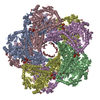
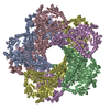
| Title | Inducible lysine decarboxylase LdcI decamer, pH 7.0 | |||||||||||||||||||||||||||
 Components Components | Inducible lysine decarboxylase | |||||||||||||||||||||||||||
 Keywords Keywords | LYASE / Acid stress-inducible / decarboxylase / LAOdc | |||||||||||||||||||||||||||
| Function / homology |  Function and homology information Function and homology informationlysine decarboxylase / lysine decarboxylase activity / : / guanosine tetraphosphate binding / identical protein binding / cytoplasm Similarity search - Function | |||||||||||||||||||||||||||
| Biological species |  | |||||||||||||||||||||||||||
| Method | ELECTRON MICROSCOPY / single particle reconstruction / cryo EM / Resolution: 2.7 Å | |||||||||||||||||||||||||||
 Authors Authors | Jessop, M. / Felix, J. / Desfosses, A. / Effantin, G. / Gutsche, I. | |||||||||||||||||||||||||||
| Funding support | 3items
| |||||||||||||||||||||||||||
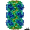
 Citation Citation | Journal: Proc Natl Acad Sci U S A / Year: 2021 Title: Supramolecular assembly of the LdcI upon acid stress. Authors: Matthew Jessop / Clarissa Liesche / Jan Felix / Ambroise Desfosses / Megghane Baulard / Virgile Adam / Angélique Fraudeau / Karine Huard / Grégory Effantin / Jean-Philippe Kleman / Maria ...Authors: Matthew Jessop / Clarissa Liesche / Jan Felix / Ambroise Desfosses / Megghane Baulard / Virgile Adam / Angélique Fraudeau / Karine Huard / Grégory Effantin / Jean-Philippe Kleman / Maria Bacia-Verloop / Dominique Bourgeois / Irina Gutsche /  Abstract: Pathogenic and commensal bacteria often have to resist the harsh acidity of the host stomach. The inducible lysine decarboxylase LdcI buffers the cytosol and the local extracellular environment to ...Pathogenic and commensal bacteria often have to resist the harsh acidity of the host stomach. The inducible lysine decarboxylase LdcI buffers the cytosol and the local extracellular environment to ensure enterobacterial survival at low pH. Here, we investigate the acid stress-response regulation of LdcI by combining biochemical and biophysical characterization with negative stain and cryoelectron microscopy (cryo-EM) and wide-field and superresolution fluorescence imaging. Due to deleterious effects of fluorescent protein fusions on native LdcI decamers, we opt for three-dimensional localization of nanobody-labeled endogenous wild-type LdcI in acid-stressed cells and show that it organizes into distinct patches at the cell periphery. Consistent with recent hypotheses that in vivo clustering of metabolic enzymes often reflects their polymerization as a means of stimulus-induced regulation, we show that LdcI assembles into filaments in vitro at physiologically relevant low pH. We solve the structures of these filaments and of the LdcI decamer formed at neutral pH by cryo-EM and reveal the molecular determinants of LdcI polymerization, confirmed by mutational analysis. Finally, we propose a model for LdcI function inside the enterobacterial cell, providing a structural and mechanistic basis for further investigation of the role of its supramolecular organization in the acid stress response. | |||||||||||||||||||||||||||
| History |
|
- Structure visualization
| Movie |
Movie viewer |
|---|---|
| Structure viewer | Molecule: MolmilJmol/JSmol |
- Downloads & links
-Download
| PDBx/mmCIF format | 6yn5.cif.gz | 1.2 MB | Display | PDBx/mmCIF format |
|---|---|---|---|---|
| PDB format | pdb6yn5.ent.gz | 1005.6 KB | Display | PDB format |
| PDBx/mmJSON format | 6yn5.json.gz | Tree view | PDBx/mmJSON format | |
| Others |  Other downloads Other downloads |
-Validation report
| Arichive directory | https://data.pdbj.org/pub/pdb/validation_reports/yn/6yn5ftp://data.pdbj.org/pub/pdb/validation_reports/yn/6yn5 | HTTPS FTP |
|---|
-Related structure data
| Related structure data | 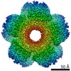 10849MC  6yn6C M: map data used to model this data C: citing same article ( |
|---|---|
| Similar structure data |

-Links
 PDBj
PDBj- Assembly
| Deposited unit | 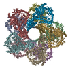
|
|---|---|
| 1 |
|
-Components
| #1: Protein | Mass: 81110.570 Da / Num. of mol.: 10 Source method: isolated from a genetically manipulated source Source: (gene. exp.) Strain: K12 / Gene: cadA, ldcI, b4131, JW4092 Production host: Variant (production host): delta_relA delta_spoT / References: UniProt: P0A9H3, lysine decarboxylase Has ligand of interest | Y | Has protein modification | Y | |
|---|
-Experimental details
-Experiment
| Experiment | Method: ELECTRON MICROSCOPY |
|---|---|
| EM experiment | Aggregation state: PARTICLE / 3D reconstruction method: single particle reconstruction |
- Sample preparation
| Component | Name: E. coli inducible lysine decarboxylase at pH 7.0 / Type: ORGANELLE OR CELLULAR COMPONENT / Entity ID: all / Source: RECOMBINANT |
|---|---|
| Molecular weight | Value: 0.82 MDa / Experimental value: YES |
| Source (natural) | Organism: |
| Source (recombinant) | Organism: |
| Buffer solution | pH: 7 |
| Specimen | Conc.: 0.25 mg/ml / Embedding applied: NO / Shadowing applied: NO / Staining applied: NO / Vitrification applied: YES |
| Specimen support | Grid material: COPPER / Grid mesh size: 300 divisions/in. / Grid type: Quantifoil R2/1 |
| Vitrification | Instrument: FEI VITROBOT MARK IV / Cryogen name: ETHANE / Humidity: 100 % |
- Electron microscopy imaging
| Microscopy | Model: TFS GLACIOS |
|---|---|
| Electron gun | Electron source:  FIELD EMISSION GUN / Accelerating voltage: 200 kV / Illumination mode: FLOOD BEAM FIELD EMISSION GUN / Accelerating voltage: 200 kV / Illumination mode: FLOOD BEAM |
| Electron lens | Mode: BRIGHT FIELD |
| Image recording | Average exposure time: 1.5 sec. / Electron dose: 45 e/Å2 / Film or detector model: FEI FALCON II (4k x 4k) / Num. of real images: 2772 |
| Image scans | Movie frames/image: 29 / Used frames/image: 3-29 |
- Processing
| Software |
| ||||||||||||||||||||||||||||||||||||
|---|---|---|---|---|---|---|---|---|---|---|---|---|---|---|---|---|---|---|---|---|---|---|---|---|---|---|---|---|---|---|---|---|---|---|---|---|---|
| EM software |
| ||||||||||||||||||||||||||||||||||||
| CTF correction | Type: PHASE FLIPPING AND AMPLITUDE CORRECTION | ||||||||||||||||||||||||||||||||||||
| Particle selection | Num. of particles selected: 796000 | ||||||||||||||||||||||||||||||||||||
| Symmetry | Point symmetry: D5 (2x5 fold dihedral) | ||||||||||||||||||||||||||||||||||||
| 3D reconstruction | Resolution: 2.7 Å / Resolution method: FSC 0.143 CUT-OFF / Num. of particles: 229000 / Symmetry type: POINT | ||||||||||||||||||||||||||||||||||||
| Atomic model building | B value: 173 / Space: REAL | ||||||||||||||||||||||||||||||||||||
| Atomic model building | PDB-ID: 3N75 Accession code: 3N75 / Source name: PDB / Type: experimental model | ||||||||||||||||||||||||||||||||||||
| Refinement | Cross valid method: NONE Stereochemistry target values: GeoStd + Monomer Library + CDL v1.2 | ||||||||||||||||||||||||||||||||||||
| Displacement parameters | Biso mean: 41.65 Å2 | ||||||||||||||||||||||||||||||||||||
| Refine LS restraints |
|