 Movie
Movie Controller


+ Open data

- Basic information
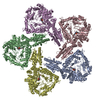
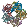
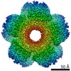
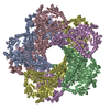
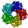
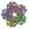
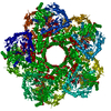
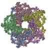
| Entry | Database: PDB / ID: 6q6i | |||||||||||||||||||||
|---|---|---|---|---|---|---|---|---|---|---|---|---|---|---|---|---|---|---|---|---|---|---|
| Title | Lysine decarboxylase A from Pseudomonas aeruginosa | |||||||||||||||||||||
 Components Components | Biodegradative arginine decarboxylase | |||||||||||||||||||||
 Keywords Keywords | OXIDOREDUCTASE / bacterial stress response | |||||||||||||||||||||
| Function / homology |  Function and homology information Function and homology informationarginine decarboxylase / arginine decarboxylase activity / lysine decarboxylase / lysine decarboxylase activity / amino acid metabolic process / L-arginine catabolic process / pyridoxal phosphate binding / cytosol / cytoplasm Similarity search - Function | |||||||||||||||||||||
| Biological species |   Pseudomonas aeruginosa (bacteria) Pseudomonas aeruginosa (bacteria) | |||||||||||||||||||||
| Method | ELECTRON MICROSCOPY / single particle reconstruction / cryo EM / Resolution: 3.7 Å | |||||||||||||||||||||
 Authors Authors | Kandiah, E. / Gutsche, I. | |||||||||||||||||||||
| Funding support |  France, 2items France, 2items
| |||||||||||||||||||||
 Citation Citation | Journal: Structure / Year: 2019 Title: Structure, Function, and Evolution of the Pseudomonas aeruginosa Lysine Decarboxylase LdcA. Authors: Eaazhisai Kandiah / Diego Carriel / Pierre Simon Garcia / Jan Felix / Manuel Banzhaf / George Kritikos / Maria Bacia-Verloop / Céline Brochier-Armanet / Sylvie Elsen / Irina Gutsche /  Abstract: The only enzyme responsible for cadaverine production in the major multidrug-resistant human pathogen Pseudomonas aeruginosa is the lysine decarboxylase LdcA. This enzyme modulates the general ...The only enzyme responsible for cadaverine production in the major multidrug-resistant human pathogen Pseudomonas aeruginosa is the lysine decarboxylase LdcA. This enzyme modulates the general polyamine homeostasis, promotes growth, and reduces bacterial persistence during carbenicillin treatment. Here we present a 3.7-Å resolution cryoelectron microscopy structure of LdcA. We introduce an original approach correlating phylogenetic signal with structural information and reveal possible recombination among LdcA and arginine decarboxylase subfamilies within structural domain boundaries. We show that LdcA is involved in full virulence in an insect pathogenesis model. Furthermore, unlike its enterobacterial counterparts, LdcA is regulated neither by the stringent response alarmone ppGpp nor by the AAA+ ATPase RavA. Instead, the P. aeruginosa ravA gene seems to play a defensive role. Altogether, our study identifies LdcA as an important player in P. aeruginosa physiology and virulence and as a potential drug target. | |||||||||||||||||||||
| History |
|
- Structure visualization
| Movie |
Movie viewer |
|---|---|
| Structure viewer | Molecule: MolmilJmol/JSmol |
- Downloads & links
-Download
| PDBx/mmCIF format | 6q6i.cif.gz | 140.1 KB | Display | PDBx/mmCIF format |
|---|---|---|---|---|
| PDB format | pdb6q6i.ent.gz | 107.8 KB | Display | PDB format |
| PDBx/mmJSON format | 6q6i.json.gz | Tree view | PDBx/mmJSON format | |
| Others |  Other downloads Other downloads |
-Validation report
| Arichive directory | https://data.pdbj.org/pub/pdb/validation_reports/q6/6q6iftp://data.pdbj.org/pub/pdb/validation_reports/q6/6q6i | HTTPS FTP |
|---|
-Related structure data
| Related structure data |  4468MC M: map data used to model this data C: citing same article ( |
|---|---|
| Similar structure data |
-Links
 PDBj
PDBj- Assembly
| Deposited unit | 
|
|---|---|
| 1 | x 10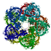
|
-Components
| #1: Protein | Mass: 82854.148 Da / Num. of mol.: 1 / Mutation: Q31H Source method: isolated from a genetically manipulated source Source: (gene. exp.) Pseudomonas aeruginosa (bacteria)Gene: adiA, adiA_2, CAZ10_25795, CGU42_31920, DY979_03865, DZ962_05275, EB236_18185, EGV95_19425, EGY23_25530, IPC3_20945, IPC669_04165, PA34_018150, PAERUG_E15_London_28_01_14_10034, PAMH19_5219, RW109_RW109_04238 Production host: References: UniProt: A0A071KXD7, UniProt: Q9I2S7*PLUS, arginine decarboxylase, lysine decarboxylase |
|---|---|
| #2: Chemical | ChemComp-PLP /   Mass: 247.142 Da / Num. of mol.: 1 / Source method: obtained synthetically / Formula: C8H10NO6P Mass: 247.142 Da / Num. of mol.: 1 / Source method: obtained synthetically / Formula: C8H10NO6P |
| Has ligand of interest | Y |
| Has protein modification | N |
-Experimental details
-Experiment
| Experiment | Method: ELECTRON MICROSCOPY |
|---|---|
| EM experiment | Aggregation state: PARTICLE / 3D reconstruction method: single particle reconstruction |
- Sample preparation
| Component | Name: Lysine decarboxylase / Type: COMPLEX / Entity ID: #1 / Source: RECOMBINANT |
|---|---|
| Molecular weight | Value: 0.8 MDa / Experimental value: NO |
| Source (natural) | Organism: Pseudomonas aeruginosa (bacteria) |
| Source (recombinant) | Organism: |
| Buffer solution | pH: 8 |
| Specimen | Embedding applied: NO / Shadowing applied: NO / Staining applied: NO / Vitrification applied: YES |
| Vitrification | Instrument: FEI VITROBOT MARK IV / Cryogen name: ETHANE |
- Electron microscopy imaging
| Experimental equipment |  Model: Tecnai Polara / Image courtesy: FEI Company |
|---|---|
| Microscopy | Model: FEI POLARA 300 |
| Electron gun | Electron source:  FIELD EMISSION GUN / Accelerating voltage: 300 kV / Illumination mode: FLOOD BEAM FIELD EMISSION GUN / Accelerating voltage: 300 kV / Illumination mode: FLOOD BEAM |
| Electron lens | Mode: BRIGHT FIELD |
| Image recording | Electron dose: 40 e/Å2 / Detector mode: SUPER-RESOLUTION / Film or detector model: GATAN K2 SUMMIT (4k x 4k) |
| Image scans | Movie frames/image: 40 |
- Processing
| Software | Name: PHENIX / Version: 1.11.1_2575: / Classification: refinement | ||||||||||||||||||||||||
|---|---|---|---|---|---|---|---|---|---|---|---|---|---|---|---|---|---|---|---|---|---|---|---|---|---|
| EM software |
| ||||||||||||||||||||||||
| CTF correction | Type: PHASE FLIPPING AND AMPLITUDE CORRECTION | ||||||||||||||||||||||||
| Symmetry | Point symmetry: D5 (2x5 fold dihedral) | ||||||||||||||||||||||||
| 3D reconstruction | Resolution: 3.7 Å / Resolution method: FSC 0.143 CUT-OFF / Num. of particles: 66193 / Symmetry type: POINT | ||||||||||||||||||||||||
| Atomic model building | Protocol: OTHER / Space: REAL | ||||||||||||||||||||||||
| Refine LS restraints |
|
