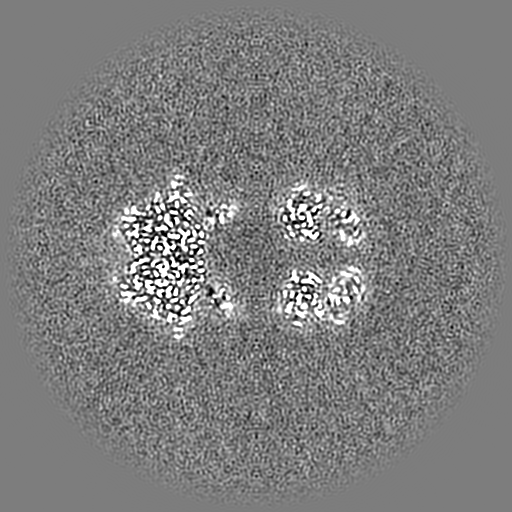
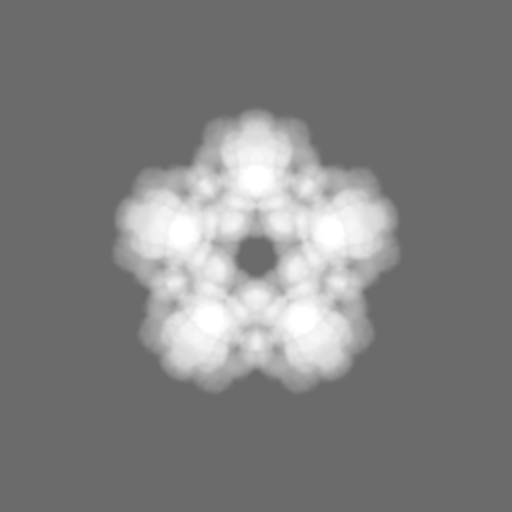
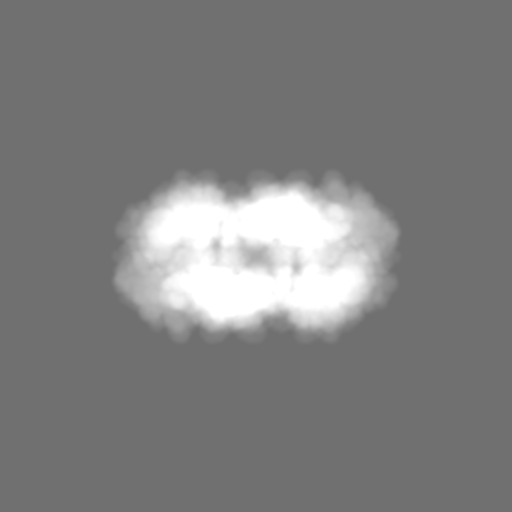
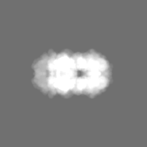
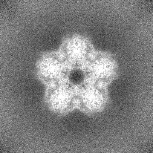
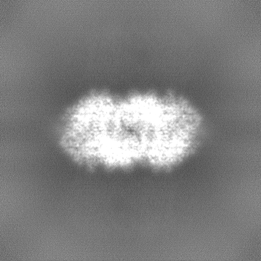
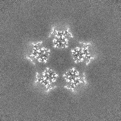
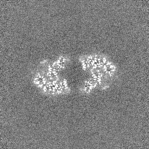
Journal: Commun Biol / Year: 2022 Title: Structural and biochemical characterisation of the Providencia stuartii arginine decarboxylase shows distinct polymerisation and regulation. Authors: Matthew Jessop / Karine Huard / Ambroise Desfosses / Guillaume Tetreau / Diego Carriel / Maria Bacia-Verloop / Caroline Mas / Philippe Mas / Angélique Fraudeau / Jacques-Philippe Colletier / Irina Gutsche / Abstract: Bacterial homologous lysine and arginine decarboxylases play major roles in the acid stress response, physiology, antibiotic resistance and virulence. The Escherichia coli enzymes are considered as ...Bacterial homologous lysine and arginine decarboxylases play major roles in the acid stress response, physiology, antibiotic resistance and virulence. The Escherichia coli enzymes are considered as their archetypes. Whereas acid stress triggers polymerisation of the E. coli lysine decarboxylase LdcI, such behaviour has not been observed for the arginine decarboxylase Adc. Here we show that the Adc from a multidrug-resistant human pathogen Providencia stuartii massively polymerises into filaments whose cryo-EM structure reveals pronounced differences between Adc and LdcI assembly mechanisms. While the structural determinants of Adc polymerisation are conserved only in certain Providencia and Burkholderia species, acid stress-induced polymerisation of LdcI appears general for enterobacteria. Analysis of the expression, activity and oligomerisation of the P. stuartii Adc further highlights the distinct properties of this unusual protein and lays a platform for future investigation of the role of supramolecular assembly in the superfamily or arginine and lysine decarboxylases.
Cryogen name: NITROGEN / Chamber humidity: 100 % / Instrument: FEI VITROBOT MARK IV
-
Electron microscopy
Microscope
FEI TITAN KRIOS
Specialist optics
Energy filter - Name: GIF Bioquantum / Energy filter - Slit width: 20 eV
Image recording
Film or detector model: GATAN K2 SUMMIT (4k x 4k) / Detector mode: COUNTING / Number real images: 10023 / Average exposure time: 3.0 sec. / Average electron dose: 40.0 e/Å2
Electron beam
Acceleration voltage: 300 kV / Electron source: FIELD EMISSION GUN
In the structure databanks used in Yorodumi, some data are registered as the other names, "COVID-19 virus" and "2019-nCoV". Here are the details of the virus and the list of structure data.
Jan 31, 2019. EMDB accession codes are about to change! (news from PDBe EMDB page)
EMDB accession codes are about to change! (news from PDBe EMDB page)
The allocation of 4 digits for EMDB accession codes will soon come to an end. Whilst these codes will remain in use, new EMDB accession codes will include an additional digit and will expand incrementally as the available range of codes is exhausted. The current 4-digit format prefixed with “EMD-” (i.e. EMD-XXXX) will advance to a 5-digit format (i.e. EMD-XXXXX), and so on. It is currently estimated that the 4-digit codes will be depleted around Spring 2019, at which point the 5-digit format will come into force.
The EM Navigator/Yorodumi systems omit the EMD- prefix.
Related info.:Q: What is EMD? / ID/Accession-code notation in Yorodumi/EM Navigator
Yorodumi is a browser for structure data from EMDB, PDB, SASBDB, etc.
This page is also the successor to EM Navigator detail page, and also detail information page/front-end page for Omokage search.
The word "yorodu" (or yorozu) is an old Japanese word meaning "ten thousand". "mi" (miru) is to see.
Related info.:EMDB / PDB / SASBDB / Comparison of 3 databanks / Yorodumi Search / Aug 31, 2016. New EM Navigator & Yorodumi / Yorodumi Papers / Jmol/JSmol / Function and homology information / Changes in new EM Navigator and Yorodumi
 Movie
Movie Controller

 Yorodumi
 Open data
 Basic information
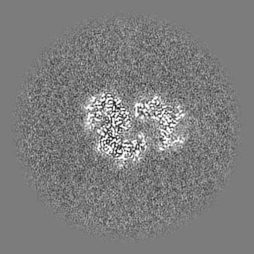
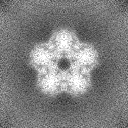
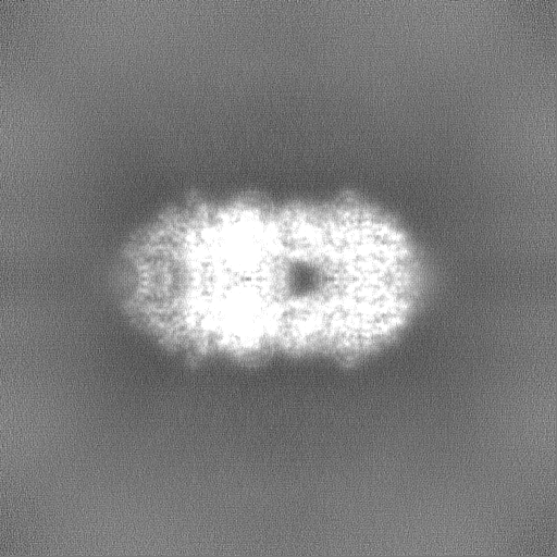
 Map data
 Sample
 Function and homology information
 Providencia stuartii (bacteria)
 Authors
 France, 2 items
  Citation
 Structure visualization
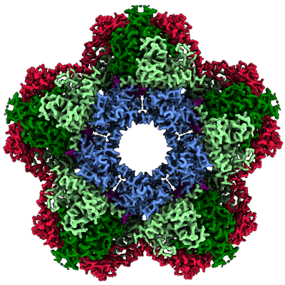
 Downloads & links
 emd_13261.png
 http://ftp.pdbj.org/pub/emdb/structures/EMD-13261

 Z (Sec.)
 Y (Row.)
 X (Col.)


 Sample components
 Processing
 Electron microscopy
 FIELD EMISSION GUN

