 Movie
Movie Controller
 Structure viewers
 About EMN search


-Search query
-Search result
Showing 1 - 50 of 92 items for (author: cambillau & c)

EMDB-57915: 
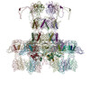
Siphohage OE33PA upon binding to its Gram+ host cell surface (view 1)
Method: electron tomography / : Goulet A, Ptchelkine D

EMDB-57916:
Siphohage OE33PA upon binding to its Gram+ host cell surface (view 2)
Method: electron tomography / : Goulet A, Ptchelkine D

EMDB-57918:
Siphohage OE33PA upon binding to its Gram+ host cell surface (view 3)
Method: electron tomography / : Goulet A, Ptchelkine D

EMDB-57919:
Siphohage OE33PA upon binding to its Gram+ host cell surface (view 4)
Method: electron tomography / : Goulet A, Ptchelkine D

EMDB-57920:
Siphohage OE33PA upon binding to its Gram+ host cell surface (view 5)
Method: electron tomography / : Goulet A, Ptchelkine D

EMDB-57921:
Siphohage OE33PA upon binding to its Gram+ host cell surface (view 6)
Method: electron tomography / : Goulet A, Ptchelkine D

EMDB-57922:
Siphohage OE33PA upon binding to its Gram+ host cell surface (view 7)
Method: electron tomography / : Goulet A, Ptchelkine D

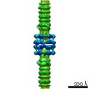
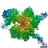
EMDB-58149:
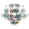
Capsid of the Oenococcus oeni phage OE33PA
Method: single particle / : Goulet A, Cambillau C

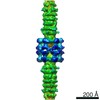
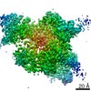
EMDB-58150:
Capsid-connector assembly of the phage OE33PA
Method: single particle / : Goulet A, Cambillau C

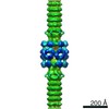
EMDB-58151:
Connector and Major Tail Protein of the phage OE33PA
Method: single particle / : Goulet A, Cambillau C

EMDB-58152:
Portal-adaptor asssembly of the phage OE33PA
Method: single particle / : Goulet A, Cambillau C

EMDB-58153:
Stopper and tail terminator assembly of the phage OE33PA
Method: single particle / : Goulet A, Cambillau C

EMDB-58154:
DNA in the capsid-tail connector of the phage OE33PA (classification focused on the adaptor-stopper region)
Method: single particle / : Goulet A, Cambillau C

EMDB-58155:
DNA in the capsid-tail connector of the phage OE33PA (classification focused on the capsid-portal junction)
Method: single particle / : goulet A, cambillau C

EMDB-58156:
Adhesion device of the phage OE33PA (unsymmetrized 3D reconstruction)
Method: single particle / : Goulet A, Cambillau C

EMDB-58157:
adhesion device (C3 symmetrized reconstruction) of the phage OE33PA
Method: single particle / : Goulet A, Cambillau C

EMDB-58158:
C6 symmetrized reconstruction of the adhesion device of the phage OE33PA
Method: single particle / : Goulet A, Cambillau C

EMDB-58159:
RBP trimer bound to a monomer of the distal tail protein of the phage OE33PA
Method: single particle / : Goulet A, Cambillau C

EMDB-58160:
Part of the tail tube of the phage OE33PA
Method: single particle / : Goulet A, Cambillau C

EMDB-58161:
Conformational variability of the phage OE33PA adhesion device (State 1)
Method: single particle / : Goulet A, Cambillau C

EMDB-58162:
Conformational variability of the phage OE33PA adhesion device (State 2)
Method: single particle / : Goulet A, Cambillau C

PDB-30ys:
Capsid of the Oenococcus oeni phage OE33PA
Method: single particle / : Goulet A, Cambillau C

PDB-30yt:
Capsid-connector assembly of the phage OE33PA
Method: single particle / : Goulet A, Cambillau C

PDB-30yu:
Connector and Major Tail Protein of the phage OE33PA
Method: single particle / : Goulet A, Cambillau C

PDB-30yv:
Portal-adaptor asssembly of the phage OE33PA
Method: single particle / : Goulet A, Cambillau C

PDB-30yw:
Stopper and tail terminator assembly of the phage OE33PA
Method: single particle / : Goulet A, Cambillau C

PDB-30yx:
adhesion device (C3 symmetrized reconstruction) of the phage OE33PA
Method: single particle / : Goulet A, Cambillau C

PDB-30yy:
C6 symmetrized reconstruction of the adhesion device of the phage OE33PA
Method: single particle / : Goulet A, Cambillau C

PDB-30yz:
RBP trimer bound to a monomer of the distal tail protein of the phage OE33PA
Method: single particle / : Goulet A, Cambillau C

PDB-30za:
Part of the tail tube of the phage OE33PA
Method: single particle / : Goulet A, Cambillau C

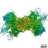
EMDB-70849:
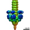
C33 reconstruction for the PorKN (single) ring complex of Type IX Secretion System (T9SS)
Method: single particle / : Liu X, Song L, Hu B

EMDB-70850:
C34 reconstruction for the PorKN (single) ring complex of Type IX Secretion System (T9SS)
Method: single particle / : Liu X, Song L, Hu B

EMDB-70851:
C32 reconstruction for the PorKN (single) ring complex of Type IX Secretion System (T9SS)
Method: single particle / : Liu X, Song L, Hu B

EMDB-70853:
C33 reconstruction for the PorKN (double) ring complex of Type IX Secretion System (T9SS)
Method: single particle / : Liu X, Song L, Hu B

EMDB-70854:
C34 reconstruction for the PorKN (double) ring complex of Type IX Secretion System (T9SS)
Method: single particle / : Liu X, Song L, Hu B

EMDB-70856:
C32 reconstruction for the PorKN (double) ring complex of Type IX Secretion System (T9SS)
Method: single particle / : Liu X, Song L, Hu B

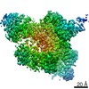
EMDB-70857:
Cryo-EM structure of the T9SS PORkN ring complex of P. Gingivalis
Method: single particle / : Liu X, Song L, Zheng L, Hu B

PDB-9ots:
Cryo-EM structure of the T9SS PORkN ring complex of P. Gingivalis
Method: single particle / : Liu X, Song L, Zheng L, Hu B
EMDB-11224:
negative staining 3D reconstruction of p2 virion baseplate in activated conformation (3D class with open Tal trimer)
Method: single particle / : Spinelli S, Cambillau C, Goulet A
EMDB-11225:
negative staining 3D reconstruction of p2 virion baseplate in activated conformation (3D class with closed Tal trimer)
Method: single particle / : Spinelli S, Cambillau C
EMDB-11226:
Negative staining 3D reconstruction of p2 virion baseplate in activated conformation
Method: single particle / : Spinelli S, Cambillau C
PDB-6zig:
Topological model of the p2 virion baseplate in activated conformation (closed Tal trimer)
Method: single particle / : Spinelli S, Cambillau C, Goulet A
PDB-6zih:
Topological model of p2 virion baseplate in activated conformation
Method: single particle / : Spinelli S, Cambillau C, Goulet A
PDB-6zjj:
Topological model of p2 virion baseplate in resting conformation
Method: single particle / : Spinelli S, Cambillau C, Goulet A
EMDB-10913:
Structure of the phage STP1 host adhesion device (baseplate) by negative staining
Method: single particle / : Lavelle K, Goulet A, McDonnell B, Spinelli S, van Sinderen D, Mahony J, Cambillau C
EMDB-4900:
Cryo-EM structure of St1Cas9-sgRNA-tDNA20-AcrIIA6 monomeric assembly.
Method: single particle / : Swuec P
EMDB-4901:
Cryo-EM structure of St1Cas9-sgRNA-tDNA20-AcrIIA6 dimeric assembly.
Method: single particle / : Swuec P
EMDB-4902:
Cryo-EM structure of St1Cas9-sgRNA-tDNA59-ntPAM complex.
Method: single particle / : Swuec P
EMDB-4904:
Cryo-EM structure of St1Cas9-sgRNA-AcrIIA6-tDNA59-ntPAM complex.
Method: single particle / : Swuec P

PDB-6rj9:
Cryo-EM structure of St1Cas9-sgRNA-tDNA20-AcrIIA6 monomeric assembly.
Method: single particle / : Goulet A, Chaves-Sanjuan A, Cambillau C
Pages:
 wwPDB to switch to version 3 of the EMDB data model
