 Movie
Movie Controller

[English] 日本語
 Yorodumi
- PDB-6n38: Structure of the type VI secretion system TssK-TssF-TssG baseplat... -
+ Open data

- Basic information
| Entry | Database: PDB / ID: 6n38 | ||||||
|---|---|---|---|---|---|---|---|
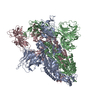
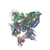
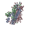
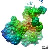
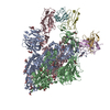
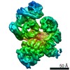
| Title | Structure of the type VI secretion system TssK-TssF-TssG baseplate subcomplex revealed by cryo-electron microscopy - full map sharpened | ||||||
 Components Components |
| ||||||
 Keywords Keywords | PROTEIN TRANSPORT / Type VI secretion system / enteroaggregative E. coli / protein secretion / baseplate / Structural Genomics / Seattle Structural Genomics Center for Infectious Disease / SSGCID | ||||||
| Function / homology | Type VI secretion system TssF / Type VI secretion system, TssF / Type VI secretion, TssG-like / Type VI secretion, TssG / Bacterial Type VI secretion, VC_A0110, EvfL, ImpJ, VasE / Type VI secretion system TssK / Type VI secretion protein / Type VI secretion protein / Type VI secretion protein Function and homology information Function and homology information | ||||||
| Biological species | Escherichia coli O44:H18 | ||||||
| Method | ELECTRON MICROSCOPY / single particle reconstruction / cryo EM / Resolution: 3.7 Å | ||||||
 Authors Authors | Park, Y.J. / Lacourse, K.D. / Cambillau, C. / Seattle Structural Genomics Center for Infectious Disease (SSGCID) / DiMaio, F. / Mougous, J.D. / Veesler, D. | ||||||
| Funding support |  United States, 1items United States, 1items
| ||||||
 Citation Citation | Journal: Nat Commun / Year: 2018 Title: Structure of the type VI secretion system TssK-TssF-TssG baseplate subcomplex revealed by cryo-electron microscopy. Authors: Young-Jun Park / Kaitlyn D Lacourse / Christian Cambillau / Frank DiMaio / Joseph D Mougous / David Veesler /  Abstract: Type VI secretion systems (T6SSs) translocate effectors into target cells and are made of a contractile sheath and a tube docked onto a multi-protein transmembrane complex via a baseplate. Although ...Type VI secretion systems (T6SSs) translocate effectors into target cells and are made of a contractile sheath and a tube docked onto a multi-protein transmembrane complex via a baseplate. Although some information is available about the mechanisms of tail contraction leading to effector delivery, the detailed architecture and function of the baseplate remain unknown. Here, we report the 3.7 Å resolution cryo-electron microscopy reconstruction of an enteroaggregative Escherichia coli baseplate subcomplex assembled from TssK, TssF and TssG. The structure reveals two TssK trimers interact with a locally pseudo-3-fold symmetrical complex comprising two copies of TssF and one copy of TssG. TssF and TssG are structurally related to each other and to components of the phage T4 baseplate and of the type IV secretion system, strengthening the evolutionary relationships among these macromolecular machines. These results, together with bacterial two-hybrid assays, provide a structural framework to understand the T6SS baseplate architecture. | ||||||
| History |
|
- Structure visualization
| Movie |
Movie viewer |
|---|---|
| Structure viewer | Molecule: MolmilJmol/JSmol |
- Downloads & links
-Download
| PDBx/mmCIF format | 6n38.cif.gz | 591.6 KB | Display | PDBx/mmCIF format |
|---|---|---|---|---|
| PDB format | pdb6n38.ent.gz | 484.2 KB | Display | PDB format |
| PDBx/mmJSON format | 6n38.json.gz | Tree view | PDBx/mmJSON format | |
| Others |  Other downloads Other downloads |
-Validation report
| Arichive directory | https://data.pdbj.org/pub/pdb/validation_reports/n3/6n38ftp://data.pdbj.org/pub/pdb/validation_reports/n3/6n38 | HTTPS FTP |
|---|
-Related structure data
| Related structure data | 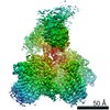 9341MC 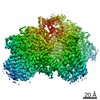 9342C 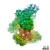 9343C M: map data used to model this data C: citing same article ( |
|---|---|
| Similar structure data |

-Links
 PDBj
PDBj- Assembly
| Deposited unit | 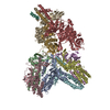
|
|---|---|
| 1 |
|
-Components
| #1: Protein | Mass: 66557.125 Da / Num. of mol.: 2 Source method: isolated from a genetically manipulated source Source: (gene. exp.)  Strain: 042 / EAEC / Gene: EC042_4542 / Production host: #2: Protein | | Mass: 33787.875 Da / Num. of mol.: 1 / Fragment: UNP residues 31-333 Source method: isolated from a genetically manipulated source Source: (gene. exp.) Strain: 042 / EAEC / Gene: EC042_4543 / Production host: #3: Protein/peptide | Mass: 1805.216 Da / Num. of mol.: 2 Source method: isolated from a genetically manipulated source Source: (gene. exp.) Strain: strain 042 / EAEC / Production host: #4: Protein | Mass: 36577.680 Da / Num. of mol.: 6 / Fragment: neck and shoulder domains (UNP residues 1-316) Source method: isolated from a genetically manipulated source Source: (gene. exp.) Strain: 042 / EAEC / Gene: EC042_4526 / Production host: |
|---|
-Experimental details
-Experiment
| Experiment | Method: ELECTRON MICROSCOPY |
|---|---|
| EM experiment | Aggregation state: PARTICLE / 3D reconstruction method: single particle reconstruction |
- Sample preparation
| Component | Name: TssK-TssF-TssG T6SS baseplate subcomplex / Type: COMPLEX / Entity ID: all / Source: RECOMBINANT |
|---|---|
| Molecular weight | Experimental value: NO |
| Source (natural) | Organism: |
| Source (recombinant) | Organism: |
| Buffer solution | pH: 7.8 |
| Specimen | Conc.: 0.12 mg/ml / Embedding applied: NO / Shadowing applied: NO / Staining applied: NO / Vitrification applied: YES |
| Specimen support | Grid material: COPPER / Grid mesh size: 400 divisions/in. / Grid type: C-flat-1.2/1.3 |
| Vitrification | Cryogen name: ETHANE |
- Electron microscopy imaging
| Experimental equipment |  Model: Titan Krios / Image courtesy: FEI Company |
|---|---|
| Microscopy | Model: FEI TITAN KRIOS |
| Electron gun | Electron source:  FIELD EMISSION GUN / Accelerating voltage: 300 kV / Illumination mode: FLOOD BEAM FIELD EMISSION GUN / Accelerating voltage: 300 kV / Illumination mode: FLOOD BEAM |
| Electron lens | Mode: BRIGHT FIELD |
| Image recording | Electron dose: 40 e/Å2 / Film or detector model: GATAN K2 SUMMIT (4k x 4k) |
- Processing
| EM software |
| ||||||||||||||||
|---|---|---|---|---|---|---|---|---|---|---|---|---|---|---|---|---|---|
| CTF correction | Type: PHASE FLIPPING AND AMPLITUDE CORRECTION | ||||||||||||||||
| Symmetry | Point symmetry: C1 (asymmetric) | ||||||||||||||||
| 3D reconstruction | Resolution: 3.7 Å / Resolution method: FSC 0.143 CUT-OFF / Num. of particles: 36496 / Symmetry type: POINT |
