 Movie
Movie Controller


+ Open data

- Basic information
| Entry | Database: EMDB / ID: EMD-2337 | |||||||||
|---|---|---|---|---|---|---|---|---|---|---|
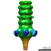
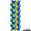
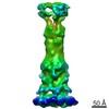
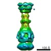
| Title | The tail of mycobacteriophage Araucaria | |||||||||
 Map data Map data | Helical reconstruction of Araucaria tail | |||||||||
 Sample Sample |
| |||||||||
 Keywords Keywords | mycobacteriophage / Araucaria / single-particle / Mycobacterium abscessus subsp. bolletii / Siphoviridae / electron microscopy / mycobacteria. | |||||||||
| Biological species | Mycobacteriophage Araucaria | |||||||||
| Method | single particle reconstruction / negative staining / Resolution: 24.0 Å | |||||||||
 Authors Authors | Sassi M / Bebeacua C / Drancourt M / Cambillau C | |||||||||
 Citation Citation | Journal: J Virol / Year: 2013 Title: The first structure of a mycobacteriophage, the Mycobacterium abscessus subsp. bolletii phage Araucaria. Authors: Mohamed Sassi / Cecilia Bebeacua / Michel Drancourt / Christian Cambillau /  Abstract: The unique characteristics of the waxy mycobacterial cell wall raise questions about specific structural features of their bacteriophages. No structure of any mycobacteriophage is available, although ...The unique characteristics of the waxy mycobacterial cell wall raise questions about specific structural features of their bacteriophages. No structure of any mycobacteriophage is available, although ∼3,500 have been described to date. To fill this gap, we embarked in a genomic and structural study of a bacteriophage from Mycobacterium abscessus subsp. bolletii, a member of the Mycobacterium abscessus group. This opportunistic pathogen is responsible for respiratory tract infections in patients with lung disorders, particularly cystic fibrosis. M. abscessus subsp. bolletii was isolated from respiratory tract specimens, and bacteriophages were observed in the cultures. We report here the genome annotation and characterization of the M. abscessus subsp. bolletii prophage Araucaria, as well as the first single-particle electron microscopy reconstruction of the whole virion. Araucaria belongs to Siphoviridae and possesses a 64-kb genome containing 89 open reading frames (ORFs), among which 27 could be annotated with certainty. Although its capsid and connector share close similarity with those of several phages from Gram-negative (Gram(-)) or Gram(+) bacteria, its most distinctive characteristic is the helical tail decorated by radial spikes, possibly host adhesion devices, according to which the phage name was chosen. Its host adsorption device, at the tail tip, assembles features observed in phages binding to protein receptors, such as phage SPP1. All together, these results suggest that Araucaria may infect its mycobacterial host using a mechanism involving adhesion to cell wall saccharides and protein, a feature that remains to be further explored. | |||||||||
| History |
|
- Structure visualization
| Movie |
 Movie viewer Movie viewer |
|---|---|
| Structure viewer | EM map: SurfViewMolmilJmol/JSmol |
| Supplemental images |
 UCSF Chimera
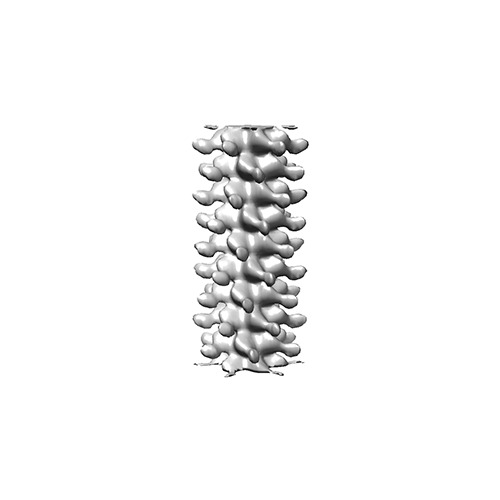
- Downloads & links
-EMDB archive
| Map data | emd_2337.map.gz | 539.5 KB | EMDB map data format | |
|---|---|---|---|---|
| Header (meta data) | emd-2337-v30.xmlemd-2337.xml | 8.7 KB 8.7 KB | Display Display | EMDB header |
| Images | EMD-2337.tif | 58.1 KB | ||
| Archive directory |  http://ftp.pdbj.org/pub/emdb/structures/EMD-2337ftp://ftp.pdbj.org/pub/emdb/structures/EMD-2337 http://ftp.pdbj.org/pub/emdb/structures/EMD-2337ftp://ftp.pdbj.org/pub/emdb/structures/EMD-2337 | HTTPS FTP |
-Related structure data

-Links
| EMDB pages | EMDB (EBI/PDBe) / EMDataResource |
|---|
-Map
| File | Download / File: emd_2337.map.gz / Format: CCP4 / Size: 1.9 MB / Type: IMAGE STORED AS FLOATING POINT NUMBER (4 BYTES) | ||||||||||||||||||||||||||||||||||||||||||||||||||||||||||||||||||||
|---|---|---|---|---|---|---|---|---|---|---|---|---|---|---|---|---|---|---|---|---|---|---|---|---|---|---|---|---|---|---|---|---|---|---|---|---|---|---|---|---|---|---|---|---|---|---|---|---|---|---|---|---|---|---|---|---|---|---|---|---|---|---|---|---|---|---|---|---|---|
| Annotation | Helical reconstruction of Araucaria tail | ||||||||||||||||||||||||||||||||||||||||||||||||||||||||||||||||||||
| Projections & slices | Image control
Images are generated by Spider. | ||||||||||||||||||||||||||||||||||||||||||||||||||||||||||||||||||||
| Voxel size | X=Y=Z: 4.95 Å | ||||||||||||||||||||||||||||||||||||||||||||||||||||||||||||||||||||
| Density |
| ||||||||||||||||||||||||||||||||||||||||||||||||||||||||||||||||||||
| Symmetry | Space group: 1 | ||||||||||||||||||||||||||||||||||||||||||||||||||||||||||||||||||||
| Details | EMDB XML:
CCP4 map header:
| ||||||||||||||||||||||||||||||||||||||||||||||||||||||||||||||||||||
 Z (Sec.)
 Y (Row.)
 X (Col.)


-Supplemental data
- Sample components
-Entire : Mycobacteriophage Araucaria Tail
| Entire | Name: Mycobacteriophage Araucaria Tail |
|---|---|
| Components |
|
-Supramolecule #1000: Mycobacteriophage Araucaria Tail
| Supramolecule | Name: Mycobacteriophage Araucaria Tail / type: sample / ID: 1000 Details: Tail particles were selected from a sample containing whole phages and submitted to Helical Processing. Oligomeric state: C6 Helical / Number unique components: 1 |
|---|
-Supramolecule #1: Mycobacteriophage Araucaria
| Supramolecule | Name: Mycobacteriophage Araucaria / type: virus / ID: 1 / Name.synonym: Araucaria / Sci species name: Mycobacteriophage Araucaria / Virus type: VIRION / Virus isolate: SPECIES / Virus enveloped: No / Virus empty: No / Syn species name: Araucaria |
|---|---|
| Host (natural) | Organism:  Mycobacterium abscessus subsp. bolletii (bacteria) Mycobacterium abscessus subsp. bolletii (bacteria)Strain: CIP108541T / synonym: BACTERIA(EUBACTERIA) |
| Virus shell | Shell ID: 1 / Name: Capsid T7 / Diameter: 600 Å / T number (triangulation number): 7 |
-Experimental details
-Structure determination
| Method | negative staining |
|---|---|
 Processing Processing | single particle reconstruction |
| Aggregation state | particle |
-Sample preparation
| Buffer | pH: 7 / Details: PBS Buffer |
|---|---|
| Staining | Type: NEGATIVE Details: Grids with adsorbed viral particles were stained with 2% uranyl acetate for 20 seconds. |
| Grid | Details: 300 copper mesh grids with a film of colodion and carbon |
| Vitrification | Cryogen name: NONE / Instrument: OTHER |
- Electron microscopy
| Microscope | FEI TECNAI 12 |
|---|---|
| Alignment procedure | Legacy - Astigmatism: Objective lens astigmatism was corrected at 150,000 times magnification |
| Date | Jul 1, 2012 |
| Image recording | Category: CCD / Film or detector model: GENERIC CCD (2k x 2k) / Number real images: 1500 / Average electron dose: 10 e/Å2 |
| Electron beam | Acceleration voltage: 120 kV / Electron source: LAB6 |
| Electron optics | Calibrated magnification: 48500 / Illumination mode: FLOOD BEAM / Imaging mode: BRIGHT FIELD / Cs: 2 mm / Nominal defocus max: 1.5 µm / Nominal defocus min: 1.5 µm / Nominal magnification: 48500 |
| Sample stage | Specimen holder: Room temperature holder / Specimen holder model: SIDE ENTRY, EUCENTRIC |
-Image processing
| Details | Particles were processed using a Helical approach imposing C6 symmetry. |
|---|---|
| Final reconstruction | Applied symmetry - Point group: C6 (6 fold cyclic) / Algorithm: OTHER / Resolution.type: BY AUTHOR / Resolution: 24.0 Å / Resolution method: OTHER Software - Name: EMAN2, Spider, Brandeis, Helical, Package, IHRSR Number images used: 2565 |
| Final angle assignment | Details: C6 Helical |
