 Movie
Movie Controller
 Structure viewers
 About EMN search


-Search query
-Search result
Showing 1 - 50 of 749 items for (author: brown & n)
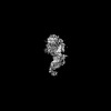
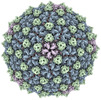
EMDB-44389: 
Cryo-EM structure of the ZBTB5 BTB domain filament
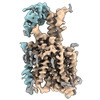
EMDB-44391:
Cryo-EM structure of the ZBTB9 BTB domain filament
PDB-9b9r:
Cryo-EM structure of the ZBTB5 BTB domain filament
PDB-9b9v:
Cryo-EM structure of the ZBTB9 BTB domain filament

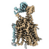
EMDB-43762:
Aca2 from Pectobacterium phage ZF40 bound to RNA

PDB-8w35:
Aca2 from Pectobacterium phage ZF40 bound to RNA
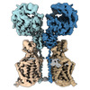
EMDB-16825:
Structure of human terminal uridylyltransferase 7 (hTUT7/ZCCHC6)
EMDB-17084:
Structure of human terminal uridylyltransferase 7 (hTUT7/ZCCHC6) bound with pre-let7g miRNA and UTPalphaS
EMDB-17086:
Human terminal uridylyltransferase 7 (TUT7/ZCCHC6) bound with pre-let7g miRNA and Lin28A - complex 1
EMDB-17087:
Human terminal uridylyltransferase 7 (TUT7/ZCCHC6) bound with pre-let7g miRNA and Lin28A - complex 2
EMDB-17164:
Structure of human terminal uridylyltransferase 4 (TUT4, ZCCHC11) in complex with pre-let7g miRNA and Lin28A
EMDB-17377:
Structure of human SIT1 (focussed map / refinement)

EMDB-17378:
Structure of human SIT1:ACE2 complex (open PD conformation)

EMDB-17379:
Structure of human SIT1:ACE2 complex (closed PD conformation)
EMDB-17380:
Structure of human SIT1 bound to L-pipecolate (focussed map / refinement)
EMDB-17381:
Structure of human SIT1:ACE2 complex (open PD conformation) bound to L-pipecolate
EMDB-17382:
Structure of human SIT1:ACE2 complex (closed PD conformation) bound to L-pipecolate

PDB-8p2w:
Structure of human SIT1 (focussed map / refinement)

PDB-8p2x:
Structure of human SIT1:ACE2 complex (open PD conformation)

PDB-8p2y:
Structure of human SIT1:ACE2 complex (closed PD conformation)

PDB-8p2z:
Structure of human SIT1 bound to L-pipecolate (focussed map / refinement)
PDB-8p30:
Structure of human SIT1:ACE2 complex (open PD conformation) bound to L-pipecolate

PDB-8p31:
Structure of human SIT1:ACE2 complex (closed PD conformation) bound to L-pipecolate
EMDB-17197:
Human TPC2 in Complex with Antagonist (S)-SG-094
EMDB-19108:
Human TPC2 in Complex withAntagonist (R)-SG-094

PDB-8ouo:
Human TPC2 in Complex with Antagonist (S)-SG-094

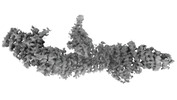
EMDB-41271:
Consensus map of 96nm repeat of human respiratory doublet microtubule, RS1-2 region

EMDB-50185:
KS + AT di-domain of polyketide synthase 13 in Mycobacterium tuberculosis

EMDB-41152:
Cryo-EM Structure of Spike Glycoprotein from Civet Coronavirus SZ3 in Closed Conformation

PDB-8tc5:
Cryo-EM Structure of Spike Glycoprotein from Civet Coronavirus SZ3 in Closed Conformation
EMDB-41149:
Cryo-EM Structure of Spike Glycoprotein from Bat Coronavirus WIV1 in Closed Conformation
EMDB-41150:
Cryo-EM Structure of Spike Glycoprotein from Civet Coronavirus 007 in Closed Conformation
PDB-8tc0:
Cryo-EM Structure of Spike Glycoprotein from Bat Coronavirus WIV1 in Closed Conformation

PDB-8tc1:
Cryo-EM Structure of Spike Glycoprotein from Civet Coronavirus 007 in Closed Conformation
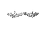
EMDB-40480:
TUBB4B and TUBA1A Heterodimer from Human Respiratory Doublet Microtubules
PDB-8sh7:
TUBB4B and TUBA1A Heterodimer from Human Respiratory Doublet Microtubules
EMDB-43889:
Chlamydomonas reinhardtii mastigoneme (constituent map 1)
EMDB-43890:
Chlamydomonas reinhardtii mastigoneme (constituent map 2)

EMDB-43891:
Chlamydomonas reinhardtii mastigoneme (constituent map 3)
EMDB-43892:
Composite cryo-EM map of the Chlamydomonas reinhardtii mastigoneme

PDB-9b4h:
Chlamydomonas reinhardtii mastigoneme filament
EMDB-41363:
Cryo-EM structure of DDB1deltaB-DDA1-DCAF5

PDB-8tl6:
Cryo-EM structure of DDB1deltaB-DDA1-DCAF5
EMDB-43083:
S. aureus TarL H300N in complex with CDP-ribitol (single tetramer)
EMDB-43084:
S. aureus TarL H300N in complex with CDP-ribitol (two tetramers with CHAPS micelle)
PDB-8va1:
S. aureus TarL H300N in complex with CDP-ribitol (single tetramer)
EMDB-40271:
Mycobacterium phage Adjutor

EMDB-42135:
CryoEM structure of Sec7 autoinhibited conformation
EMDB-42182:
Focused map of Sec7 monomer autoinhibited conformation
EMDB-42183:
Consensus map of Sec7 dimer autoinhibited conformation
Pages:
 wwPDB to switch to version 3 of the EMDB data model
