 ムービー
ムービー コントローラー
 構造ビューア
 EMN検索について


-検索条件
-検索結果
検索 (著者・登録者: brown & d)の結果656件中、1から50件目までを表示しています
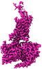
EMDB-17377: 
Structure of human SIT1 (focussed map / refinement)

EMDB-17378:
Structure of human SIT1:ACE2 complex (open PD conformation)

EMDB-17379:
Structure of human SIT1:ACE2 complex (closed PD conformation)
EMDB-17380:
Structure of human SIT1 bound to L-pipecolate (focussed map / refinement)
EMDB-17381:
Structure of human SIT1:ACE2 complex (open PD conformation) bound to L-pipecolate
EMDB-17382:
Structure of human SIT1:ACE2 complex (closed PD conformation) bound to L-pipecolate

PDB-8p2w:
Structure of human SIT1 (focussed map / refinement)

PDB-8p2x:
Structure of human SIT1:ACE2 complex (open PD conformation)

PDB-8p2y:
Structure of human SIT1:ACE2 complex (closed PD conformation)

PDB-8p2z:
Structure of human SIT1 bound to L-pipecolate (focussed map / refinement)
PDB-8p30:
Structure of human SIT1:ACE2 complex (open PD conformation) bound to L-pipecolate

PDB-8p31:
Structure of human SIT1:ACE2 complex (closed PD conformation) bound to L-pipecolate
EMDB-17197:
Human TPC2 in Complex with Antagonist (S)-SG-094
EMDB-19108:
Human TPC2 in Complex withAntagonist (R)-SG-094

PDB-8ouo:
Human TPC2 in Complex with Antagonist (S)-SG-094

EMDB-41271:
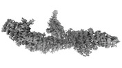
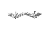
Consensus map of 96nm repeat of human respiratory doublet microtubule, RS1-2 region

EMDB-41152:
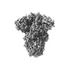
Cryo-EM Structure of Spike Glycoprotein from Civet Coronavirus SZ3 in Closed Conformation

PDB-8tc5:
Cryo-EM Structure of Spike Glycoprotein from Civet Coronavirus SZ3 in Closed Conformation
EMDB-41149:
Cryo-EM Structure of Spike Glycoprotein from Bat Coronavirus WIV1 in Closed Conformation
EMDB-41150:
Cryo-EM Structure of Spike Glycoprotein from Civet Coronavirus 007 in Closed Conformation
PDB-8tc0:
Cryo-EM Structure of Spike Glycoprotein from Bat Coronavirus WIV1 in Closed Conformation

PDB-8tc1:
Cryo-EM Structure of Spike Glycoprotein from Civet Coronavirus 007 in Closed Conformation
EMDB-40480:
TUBB4B and TUBA1A Heterodimer from Human Respiratory Doublet Microtubules
PDB-8sh7:
TUBB4B and TUBA1A Heterodimer from Human Respiratory Doublet Microtubules
EMDB-43889:
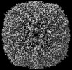
Chlamydomonas reinhardtii mastigoneme (constituent map 1)
EMDB-43890:
Chlamydomonas reinhardtii mastigoneme (constituent map 2)

EMDB-43891:
Chlamydomonas reinhardtii mastigoneme (constituent map 3)
EMDB-43892:
Composite cryo-EM map of the Chlamydomonas reinhardtii mastigoneme

PDB-9b4h:
Chlamydomonas reinhardtii mastigoneme filament
EMDB-41363:
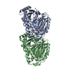
Cryo-EM structure of DDB1deltaB-DDA1-DCAF5

PDB-8tl6:
Cryo-EM structure of DDB1deltaB-DDA1-DCAF5
EMDB-43083:
S. aureus TarL H300N in complex with CDP-ribitol (single tetramer)
EMDB-43084:
S. aureus TarL H300N in complex with CDP-ribitol (two tetramers with CHAPS micelle)
PDB-8va1:
S. aureus TarL H300N in complex with CDP-ribitol (single tetramer)
EMDB-40271:
Mycobacterium phage Adjutor

EMDB-42135:
CryoEM structure of Sec7 autoinhibited conformation
EMDB-42182:
Focused map of Sec7 monomer autoinhibited conformation
EMDB-42183:
Consensus map of Sec7 dimer autoinhibited conformation

PDB-8ucq:
CryoEM structure of Sec7 autoinhibited conformation

EMDB-43224:
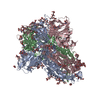
Structure of the HKU1 RBD bound to the human TMPRSS2 receptor

PDB-8vgt:
Structure of the HKU1 RBD bound to the human TMPRSS2 receptor
EMDB-41933:
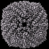
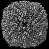
CryoEM map of horse spleen apoferritin determined as a reference for benchmarking square and rectangular apertures for cryo-EM

EMDB-41936:
CryoEM map of horse spleen apoferritin determined as a reference for benchmarking square and rectangular apertures for cryo-EM (Falcon IV round beam)
EMDB-41937:
CryoEM map of horse spleen apoferritin determined for benchmarking square and rectangular apertures for cryo-EM (Falcon IV square beam)
EMDB-41938:
CryoEM map of horse spleen apoferritin determined for benchmarking square and rectangular apertures for cryo-EM (Gatan K3 rectangular beam)
EMDB-42882:
Amylin 1 receptor bound to salmon calcitonin (Amy1R:sCT) reconstructed from cryo-EM datasets recorded using a rectangular aperture

EMDB-19067:
Cryo-EM structure of P. urativorans 70S ribosome in complex with hibernation factors Balon and RaiA (structure 1).

EMDB-19076:
Cryo-EM structure of P. urativorans 70S ribosome in complex with hibernation factor Balon, mRNA and P-site tRNA (structure 2).

EMDB-19077:
Cryo-EM structure of P. urativorans 70S ribosome in complex with hibernation factor Balon and EF-Tu(GDP) (structure 3).

PDB-8rd8:
Cryo-EM structure of P. urativorans 70S ribosome in complex with hibernation factors Balon and RaiA (structure 1).
ページ:
 wwPDBはEMDBデータモデルのバージョン3へ移行します
