Loading
PDBj
MenuPDBj@FacebookPDBj@TwitterPDBj@YouTubewwPDB FoundationwwPDB
RCSB PDBPDBeBMRBAdv. SearchSearch help

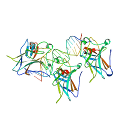
1TUP
DownloadVisualize
BU of 1tup by Molmil
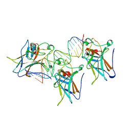
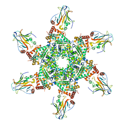
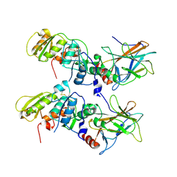
TUMOR SUPPRESSOR P53 COMPLEXED WITH DNA
Descriptor: DNA (5'-D(*AP*TP*AP*AP*TP*TP*GP*GP*GP*CP*AP*AP*GP*TP*CP*TP*A P*GP*GP*AP*A)-3'), DNA (5'-D(*TP*TP*TP*CP*CP*TP*AP*GP*AP*CP*TP*TP*GP*CP*CP*CP*A P*AP*TP*TP*A)-3'), PROTEIN (P53 TUMOR SUPPRESSOR ), ...
Authors:Cho, Y, Gorina, S, Jeffrey, P.D, Pavletich, N.P.
Deposit date:1995-07-11
Release date:1995-07-11
Last modified:2024-02-14
Method:X-RAY DIFFRACTION (2.2 Å)
Cite:Crystal structure of a p53 tumor suppressor-DNA complex: understanding tumorigenic mutations.
Science, 265, 1994
1TSR
DownloadVisualize
BU of 1tsr by Molmil
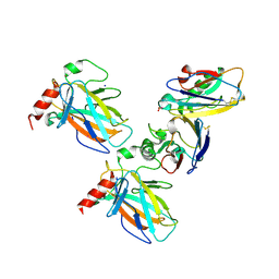
P53 CORE DOMAIN IN COMPLEX WITH DNA
Descriptor: DNA (5'-D(*AP*TP*AP*AP*TP*TP*GP*GP*GP*CP*AP*AP*GP*TP*CP*TP*A P*GP*GP*AP*A)-3'), DNA (5'-D(*TP*TP*TP*CP*CP*TP*AP*GP*AP*CP*TP*TP*GP*CP*CP*CP*A P*AP*TP*TP*A)-3'), PROTEIN (P53 TUMOR SUPPRESSOR), ...
Authors:Cho, Y, Gorina, S, Jeffrey, P, Pavletich, N.
Deposit date:1995-07-28
Release date:1996-01-29
Last modified:2024-02-14
Method:X-RAY DIFFRACTION (2.2 Å)
Cite:Crystal structure of a p53 tumor suppressor-DNA complex: understanding tumorigenic mutations.
Science, 265, 1994
1C26
DownloadVisualize
BU of 1c26 by Molmil
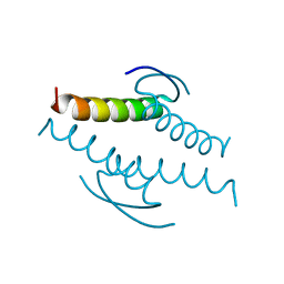
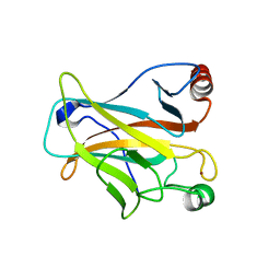
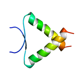
CRYSTAL STRUCTURE OF P53 TETRAMERIZATION DOMAIN
Descriptor: P53 TUMOR SUPPRESSOR
Authors:Jeffrey, P.D, Gorina, S, Pavletich, N.P.
Deposit date:1999-07-22
Release date:1999-07-27
Last modified:2024-02-07
Method:X-RAY DIFFRACTION (1.7 Å)
Cite:Crystal structure of the tetramerization domain of the p53 tumor suppressor at 1.7 angstroms.
Science, 267, 1995
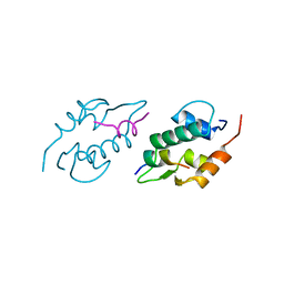
1YCS
DownloadVisualize
BU of 1ycs by Molmil
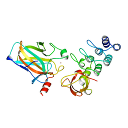
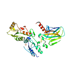
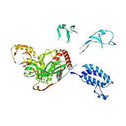
P53-53BP2 COMPLEX
Descriptor: 53BP2, P53, ZINC ION
Authors:Gorina, S, Pavletich, N.P.
Deposit date:1996-09-30
Release date:1997-11-19
Last modified:2024-02-14
Method:X-RAY DIFFRACTION (2.2 Å)
Cite:Structure of the p53 tumor suppressor bound to the ankyrin and SH3 domains of 53BP2.
Science, 274, 1996
3DAC
DownloadVisualize
BU of 3dac by Molmil
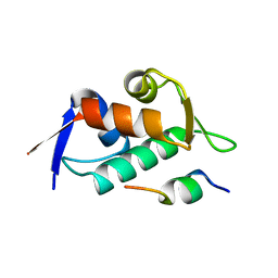
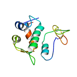
Structure of the human Mdmx protein bound to the p53 tumor suppressor transactivation domain
Descriptor: Cellular tumor antigen p53, Mdm4 protein
Authors:Popowicz, G.M, Czarna, A, Holak, T.A.
Deposit date:2008-05-29
Release date:2008-09-02
Last modified:2023-11-01
Method:X-RAY DIFFRACTION (1.8 Å)
Cite:Structure of the human Mdmx protein bound to the p53 tumor suppressor transactivation domain.
Cell Cycle, 7, 2008
3DAB
DownloadVisualize
BU of 3dab by Molmil
Structure of the human Mdmx protein bound to the p53 tumor suppressor transactivation domain
Descriptor: Cellular tumor antigen p53, Mdm4 protein
Authors:Popowicz, G.M, Czarna, A, Holak, T.A.
Deposit date:2008-05-29
Release date:2008-09-02
Last modified:2023-11-01
Method:X-RAY DIFFRACTION (1.9 Å)
Cite:Structure of the human Mdmx protein bound to the p53 tumor suppressor transactivation domain.
Cell Cycle, 7, 2008
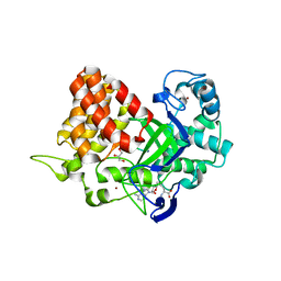
3TG5
DownloadVisualize
BU of 3tg5 by Molmil
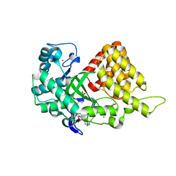
Structure of SMYD2 in complex with p53 and SAH
Descriptor: Cellular tumor antigen p53, GLYCEROL, N-lysine methyltransferase SMYD2, ...
Authors:Zhao, K, Wang, L.
Deposit date:2011-08-17
Release date:2011-08-31
Last modified:2023-11-01
Method:X-RAY DIFFRACTION (2.3 Å)
Cite:Structure of human SMYD2 reveals the basis of p53 tumor suppressor methylation
J.Biol.Chem., 2011
1YCQ
DownloadVisualize
BU of 1ycq by Molmil
XENOPUS LAEVIS MDM2 BOUND TO THE TRANSACTIVATION DOMAIN OF HUMAN P53
Descriptor: MDM2, P53
Authors:Kussie, P.H, Pavletich, N.P.
Deposit date:1996-09-30
Release date:1997-11-19
Last modified:2024-02-14
Method:X-RAY DIFFRACTION (2.3 Å)
Cite:Structure of the MDM2 oncoprotein bound to the p53 tumor suppressor transactivation domain.
Science, 274, 1996
1YCR
DownloadVisualize
BU of 1ycr by Molmil
MDM2 BOUND TO THE TRANSACTIVATION DOMAIN OF P53
Descriptor: MDM2, P53
Authors:Kussie, P.H, Pavletich, N.P.
Deposit date:1996-09-30
Release date:1997-11-19
Last modified:2024-02-14
Method:X-RAY DIFFRACTION (2.6 Å)
Cite:Structure of the MDM2 oncoprotein bound to the p53 tumor suppressor transactivation domain.
Science, 274, 1996
2H1L
DownloadVisualize
BU of 2h1l by Molmil
The Structure of the Oncoprotein SV40 Large T Antigen and p53 Tumor Suppressor Complex
Descriptor: Cellular tumor antigen p53, Large T antigen, ZINC ION
Authors:Lilyestrom, W, Klein, M.G, Chen, X.S.
Deposit date:2006-05-16
Release date:2006-09-12
Last modified:2023-08-30
Method:X-RAY DIFFRACTION (3.16 Å)
Cite:Crystal structure of SV40 large T-antigen bound to p53: interplay between a viral oncoprotein and a cellular tumor suppressor.
Genes Dev., 20, 2006
7YGI
DownloadVisualize
BU of 7ygi by Molmil
Crystal structure of p53 DBD domain in complex with azurin
Descriptor: Azurin, Cellular tumor antigen p53, PHOSPHATE ION, ...
Authors:Jiang, W.X, Zuo, J.Q, Hu, J.J, Chen, X.Q, Ma, L.X, Liu, Z, Xing, Q.
Deposit date:2022-07-11
Release date:2023-02-08
Method:X-RAY DIFFRACTION (2.1 Å)
Cite:Structural basis of bacterial effector protein azurin targeting tumor suppressor p53 and inhibiting its ubiquitination.
Commun Biol, 6, 2023
2OCJ
DownloadVisualize
BU of 2ocj by Molmil
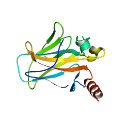
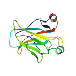
Human p53 core domain in the absence of DNA
Descriptor: P53 TUMOR SUPPRESSOR, ZINC ION
Authors:Wang, Y, Rosengarth, A, Luecke, H.
Deposit date:2006-12-20
Release date:2007-03-06
Last modified:2023-08-30
Method:X-RAY DIFFRACTION (2.05 Å)
Cite:Structure of the human p53 core domain in the absence of DNA.
Acta Crystallogr.,Sect.D, 63, 2007
1GZH
DownloadVisualize
BU of 1gzh by Molmil
Crystal structure of the BRCT domains of human 53BP1 bound to the p53 tumor supressor
Descriptor: CELLULAR TUMOR ANTIGEN P53, SULFATE ION, TUMOR SUPPRESSOR P53-BINDING PROTEIN 1, ...
Authors:Derbyshire, D.J, Doherty, A.J.
Deposit date:2002-05-22
Release date:2002-06-27
Last modified:2023-12-13
Method:X-RAY DIFFRACTION (2.6 Å)
Cite:Crystal Structure of Human 53BP1 Brct Domains Bound to P53 Tumour Suppressor
Embo J., 21, 2002
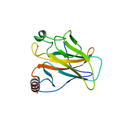
1UOL
DownloadVisualize
BU of 1uol by Molmil
Crystal structure of the human p53 core domain mutant M133L/V203A/N239Y/N268D at 1.9 A resolution.
Descriptor: CELLULAR TUMOR ANTIGEN P53, ZINC ION
Authors:Joerger, A.C, Allen, M.D, Fersht, A.R.
Deposit date:2003-09-19
Release date:2003-10-16
Last modified:2011-07-13
Method:X-RAY DIFFRACTION (1.9 Å)
Cite:Crystal Structure of a Superstable Mutant of Human P53 Core Domain. Insights Into the Mechanism of Rescuing Oncogenic Mutations
J.Biol.Chem., 279, 2004
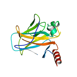
2BIP
DownloadVisualize
BU of 2bip by Molmil
human p53 core domain mutant M133L-H168R-V203A-N239Y-R249S-N268D
Descriptor: CELLULAR TUMOR ANTIGEN P53, ZINC ION
Authors:Joerger, A.C, Fersht, A.R.
Deposit date:2005-01-25
Release date:2005-01-26
Last modified:2023-12-13
Method:X-RAY DIFFRACTION (1.8 Å)
Cite:Structures of P53 Cancer Mutants and Mechanism of Rescue by Second-Site Suppressor Mutations
J.Biol.Chem., 280, 2005
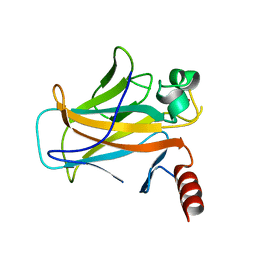
2BIQ
DownloadVisualize
BU of 2biq by Molmil
human p53 core domain mutant T123A-M133L-H168R-V203A-N239Y-R249S- N268D
Descriptor: CELLULAR TUMOR ANTIGEN P53, ZINC ION
Authors:Joerger, A.C, Fersht, A.R.
Deposit date:2005-01-25
Release date:2005-01-26
Last modified:2023-12-13
Method:X-RAY DIFFRACTION (1.8 Å)
Cite:Structures of P53 Cancer Mutants and Mechanism of Rescue by Second-Site Suppressor Mutations
J.Biol.Chem., 280, 2005
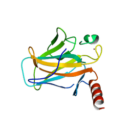
2BIN
DownloadVisualize
BU of 2bin by Molmil
human p53 core domain mutant M133L-H168R-V203A-N239Y-N268D
Descriptor: CELLULAR TUMOR ANTIGEN P53, ZINC ION
Authors:Joerger, A.C, Fersht, A.R.
Deposit date:2005-01-25
Release date:2005-01-26
Last modified:2023-12-13
Method:X-RAY DIFFRACTION (1.9 Å)
Cite:Structures of P53 Cancer Mutants and Mechanism of Rescue by Second-Site Suppressor Mutations
J.Biol.Chem., 280, 2005
2BIO
DownloadVisualize
BU of 2bio by Molmil
human p53 core domain mutant M133L-V203A-N239Y-R249S-N268D
Descriptor: CELLULAR TUMOR ANTIGEN P53, ZINC ION
Authors:Joerger, A.C, Fersht, A.R.
Deposit date:2005-01-25
Release date:2005-01-26
Last modified:2023-12-13
Method:X-RAY DIFFRACTION (1.9 Å)
Cite:Structures of P53 Cancer Mutants and Mechanism of Rescue by Second-Site Suppressor Mutations
J.Biol.Chem., 280, 2005
2BIM
DownloadVisualize
BU of 2bim by Molmil
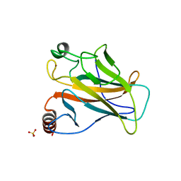
human p53 core domain mutant M133L-V203A-N239Y-N268D-R273H
Descriptor: CELLULAR TUMOR ANTIGEN P53, SULFATE ION, ZINC ION
Authors:Joerger, A.C, Fersht, A.R.
Deposit date:2005-01-25
Release date:2005-01-26
Last modified:2023-12-13
Method:X-RAY DIFFRACTION (1.98 Å)
Cite:Structures of P53 Cancer Mutants and Mechanism of Rescue by Second-Site Suppressor Mutations
J.Biol.Chem., 280, 2005
5XZC
DownloadVisualize
BU of 5xzc by Molmil
Cryo-EM structure of p300-p53 protein complex
Descriptor: Cellular tumor antigen p53, Histone acetyltransferase p300
Authors:Ghosh, R, Roy, S, Sengupta, J.
Deposit date:2017-07-12
Release date:2019-01-23
Last modified:2024-03-27
Method:ELECTRON MICROSCOPY (10.7 Å)
Cite:Tumor suppressor p53-mediated structural reorganization of the transcriptional coactivator p300.
Biochemistry, 2019
3TG4
DownloadVisualize
BU of 3tg4 by Molmil
Structure of SMYD2 in complex with SAM
Descriptor: GLYCEROL, N-lysine methyltransferase SMYD2, S-ADENOSYLMETHIONINE, ...
Authors:Zhao, K, Wang, L.
Deposit date:2011-08-17
Release date:2011-08-31
Last modified:2023-11-01
Method:X-RAY DIFFRACTION (2 Å)
Cite:Structure of human SMYD2 reveals the basis of p53 tumor suppressor methylation
J.Biol.Chem., 2011
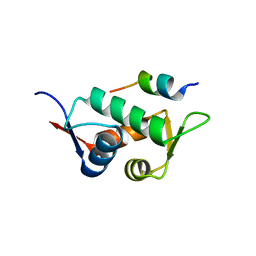
2M86
DownloadVisualize
BU of 2m86 by Molmil
Solution structure of Hdm2 with engineered cyclotide
Descriptor: E3 ubiquitin-protein ligase Mdm2, MCo-PMI
Authors:Majumder, S, Ji, Y, Millard, M, Borra, R, Bi, T, Elnagar, A.Y, Neamati, N, Camarero, J.A.
Deposit date:2013-05-07
Release date:2013-07-31
Last modified:2023-06-14
Method:SOLUTION NMR
Cite:In Vivo Activation of the p53 Tumor Suppressor Pathway by an Engineered Cyclotide.
J.Am.Chem.Soc., 135, 2013
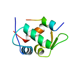
1A1U
DownloadVisualize
BU of 1a1u by Molmil
SOLUTION STRUCTURE DETERMINATION OF A P53 MUTANT DIMERIZATION DOMAIN, NMR, MINIMIZED AVERAGE STRUCTURE
Descriptor: P53
Authors:Mccoy, M.A, Stavridi, E.S, Waterman, J.L.F, Wieczorek, A, Opella, S.J, Halezonetis, T.D.
Deposit date:1997-12-16
Release date:1998-04-08
Last modified:2022-02-16
Method:SOLUTION NMR
Cite:Hydrophobic side-chain size is a determinant of the three-dimensional structure of the p53 oligomerization domain.
EMBO J., 16, 1997
1KZY
DownloadVisualize
BU of 1kzy by Molmil
Crystal Structure of the 53bp1 BRCT Region Complexed to Tumor Suppressor P53
Descriptor: CELLULAR TUMOR ANTIGEN P53, TUMOR SUPPRESSOR P53-BINDING PROTEIN 1, ZINC ION
Authors:Joo, W.S, Jeffrey, P.D, Cantor, S.B, Finnin, M.S, Livingston, D.M, Pavletich, N.P.
Deposit date:2002-02-08
Release date:2002-03-20
Last modified:2011-07-13
Method:X-RAY DIFFRACTION (2.5 Å)
Cite:Structure of the 53BP1 BRCT region bound to p53 and its comparison to the Brca1 BRCT structure.
Genes Dev., 16, 2002
3D08
DownloadVisualize
BU of 3d08 by Molmil
Human p53 core domain with hot spot mutation R249S and second-site suppressor mutation H168R
Descriptor: Cellular tumor antigen p53, ZINC ION
Authors:Suad, O, Rozenberg, H, Shimon, L.J.W, Frolow, F, Shakked, Z.
Deposit date:2008-05-01
Release date:2009-01-20
Last modified:2023-11-01
Method:X-RAY DIFFRACTION (1.4 Å)
Cite:Structural basis of restoring sequence-specific DNA binding and transactivation to mutant p53 by suppressor mutations
J.Mol.Biol., 385, 2009

218853

PDB entries from 2024-04-24

PDB statisticsPDBj update infoContact PDBjnumon