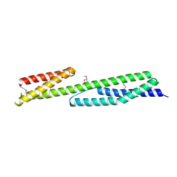
1WLX
 
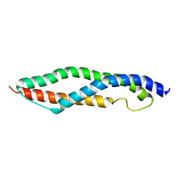 | |
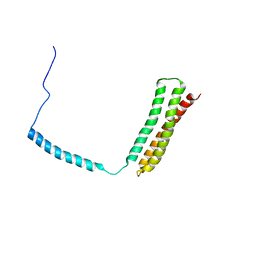
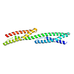
1AJ3
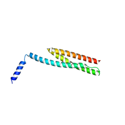
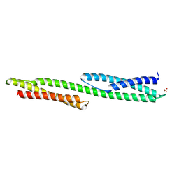
 | | SOLUTION STRUCTURE OF THE SPECTRIN REPEAT, NMR, 20 STRUCTURES | | 分子名称: | ALPHA SPECTRIN | | 著者 | Pascual, J, Pfuhl, M, Walther, D, Saraste, M, Nilges, M. | | 登録日 | 1997-05-14 | | 公開日 | 1997-07-07 | | 最終更新日 | 2022-02-16 | | 実験手法 | SOLUTION NMR | | 主引用文献 | Solution structure of the spectrin repeat: a left-handed antiparallel triple-helical coiled-coil.
J.Mol.Biol., 273, 1997
|
|
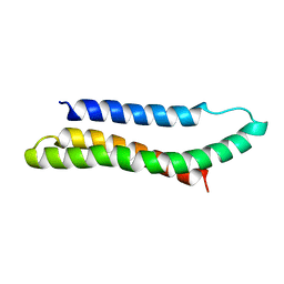
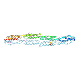
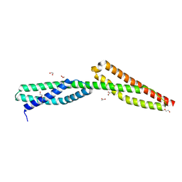
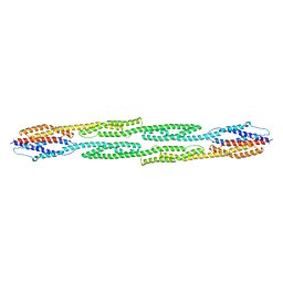
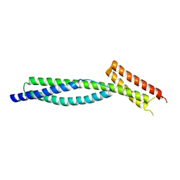
3LBX
 | | Crystal Structure of the Erythrocyte Spectrin Tetramerization Domain Complex | | 分子名称: | Spectrin alpha chain, erythrocyte, Spectrin beta chain | | 著者 | Ipsaro, J.J, Harper, S.L, Messick, T.E, Marmorstein, R, Mondragon, A, Speicher, D.W. | | 登録日 | 2010-01-08 | | 公開日 | 2010-03-09 | | 最終更新日 | 2024-02-21 | | 実験手法 | X-RAY DIFFRACTION (2.8 Å) | | 主引用文献 | Crystal structure and functional interpretation of the erythrocyte spectrin tetramerization domain complex.
Blood, 115, 2010
|
|
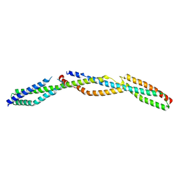
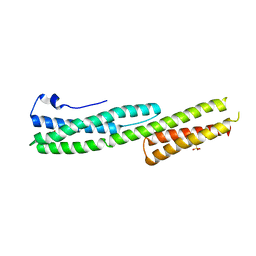
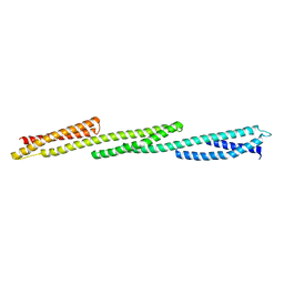
2SPC
 | | CRYSTAL STRUCTURE OF THE REPETITIVE SEGMENTS OF SPECTRIN | | 分子名称: | SPECTRIN | | 著者 | Yan, Y, Winograd, E, Viel, A, Cronin, T, Harrison, S.C, Branton, D. | | 登録日 | 1994-03-01 | | 公開日 | 1994-05-31 | | 最終更新日 | 2024-02-21 | | 実験手法 | X-RAY DIFFRACTION (1.8 Å) | | 主引用文献 | Crystal structure of the repetitive segments of spectrin.
Science, 262, 1993
|
|
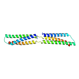
1OWA
 | | Solution Structural Studies on Human Erythrocyte Alpha Spectrin N Terminal Tetramerization Domain | | 分子名称: | Spectrin alpha chain, erythrocyte | | 著者 | Park, S, Caffrey, M.S, Johnson, M.E, Fung, L.W. | | 登録日 | 2003-03-28 | | 公開日 | 2004-03-30 | | 最終更新日 | 2022-02-23 | | 実験手法 | SOLUTION NMR | | 主引用文献 | Solution structural studies on human erythrocyte alpha-spectrin tetramerization site.
J.Biol.Chem., 278, 2003
|
|
2IAK
 | |
7A8T
 | | Crystal structure of sarcomeric protein FATZ-1 (mini-FATZ-1 construct) in complex with rod domain of alpha-actinin-2 | | 分子名称: | Alpha-actinin-2, Myozenin-1 | | 著者 | Sponga, A, Arolas, J.L, Rodriguez Chamorro, A, Mlynek, G, Hollerl, E, Schreiner, C, Pedron, M, Kostan, J, Ribeiro, E.A, Djinovic-Carugo, K. | | 登録日 | 2020-08-31 | | 公開日 | 2021-06-30 | | 最終更新日 | 2024-01-31 | | 実験手法 | X-RAY DIFFRACTION (2.69 Å) | | 主引用文献 | Order from disorder in the sarcomere: FATZ forms a fuzzy but tight complex and phase-separated condensates with alpha-actinin.
Sci Adv, 7, 2021
|
|
3UUM
 | |
5J4O
 | |
3UUN
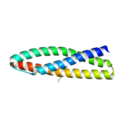 | |
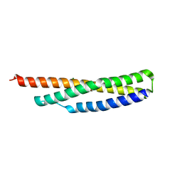
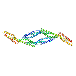
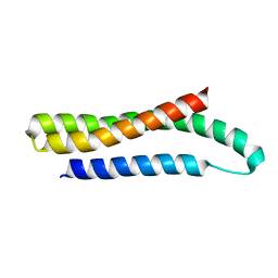
1CUN
 | | CRYSTAL STRUCTURE OF REPEATS 16 AND 17 OF CHICKEN BRAIN ALPHA SPECTRIN | | 分子名称: | PROTEIN (ALPHA SPECTRIN) | | 著者 | Grum, V.L, Li, D, MacDonald, R.I, Mondragon, A. | | 登録日 | 1999-08-20 | | 公開日 | 1999-10-06 | | 最終更新日 | 2024-02-07 | | 実験手法 | X-RAY DIFFRACTION (2 Å) | | 主引用文献 | Structures of two repeats of spectrin suggest models of flexibility.
Cell(Cambridge,Mass.), 98, 1999
|
|
3UUL
 | |
1HCI
 | |
3EDV
 | |
3EDU
 | | Crystal structure of the ankyrin-binding domain of human erythroid spectrin | | 分子名称: | Spectrin beta chain, erythrocyte | | 著者 | Simonovic, M, Stabach, P, Simonovic, I, Steitz, T.A, Morrow, J.S. | | 登録日 | 2008-09-03 | | 公開日 | 2009-02-10 | | 最終更新日 | 2024-02-21 | | 実験手法 | X-RAY DIFFRACTION (2.1 Å) | | 主引用文献 | The structure of the ankyrin-binding site of {beta}-spectrin reveals how tandem spectrin-repeats generate unique ligand-binding properties
Blood, 113, 2009
|
|
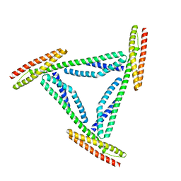
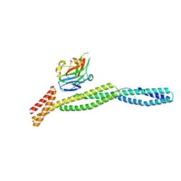
1QUU
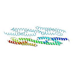 | | CRYSTAL STRUCTURE OF TWO CENTRAL SPECTRIN-LIKE REPEATS FROM ALPHA-ACTININ | | 分子名称: | HUMAN SKELETAL MUSCLE ALPHA-ACTININ 2 | | 著者 | Djinovic-Carugo, K, Young, P, Gautel, M, Saraste, M. | | 登録日 | 1999-07-03 | | 公開日 | 1999-08-20 | | 最終更新日 | 2024-02-14 | | 実験手法 | X-RAY DIFFRACTION (2.5 Å) | | 主引用文献 | Structure of the alpha-actinin rod: molecular basis for cross-linking of actin filaments.
Cell(Cambridge,Mass.), 98, 1999
|
|
3F31
 | | Crystal Structure of the N-terminal region of AlphaII-spectrin Tetramerization Domain | | 分子名称: | Spectrin alpha chain, brain | | 著者 | Mehboob, S, Santarsiero, B.D, Long, F, Witek, M, Fung, L.W. | | 登録日 | 2008-10-30 | | 公開日 | 2009-10-13 | | 最終更新日 | 2023-12-27 | | 実験手法 | X-RAY DIFFRACTION (2.3 Å) | | 主引用文献 | Crystal structure of the nonerythroid alpha-spectrin tetramerization site reveals differences between erythroid and nonerythroid spectrin tetramer formation.
J.Biol.Chem., 285, 2010
|
|
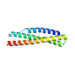
3FB2
 | | Crystal structure of the human brain alpha spectrin repeats 15 and 16. Northeast Structural Genomics Consortium target HR5563a. | | 分子名称: | Spectrin alpha chain, brain spectrin | | 著者 | Vorobiev, S.M, Su, M, Seetharaman, J, Shastry, R, Foote, E.L, Ciccosanti, C, Janjua, H, Xiao, R, Acton, T.B, Montelione, G.T, Tong, L, Hunt, J.F, Northeast Structural Genomics Consortium (NESG) | | 登録日 | 2008-11-18 | | 公開日 | 2008-11-25 | | 最終更新日 | 2023-12-27 | | 実験手法 | X-RAY DIFFRACTION (2.3 Å) | | 主引用文献 | Crystal structure of the human brain alpha spectrin repeats 15 and 16.
To be Published
|
|
1S35
 | |
3F57
 | |
1U4Q
 | | Crystal Structure of Repeats 15, 16 and 17 of Chicken Brain Alpha Spectrin | | 分子名称: | Spectrin alpha chain, brain | | 著者 | Kusunoki, H, Minasov, G, MacDonald, R.I, Mondragon, A. | | 登録日 | 2004-07-26 | | 公開日 | 2004-10-19 | | 最終更新日 | 2024-03-13 | | 実験手法 | X-RAY DIFFRACTION (2.5 Å) | | 主引用文献 | Independent Movement, Dimerization and Stability of Tandem Repeats of Chicken Brain alpha-Spectrin
J.Mol.Biol., 344, 2004
|
|
1U5P
 | | Crystal Structure of Repeats 15 and 16 of Chicken Brain Alpha Spectrin | | 分子名称: | PHOSPHATE ION, POTASSIUM ION, Spectrin alpha chain, ... | | 著者 | Kusunoki, H, Minasov, G, MacDonald, R.I, Mondragon, A. | | 登録日 | 2004-07-28 | | 公開日 | 2004-10-19 | | 最終更新日 | 2024-03-13 | | 実験手法 | X-RAY DIFFRACTION (2 Å) | | 主引用文献 | Independent Movement, Dimerization and Stability of Tandem Repeats of Chicken Brain alpha-Spectrin
J.Mol.Biol., 344, 2004
|
|
5M6S
 | |
3KBT
 | |
3KBU
 | |


