Loading
PDBj
MenuPDBj@FacebookPDBj@TwitterPDBj@YouTubewwPDB FoundationwwPDB
RCSB PDBPDBeBMRBAdv. SearchSearch help

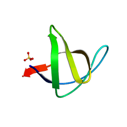
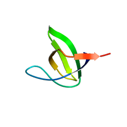
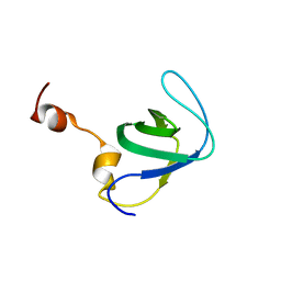
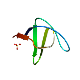
2F2W
DownloadVisualize
BU of 2f2w by Molmil
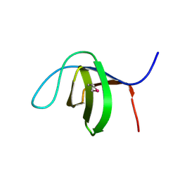
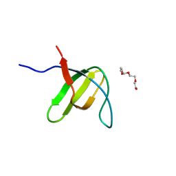
alpha-spectrin SH3 domain R21A mutant
Descriptor: SULFATE ION, Spectrin alpha chain, brain
Authors:Camara-Artigas, A, Conejero-Lara, F, Casares, S, Lopez-Mayorga, O, Vega, C.
Deposit date:2005-11-18
Release date:2006-10-31
Last modified:2023-08-23
Method:X-RAY DIFFRACTION (1.7 Å)
Cite:Cooperative propagation of local stability changes from low-stability and high-stability regions in a SH3 domain
Proteins, 67, 2007
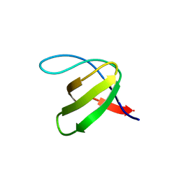
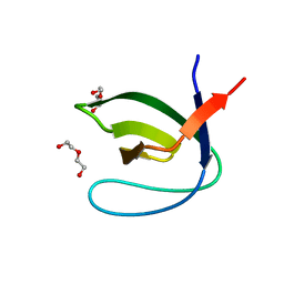
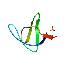
3M0T
DownloadVisualize
BU of 3m0t by Molmil
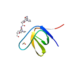
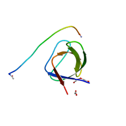
Crystal Structure of the R21D mutant of alpha-spectrin SH3 domain. Crystal obtained in ammonium sulphate at pH 9.
Descriptor: SULFATE ION, Spectrin alpha chain, brain
Authors:Camara-Artigas, A, Gavira, J.A.
Deposit date:2010-03-03
Release date:2011-01-19
Last modified:2024-02-21
Method:X-RAY DIFFRACTION (1.7 Å)
Cite:Understanding the polymorphic behaviour of a mutant of the alpha-spectrin SH3 domain by means of two 1.1 A structures
Acta Crystallogr.,Sect.D, 2011
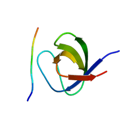
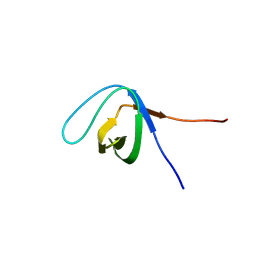
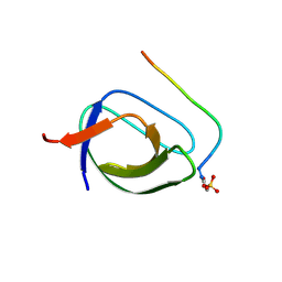
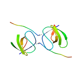
7PW0
DownloadVisualize
BU of 7pw0 by Molmil
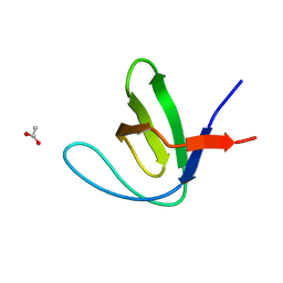
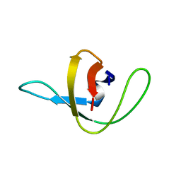
Crystal structure of the c-Src SH3 domain N112G-N113Y-T114N-E115H mutant
Descriptor: Isoform 1 of Proto-oncogene tyrosine-protein kinase Src
Authors:Camara-Artigas, A, Salinas Garcia, M.C.
Deposit date:2021-10-05
Release date:2022-09-14
Last modified:2024-01-31
Method:X-RAY DIFFRACTION (1.7 Å)
Cite:The effect of the hinge loops composition in the domain swapping of the SH3 domain
To be published
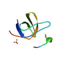
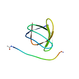
2W0Z
DownloadVisualize
BU of 2w0z by Molmil
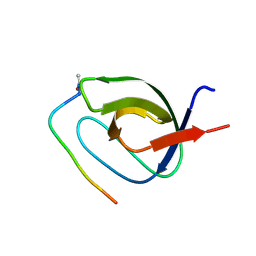
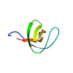
Grb2 SH3C (3)
Descriptor: GRB2-ASSOCIATED-BINDING PROTEIN 2, GROWTH FACTOR RECEPTOR-BOUND PROTEIN 2
Authors:Harkiolaki, M, Tsirka, T, Feller, S.M.
Deposit date:2008-10-13
Release date:2009-05-26
Last modified:2023-12-13
Method:X-RAY DIFFRACTION (1.7 Å)
Cite:Distinct Binding Modes of Two Epitopes in Gab2 that Interact with the Sh3C Domain of Grb2.
Structure, 17, 2009
1UJ0
DownloadVisualize
BU of 1uj0 by Molmil
Crystal Structure of STAM2 SH3 domain in complex with a UBPY-derived peptide
Descriptor: PHOSPHATE ION, deubiquitinating enzyme UBPY, signal transducing adaptor molecule (SH3 domain and ITAM motif) 2
Authors:Kaneko, T, Kumasaka, T, Ganbe, T, Sato, T, Miyazawa, K, Kitamura, N, Tanaka, N.
Deposit date:2003-07-24
Release date:2003-12-23
Last modified:2023-12-27
Method:X-RAY DIFFRACTION (1.7 Å)
Cite:Structural insight into modest binding of a non-PXXP ligand to the signal transducing adaptor molecule-2 Src homology 3 domain.
J.Biol.Chem., 278, 2003
3C0C
DownloadVisualize
BU of 3c0c by Molmil
X-ray Crystal Structure of the Rat Endophilin A2 SH3 Domain
Descriptor: Endophilin-A2
Authors:Loll, P.J, Swain, E.
Deposit date:2008-01-19
Release date:2008-03-18
Last modified:2011-07-13
Method:X-RAY DIFFRACTION (1.7 Å)
Cite:Crystal structure of the SH3 domain of rat endophilin A2
To be Published
3THK
DownloadVisualize
BU of 3thk by Molmil
Structure of SH3 chimera with a type II ligand linked to the chain C-terminal
Descriptor: BETA-MERCAPTOETHANOL, Proline-rich peptide, SULFATE ION, ...
Authors:Gabdulkhakov, A.G, Gushchina, L.V, Nikulin, A.D, Nikonov, S.V, Filimonov, V.V.
Deposit date:2011-08-19
Release date:2011-11-23
Last modified:2023-09-13
Method:X-RAY DIFFRACTION (1.7 Å)
Cite:High-Resolution Crystal Structure of Spectrin SH3 Domain Fused with a Proline-Rich Peptide.
J.Biomol.Struct.Dyn., 29, 2011
7A2U
DownloadVisualize
BU of 7a2u by Molmil
Crystal structure of the Fyn SH3 domain L112V-S114N-S115T at pH 4.5
Descriptor: ACETATE ION, Tyrosine-protein kinase Fyn
Authors:Camara-Artigas, A, Plaza-Garrido, M, Salinas-Garcia, M.C.
Deposit date:2020-08-18
Release date:2021-08-25
Last modified:2024-01-31
Method:X-RAY DIFFRACTION (1.7 Å)
Cite:Crystal structure of the Fyn SH3 domain L112V-S114N-S115T at pH 4.5
To be published
4J9H
DownloadVisualize
BU of 4j9h by Molmil
Crystal structure of the ABL-SH3 domain complexed with the designed high-affinity peptide ligand P7 at pH 8
Descriptor: P7, SULFATE ION, Tyrosine-protein kinase ABL1
Authors:Camara-Artigas, A.
Deposit date:2013-02-16
Release date:2014-01-29
Last modified:2023-09-20
Method:X-RAY DIFFRACTION (1.7 Å)
Cite:Crystal structure of the ABL-SH3 domain complexed with the designed high-affinity peptide ligand P7
To be Published
1YN8
DownloadVisualize
BU of 1yn8 by Molmil
SH3 domain of yeast NBP2
Descriptor: CALCIUM ION, NAP1-binding protein 2
Authors:Kursula, P, Kursula, I, Song, Y.H, Wilmanns, M.
Deposit date:2005-01-24
Release date:2006-05-30
Last modified:2024-03-13
Method:X-RAY DIFFRACTION (1.7 Å)
Cite:Structural analysis of the yeast SH3 domain proteome
To be Published
4J9B
DownloadVisualize
BU of 4j9b by Molmil
Crystal structure of the Abl-SH3 domain H59Q-N96T mutant
Descriptor: DI(HYDROXYETHYL)ETHER, Tyrosine-protein kinase ABL1
Authors:Camara-Artigas, A.
Deposit date:2013-02-16
Release date:2014-01-29
Last modified:2023-09-20
Method:X-RAY DIFFRACTION (1.702 Å)
Cite:Crystal structure of the Abl-SH3 domain H59Q-N96T mutant
To be Published
5XGG
DownloadVisualize
BU of 5xgg by Molmil
Crystal Structure C-terminal SH3 domain of Myosin IB from Entamoeba histolytica
Descriptor: SULFATE ION, Unconventional myosin IB
Authors:Gautam, G, Gourinath, S.
Deposit date:2017-04-13
Release date:2017-08-16
Last modified:2023-11-22
Method:X-RAY DIFFRACTION (1.72 Å)
Cite:Crystal structure of the PEG-bound SH3 domain of myosin IB from Entamoeba histolytica reveals its mode of ligand recognition
Acta Crystallogr D Struct Biol, 73, 2017
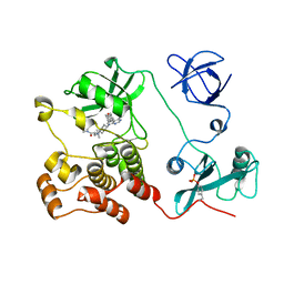
5H0H
DownloadVisualize
BU of 5h0h by Molmil
Crystal structure of HCK complexed with a pyrrolo-pyrimidine inhibitor (S)-2-(((1r,4S)-4-(4-amino-5-(4-phenoxyphenyl)-7H-pyrrolo[2,3-d]pyrimidin-7-yl)cyclohexyl)amino)-N,N,4-trimethylpentanamide
Descriptor: (2~{S})-2-[[4-[4-azanyl-5-(4-phenoxyphenyl)pyrrolo[2,3-d]pyrimidin-7-yl]cyclohexyl]amino]-~{N},~{N},4-trimethyl-pentanamide, Tyrosine-protein kinase HCK
Authors:Tomabechi, Y, Kukimoto-Niino, M, Shirouzu, M.
Deposit date:2016-10-04
Release date:2017-10-04
Last modified:2023-11-15
Method:X-RAY DIFFRACTION (1.72 Å)
Cite:Activity cliff for 7-substituted pyrrolo-pyrimidine inhibitors of HCK explained in terms of predicted basicity of the amine nitrogen.
Bioorg. Med. Chem., 25, 2017
4RUG
DownloadVisualize
BU of 4rug by Molmil
Cyrstal structure of SLIT-ROBO Rho GTPase-activating protein 2 fragment
Descriptor: SLIT-ROBO Rho GTPase-activating protein 2
Authors:Opatowsky, Y, Guez-Hadad, J.
Deposit date:2014-11-19
Release date:2015-11-04
Last modified:2024-02-28
Method:X-RAY DIFFRACTION (1.73 Å)
Cite:The Neuronal Migration Factor srGAP2 Achieves Specificity in Ligand Binding through a Two-Component Molecular Mechanism.
Structure, 23, 2015
3M0Q
DownloadVisualize
BU of 3m0q by Molmil
Crystal Structure of the R21D mutant of alpha-spectrin SH3 domain. Crystal obtained in ammonium sulphate at pH 5.
Descriptor: SULFATE ION, Spectrin alpha chain, brain
Authors:Camara-Artigas, A, Gavira, J.A.
Deposit date:2010-03-03
Release date:2011-01-19
Last modified:2023-09-06
Method:X-RAY DIFFRACTION (1.75 Å)
Cite:Understanding the polymorphic behaviour of a mutant of the alpha-spectrin SH3 domain by means of two 1.1 A structures
Acta Crystallogr.,Sect.D, 2011
2O88
DownloadVisualize
BU of 2o88 by Molmil
Crystal structure of the N114A mutant of ABL-SH3 domain complexed with a designed high-affinity peptide ligand: implications for SH3-ligand interactions
Descriptor: P41 peptide, Proto-oncogene tyrosine-protein kinase ABL1, SULFATE ION
Authors:Camara-Artigas, A.
Deposit date:2006-12-12
Release date:2007-05-01
Last modified:2023-08-30
Method:X-RAY DIFFRACTION (1.75 Å)
Cite:Crystallization by capillary counter-diffusion and structure determination of the N114A mutant of the SH3 domain of Abl tyrosine kinase complexed with a high-affinity peptide ligand.
Acta Crystallogr.,Sect.D, 63, 2007
6ATV
DownloadVisualize
BU of 6atv by Molmil
The molecular mechanisms by which NS1 of the 1918 Spanish influenza A virus hijack host protein-protein interactions
Descriptor: Adapter molecule crk, proline-rich motif in IAV-NS1
Authors:Shen, Q, Zeng, D, Zhao, B, Li, P, Cho, J.H.
Deposit date:2017-08-29
Release date:2018-08-08
Last modified:2023-10-04
Method:X-RAY DIFFRACTION (1.751 Å)
Cite:Molecular Mechanisms of Tight Binding through Fuzzy Interactions.
Biophys. J., 114, 2018
3H0H
DownloadVisualize
BU of 3h0h by Molmil
human Fyn SH3 domain R96I mutant, crystal form I
Descriptor: Proto-oncogene tyrosine-protein kinase Fyn, TETRAETHYLENE GLYCOL
Authors:Dumas, C, Strub, M.-P, Arold, S.T.
Deposit date:2009-04-09
Release date:2010-04-21
Last modified:2023-11-01
Method:X-RAY DIFFRACTION (1.76 Å)
Cite:Synergy and allostery in ligand binding by HIV-1 Nef.
Biochem.J., 478, 2021
1OEB
DownloadVisualize
BU of 1oeb by Molmil
Mona/Gads SH3C domain
Descriptor: CADMIUM ION, GRB2-RELATED ADAPTOR PROTEIN 2, LYMPHOCYTE CYTOSOLIC PROTEIN 2
Authors:Harkiolaki, M, Lewitzky, M, Gilbert, R.J.C, Jones, E.Y, Bourette, R.P, Mouchiroud, G, Sondermann, H, Moarefi, I, Feller, S.M.
Deposit date:2003-03-24
Release date:2003-04-02
Last modified:2011-07-13
Method:X-RAY DIFFRACTION (1.76 Å)
Cite:Structural Basis for SH3 Domain-Mediated High-Affinity Binding between Mona/Gads and Slp-76
Embo J., 22, 2003
5L23
DownloadVisualize
BU of 5l23 by Molmil
Crystal structure of the complex between the N-terminal SH3 domain of CrkII and a proline-rich ligand
Descriptor: Adapter molecule crk, C3G derived peptide, DI(HYDROXYETHYL)ETHER
Authors:Bhatt, V.S, Krieger, I, Sacchettini, J, Cho, J.-H.
Deposit date:2016-07-30
Release date:2016-09-07
Last modified:2023-10-04
Method:X-RAY DIFFRACTION (1.77 Å)
Cite:Crystal structure of the complex between the N-terminal SH3 domain of CrkII and a proline-rich ligand
To Be Published
1TUD
DownloadVisualize
BU of 1tud by Molmil
ALPHA-SPECTRIN SRC HOMOLOGY 3 DOMAIN, CIRCULAR PERMUTANT, CUT AT N47-D48
Descriptor: ALPHA-SPECTRIN
Authors:Viguera, A.R, Serrano, L, Wilmanns, M.
Deposit date:1996-02-29
Release date:1996-08-01
Last modified:2024-02-14
Method:X-RAY DIFFRACTION (1.77 Å)
Cite:The order of secondary structure elements does not determine the structure of a protein but does affect its folding kinetics.
J.Mol.Biol., 247, 1995
1PWT
DownloadVisualize
BU of 1pwt by Molmil
THERMODYNAMIC ANALYSIS OF ALPHA-SPECTRIN SH3 AND TWO OF ITS CIRCULAR PERMUTANTS WITH DIFFERENT LOOP LENGTHS: DISCERNING THE REASONS FOR RAPID FOLDING IN PROTEINS
Descriptor: ALPHA SPECTRIN
Authors:Martinez, J.C, Viguera, A.R, Berisio, R, Wilmanns, M, Mateo, P.L, Filmonov, V.V, Serrano, L.
Deposit date:1998-10-06
Release date:1999-05-11
Last modified:2023-08-09
Method:X-RAY DIFFRACTION (1.77 Å)
Cite:Thermodynamic analysis of alpha-spectrin SH3 and two of its circular permutants with different loop lengths: discerning the reasons for rapid folding in proteins.
Biochemistry, 38, 1999
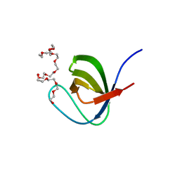
5XG9
DownloadVisualize
BU of 5xg9 by Molmil
Crystal Structure of PEG-bound SH3 domain of Myosin IB from Entamoeba histolytica
Descriptor: 1-(2-METHOXY-ETHOXY)-2-{2-[2-(2-METHOXY-ETHOXY]-ETHOXY}-ETHANE, 2,5,8,11,14,17,20,23,26,29,32,35,38,41,44,47,50,53,56,59,62,65,68,71,74,77,80-HEPTACOSAOXADOOCTACONTAN-82-OL, PENTAETHYLENE GLYCOL, ...
Authors:Gautam, G, Gourinath, S.
Deposit date:2017-04-13
Release date:2017-08-16
Last modified:2023-11-22
Method:X-RAY DIFFRACTION (1.78 Å)
Cite:Crystal structure of the PEG-bound SH3 domain of myosin IB from Entamoeba histolytica reveals its mode of ligand recognition
Acta Crystallogr D Struct Biol, 73, 2017
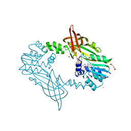
5JMQ
DownloadVisualize
BU of 5jmq by Molmil
Crystal Structure of Mus musculus Protein Arginine Methyltransferase 2 with CP3
Descriptor: 1,2-ETHANEDIOL, 9-[(5E)-7-carbamimidamido-5,6,7-trideoxy-beta-D-ribo-hept-5-enofuranosyl]-9H-purin-6-amine, CALCIUM ION, ...
Authors:Cura, V, Troffer-Charlier, N, Marechal, N, Bonnefond, L, Cavarelli, J.
Deposit date:2016-04-29
Release date:2017-05-10
Last modified:2024-01-10
Method:X-RAY DIFFRACTION (1.795 Å)
Cite:Structural studies of protein arginine methyltransferase 2 reveal its interactions with potential substrates and inhibitors.
Febs J., 284, 2017
5OB2
DownloadVisualize
BU of 5ob2 by Molmil
Crystal structure of the c-Src-SH3 domain E97T mutant in complex with the high affinity peptide APP12
Descriptor: APP12, Proto-oncogene tyrosine-protein kinase Src
Authors:Camara-Artigas, A.
Deposit date:2017-06-25
Release date:2017-07-12
Last modified:2024-01-17
Method:X-RAY DIFFRACTION (1.8 Å)
Cite:Crystal structure of the c-Src-SH3 domain E97T mutant in complex with the high affinity peptide APP12
to be published

218853

PDB entries from 2024-04-24

PDB statisticsPDBj update infoContact PDBjnumon