8AH4
 
 | |
8AH6
 | |
8AH5
 | |
8AH8
 | |
8AH7
 | |
7NES
 | |
7NER
 | |
7NET
 | |
6S7N
 | |
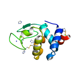
6SYC
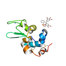
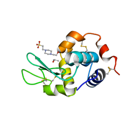
 | | Crystal structure of the lysozyme in presence of bromophenol blue at pH 6.5 | | Descriptor: | CHLORIDE ION, IMIDAZOLE, Lysozyme, ... | | Authors: | Camara-Artigas, A, Plaza-Garrido, M, Salinas-Garcia, M.C. | | Deposit date: | 2019-09-27 | | Release date: | 2020-09-09 | | Last modified: | 2024-01-24 | | Method: | X-RAY DIFFRACTION (1.38 Å) | | Cite: | Lysozyme crystals dyed with bromophenol blue: where has the dye gone?
Acta Crystallogr D Struct Biol, 76, 2020
|
|
6SYE
 | |
6SYD
 | |
6TG7
 | |
7PVT
 | |
7OL7
 | |
7OL8
 | |
7OL5
 | |
7OL6
 | |
7PVZ
 | |
7PW2
 | |
7PW0
 | |
7PVS
 | |
7PVY
 | |
7PVV
 | |
7PVQ
 | |


