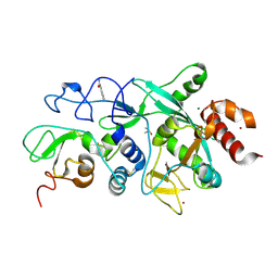
8COE
 
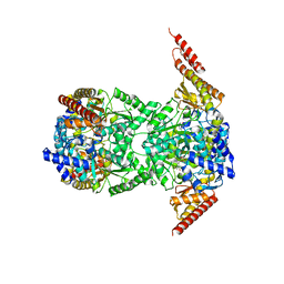
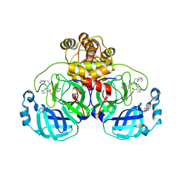
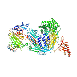 | | complement C5 in complex with the LCP0195 nanobody | | Descriptor: | 2-acetamido-2-deoxy-beta-D-glucopyranose, Complement C5 alpha chain, Complement C5 beta chain, ... | | Authors: | Andersen, G.R, Pedersen, D.V. | | Deposit date: | 2023-02-28 | | Release date: | 2024-01-03 | | Last modified: | 2024-11-13 | | Method: | X-RAY DIFFRACTION (4.2 Å) | | Cite: | Characterization of the bispecific VHH antibody gefurulimab (ALXN1720) targeting complement component 5, and designed for low volume subcutaneous administration.
Mol.Immunol., 165, 2023
|
|
8CGQ
 | |
3D48
 | |
9FWH
 | | Crystal Structure of SARS-CoV-2 NSP10-ExoN in complex with VT00019 | | Descriptor: | (4R)-4-phenyl-1,2-thiazolidine 1,1-dioxide, Guanine-N7 methyltransferase nsp14, Non-structural protein 10, ... | | Authors: | Krojer, T, Kozielski, F, Sele, C, Nyblom, M, Fisher, S.Z, Knecht, W. | | Deposit date: | 2024-06-30 | | Release date: | 2025-07-09 | | Method: | X-RAY DIFFRACTION (2.35 Å) | | Cite: | Crystal Structure of SARS-CoV-2 NSP10-ExoN in complex with VT00019
To Be Published
|
|
9FWO
 | | Crystal Structure of SARS-CoV-2 NSP10-NSP14 (ExoN) in complex with VT00216 | | Descriptor: | 1-methylpyrrole-2-carboxamide, Guanine-N7 methyltransferase nsp14, MAGNESIUM ION, ... | | Authors: | Krojer, T, Kozielski, F, Sele, C, Nyblom, M, Fisher, S.Z, Knecht, W. | | Deposit date: | 2024-06-30 | | Release date: | 2025-07-09 | | Method: | X-RAY DIFFRACTION (2.179 Å) | | Cite: | Crystal Structure of SARS-CoV-2 NSP10-NSP14 (ExoN) in complex with VT00216
To Be Published
|
|
9FWQ
 | | Crystal Structure of SARS-CoV-2 NSP10-NSP14 (ExoN) in complex with VT00218 | | Descriptor: | 5,6,7,8-tetrahydro-[1,2,4]triazolo[4,3-a]pyridine, Guanine-N7 methyltransferase nsp14, MAGNESIUM ION, ... | | Authors: | Krojer, T, Kozielski, F, Sele, C, Nyblom, M, Fisher, S.Z, Knecht, W. | | Deposit date: | 2024-06-30 | | Release date: | 2025-07-09 | | Method: | X-RAY DIFFRACTION (2.322 Å) | | Cite: | Crystal Structure of SARS-CoV-2 NSP10-NSP14 (ExoN) in complex with VT00218
To Be Published
|
|
9FWN
 | | Crystal Structure of SARS-CoV-2 NSP10-NSP14 (ExoN) in complex with VT00219 | | Descriptor: | 1-methyl-1-(phenylmethyl)urea, DIMETHYL SULFOXIDE, Guanine-N7 methyltransferase nsp14, ... | | Authors: | Krojer, T, Kozielski, F, Sele, C, Nyblom, M, Fisher, S.Z, Knecht, W. | | Deposit date: | 2024-06-30 | | Release date: | 2025-07-09 | | Method: | X-RAY DIFFRACTION (1.866 Å) | | Cite: | Crystal Structure of SARS-CoV-2 NSP10-NSP14 (ExoN) in complex with VT00219
To Be Published
|
|
9FWR
 | | Crystal Structure of SARS-CoV-2 NSP10-NSP14 (ExoN) in complex with VT00249 | | Descriptor: | (4R)-4-phenyl-1,3-oxazolidin-2-one, Guanine-N7 methyltransferase nsp14, MAGNESIUM ION, ... | | Authors: | Krojer, T, Kozielski, F, Sele, C, Nyblom, M, Fisher, S.Z, Knecht, W. | | Deposit date: | 2024-06-30 | | Release date: | 2025-07-09 | | Method: | X-RAY DIFFRACTION (2.294 Å) | | Cite: | Crystal Structure of SARS-CoV-2 NSP10-NSP14 (ExoN) in complex with VT00249
To Be Published
|
|
9FWI
 | | Ensemble model of ligand-free SARS-CoV-2 NSP10-NSP14 (ExoN) and in complex with partially bound VT00025 | | Descriptor: | (3-oxidanylazetidin-1-yl)-phenyl-methanone, DIMETHYL SULFOXIDE, Guanine-N7 methyltransferase nsp14, ... | | Authors: | Krojer, T, Kozielski, F, Sele, C, Nyblom, M, Fisher, S.Z, Knecht, W. | | Deposit date: | 2024-06-30 | | Release date: | 2025-07-09 | | Method: | X-RAY DIFFRACTION (1.533 Å) | | Cite: | Ensemble model of ligand-free SARS-CoV-2 NSP10-NSP14 (ExoN) and in complex with partially bound VT00025
To Be Published
|
|
9FWL
 | | Ensemble model of ligand-free SARS-CoV-2 NSP10-NSP14 (ExoN) and in complex with partially bound VT00025 | | Descriptor: | 3-phenylthiophene-2-carboxamide, DIMETHYL SULFOXIDE, Guanine-N7 methyltransferase nsp14, ... | | Authors: | Krojer, T, Kozielski, F, Sele, C, Nyblom, M, Fisher, S.Z, Knecht, W. | | Deposit date: | 2024-06-30 | | Release date: | 2025-07-09 | | Method: | X-RAY DIFFRACTION (2.09 Å) | | Cite: | Ensemble model of ligand-free SARS-CoV-2 NSP10-NSP14 (ExoN) and in complex with partially bound VT00025
To Be Published
|
|
9FWM
 | | Crystal Structure of SARS-CoV-2 NSP10-NSP14 (ExoN) in complex with VT00180 | | Descriptor: | 1H-indole-3-carboxamide, DIMETHYL SULFOXIDE, Guanine-N7 methyltransferase nsp14, ... | | Authors: | Krojer, T, Kozielski, F, Sele, C, Nyblom, M, Fisher, S.Z, Knecht, W. | | Deposit date: | 2024-06-30 | | Release date: | 2025-07-09 | | Method: | X-RAY DIFFRACTION (1.574 Å) | | Cite: | Crystal Structure of SARS-CoV-2 NSP10-NSP14 (ExoN) in complex with VT00180
To Be Published
|
|
9FWP
 | | Crystal Structure of SARS-CoV-2 NSP10-NSP14 (ExoN) in complex with VT00198 | | Descriptor: | Guanine-N7 methyltransferase nsp14, MAGNESIUM ION, N-methylbenzamide, ... | | Authors: | Krojer, T, Kozielski, F, Sele, C, Nyblom, M, Fisher, S.Z, Knecht, W. | | Deposit date: | 2024-06-30 | | Release date: | 2025-07-09 | | Method: | X-RAY DIFFRACTION (2.381 Å) | | Cite: | Crystal Structure of SARS-CoV-2 NSP10-NSP14 (ExoN) in complex with VT00198
To Be Published
|
|
9FWU
 | | Crystal Structure of SARS-CoV-2 NSP10-NSP14 (ExoN) in complex with VT00421 | | Descriptor: | DIMETHYL SULFOXIDE, Guanine-N7 methyltransferase nsp14, N,N-dimethyl-3-oxidanyl-benzamide, ... | | Authors: | Krojer, T, Kozielski, F, Sele, C, Nyblom, M, Fisher, S.Z, Knecht, W. | | Deposit date: | 2024-06-30 | | Release date: | 2025-07-09 | | Method: | X-RAY DIFFRACTION (1.425 Å) | | Cite: | Crystal Structure of SARS-CoV-2 NSP10-NSP14 (ExoN) in complex with VT00421
To Be Published
|
|
9FWJ
 | | Ensemble model of ligand-free SARS-CoV-2 NSP10-NSP14 (ExoN) and in complex with partially bound VT00079 | | Descriptor: | 2-methoxybenzamide, Guanine-N7 methyltransferase nsp14, MAGNESIUM ION, ... | | Authors: | Krojer, T, Kozielski, F, Sele, C, Nyblom, M, Fisher, S.Z, Knecht, W. | | Deposit date: | 2024-06-30 | | Release date: | 2025-07-09 | | Method: | X-RAY DIFFRACTION (2.424 Å) | | Cite: | Ensemble model of ligand-free SARS-CoV-2 NSP10-NSP14 (ExoN) and in complex with partially bound VT00079
To Be Published
|
|
9FWS
 | | Crystal Structure of SARS-CoV-2 NSP10-NSP14 (ExoN) in complex with VT00258 | | Descriptor: | 7-METHOXY-1H-INDAZOLE, DIMETHYL SULFOXIDE, Guanine-N7 methyltransferase nsp14, ... | | Authors: | Krojer, T, Kozielski, F, Sele, C, Nyblom, M, Fisher, S.Z, Knecht, W. | | Deposit date: | 2024-06-30 | | Release date: | 2025-07-09 | | Method: | X-RAY DIFFRACTION (1.433 Å) | | Cite: | Crystal Structure of SARS-CoV-2 NSP10-NSP14 (ExoN) in complex with VT00258
To Be Published
|
|
9FWT
 | | Ensemble model of ligand-free SARS-CoV-2 NSP10-NSP14 (ExoN) and in complex with partially bound VT00259 | | Descriptor: | DIMETHYL SULFOXIDE, Guanine-N7 methyltransferase nsp14, Non-structural protein 10, ... | | Authors: | Krojer, T, Kozielski, F, Sele, C, Nyblom, M, Fisher, S.Z, Knecht, W. | | Deposit date: | 2024-06-30 | | Release date: | 2025-07-09 | | Method: | X-RAY DIFFRACTION (1.639 Å) | | Cite: | Ensemble model of ligand-free SARS-CoV-2 NSP10-NSP14 (ExoN) and in complex with partially bound VT00259
To Be Published
|
|
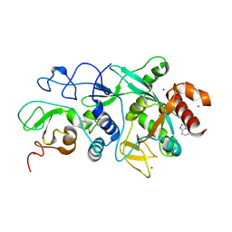
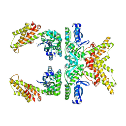
8CRG
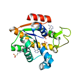 | | E. coli adenylate kinase in complex with two ADP molecules as a result of enzymatic AP4A hydrolysis | | Descriptor: | 3[N-MORPHOLINO]PROPANE SULFONIC ACID, ADENOSINE-5'-DIPHOSPHATE, Adenylate kinase | | Authors: | Oelker, M, Tischlik, S, Wolf-Watz, M, Sauer-Eriksson, A.E. | | Deposit date: | 2023-03-08 | | Release date: | 2023-06-14 | | Last modified: | 2023-10-25 | | Method: | X-RAY DIFFRACTION (1.49 Å) | | Cite: | Insights into Enzymatic Catalysis from Binding and Hydrolysis of Diadenosine Tetraphosphate by E. coli Adenylate Kinase.
Biochemistry, 62, 2023
|
|
8CPL
 | |
8CQ4
 | |
8CQ6
 | |
8F0L
 | |
9EO6
 | | SARS-CoV2 major protease in complex with a covalent inhibitor SLL11. | | Descriptor: | 3C-like proteinase nsp5, Inhibitor SLL11, POTASSIUM ION | | Authors: | Moche, M, Lennerstrand, J, Nyman, T, Strandback, E, Akaberi, D. | | Deposit date: | 2024-03-14 | | Release date: | 2024-09-04 | | Last modified: | 2024-10-16 | | Method: | X-RAY DIFFRACTION (2.11 Å) | | Cite: | Identification of novel and potent inhibitors of SARS-CoV-2 main protease from DNA-encoded chemical libraries.
Antimicrob.Agents Chemother., 68, 2024
|
|
9EOR
 | | SARS-CoV2 major protease in complex with a covalent inhibitor SLL12. | | Descriptor: | 3C-like proteinase nsp5, Inhibitor SLL12, POTASSIUM ION | | Authors: | Moche, M, Lennerstrand, J, Nyman, T, Strandback, E, Akaberi, D. | | Deposit date: | 2024-03-15 | | Release date: | 2024-09-04 | | Last modified: | 2024-10-16 | | Method: | X-RAY DIFFRACTION (2.25 Å) | | Cite: | Identification of novel and potent inhibitors of SARS-CoV-2 main protease from DNA-encoded chemical libraries.
Antimicrob.Agents Chemother., 68, 2024
|
|
4W5U
 | |
4UX6
 | | The discovery of novel, potent and highly selective inhibitors of inducible nitric oxide synthase (iNOS) | | Descriptor: | 5,6,7,8-TETRAHYDROBIOPTERIN, NITRIC OXIDE SYNTHASE, INDUCIBLE, ... | | Authors: | Cheshire, D.R, Andrews, G, Beaton, H.G, Birkinshaw, T, Boughton-Smith, N, Connolly, S, Cook, T.R, Cooper, A, Cooper, S.L, Cox, D, Dixon, J, Gensmantel, N, Hamley, P.J, Harrison, R, Hartopp, P, Kack, H, Luker, T, Mete, A, Millichip, I, Nicholls, D.J, Pimm, A.D, St-Gallay, S.A, Wallace, A.V. | | Deposit date: | 2014-08-19 | | Release date: | 2014-10-08 | | Last modified: | 2024-11-20 | | Method: | X-RAY DIFFRACTION (3 Å) | | Cite: | The Discovery of Novel, Potent and Highly Selective Inhibitors of Inducible Nitric Oxide Synthase (Inos).
Bioorg.Med.Chem.Lett., 21, 2011
|
|


