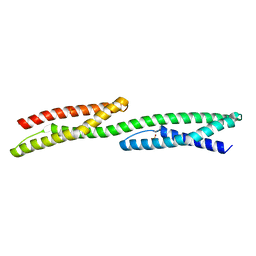
Loading
PDBj
MenuPDBj@FacebookPDBj@TwitterPDBj@YouTubewwPDB FoundationwwPDB
RCSB PDBPDBeBMRBAdv. SearchSearch help
PDB: 6 results

3EDU
DownloadVisualize
BU of 3edu by Molmil
Crystal structure of the ankyrin-binding domain of human erythroid spectrin
Descriptor: Spectrin beta chain, erythrocyte
Authors:Simonovic, M, Stabach, P, Simonovic, I, Steitz, T.A, Morrow, J.S.
Deposit date:2008-09-03
Release date:2009-02-10
Last modified:2024-02-21
Method:X-RAY DIFFRACTION (2.1 Å)
Cite:The structure of the ankyrin-binding site of {beta}-spectrin reveals how tandem spectrin-repeats generate unique ligand-binding properties
Blood, 113, 2009
1S35
DownloadVisualize
BU of 1s35 by Molmil
Crystal Structure of Repeats 8 and 9 of Human Erythroid Spectrin
Descriptor: SULFATE ION, Spectrin beta chain, erythrocyte
Authors:Kusunoki, H, MacDonald, R.I, Mondragon, A.
Deposit date:2004-01-12
Release date:2004-04-20
Last modified:2024-02-14
Method:X-RAY DIFFRACTION (2.4 Å)
Cite:Structural insights into the stability and flexibility of unusual erythroid spectrin repeats
Structure, 12, 2004
3KBT
DownloadVisualize
BU of 3kbt by Molmil
Crystal structure of the ankyrin binding domain of human erythroid beta spectrin (repeats 13-15) in complex with the spectrin binding domain of human erythroid ankyrin (ZU5-ANK)
Descriptor: Ankyrin-1, Spectrin beta chain, erythrocyte
Authors:Ipsaro, J.J, Mondragon, A.
Deposit date:2009-10-20
Release date:2010-02-02
Last modified:2024-02-21
Method:X-RAY DIFFRACTION (2.75 Å)
Cite:Structural basis for spectrin recognition by ankyrin.
Blood, 115, 2010
3KBU
DownloadVisualize
BU of 3kbu by Molmil
Crystal structure of the ankyrin binding domain of human erythroid beta spectrin (repeats 13-15) in complex with the spectrin binding domain of human erythroid ankyrin (ZU5-ANK), EMTS derivative
Descriptor: Ankyrin-1, MERCURY (II) ION, Spectrin beta chain, ...
Authors:Ipsaro, J.J, Mondragon, A.
Deposit date:2009-10-20
Release date:2010-02-02
Last modified:2023-09-06
Method:X-RAY DIFFRACTION (2.75 Å)
Cite:Structural basis for spectrin recognition by ankyrin.
Blood, 115, 2010
3LBX
DownloadVisualize
BU of 3lbx by Molmil
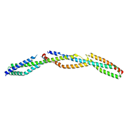
Crystal Structure of the Erythrocyte Spectrin Tetramerization Domain Complex
Descriptor: Spectrin alpha chain, erythrocyte, Spectrin beta chain
Authors:Ipsaro, J.J, Harper, S.L, Messick, T.E, Marmorstein, R, Mondragon, A, Speicher, D.W.
Deposit date:2010-01-08
Release date:2010-03-09
Last modified:2024-02-21
Method:X-RAY DIFFRACTION (2.8 Å)
Cite:Crystal structure and functional interpretation of the erythrocyte spectrin tetramerization domain complex.
Blood, 115, 2010
3F57
DownloadVisualize
BU of 3f57 by Molmil
Crystal structure of human erythroid beta spectrin repeats 14 and 15 (ankyrin binding domain)
Descriptor: Spectrin beta chain, erythrocyte
Authors:Ipsaro, J.J, Mondragon, A.
Deposit date:2008-11-03
Release date:2009-02-24
Last modified:2023-12-27
Method:X-RAY DIFFRACTION (2.9 Å)
Cite:Structures of the spectrin-ankyrin interaction binding domains.
Blood, 113, 2009

224931

PDB entries from 2024-09-11

PDB statisticsPDBj update infoContact PDBjnumon