Loading
PDBj
MenuPDBj@FacebookPDBj@TwitterPDBj@YouTubewwPDB FoundationwwPDB
RCSB PDBPDBeBMRBAdv. SearchSearch help

4GOF
DownloadVisualize
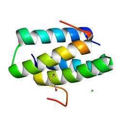
BU of 4gof by Molmil
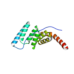
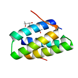
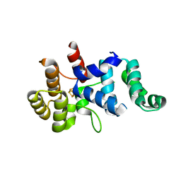
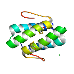
Crystal structure of the SGTA homodimerization domain with covalent modifications to both C38
Descriptor: BETA-MERCAPTOETHANOL, CHLORIDE ION, Small glutamine-rich tetratricopeptide repeat-containing protein alpha
Authors:Chartron, J.W, VanderVelde, D.G, Clemons Jr, W.M.
Deposit date:2012-08-19
Release date:2012-11-21
Last modified:2013-01-16
Method:X-RAY DIFFRACTION (1.35 Å)
Cite:Structures of the Sgt2/SGTA Dimerization Domain with the Get5/UBL4A UBL Domain Reveal an Interaction that Forms a Conserved Dynamic Interface.
Cell Rep, 2, 2012
4GOE
DownloadVisualize
BU of 4goe by Molmil
Crystal structure of the SGTA homodimerization domain with a covalent modification of a single C38
Descriptor: BETA-MERCAPTOETHANOL, CHLORIDE ION, Small glutamine-rich tetratricopeptide repeat-containing protein alpha
Authors:Chartron, J.W, VanderVelde, D.G, Clemons Jr, W.M.
Deposit date:2012-08-19
Release date:2012-11-21
Last modified:2024-03-13
Method:X-RAY DIFFRACTION (1.45 Å)
Cite:Structures of the Sgt2/SGTA Dimerization Domain with the Get5/UBL4A UBL Domain Reveal an Interaction that Forms a Conserved Dynamic Interface.
Cell Rep, 2, 2012
4J8D
DownloadVisualize
BU of 4j8d by Molmil
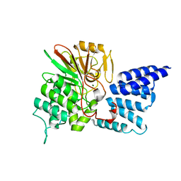
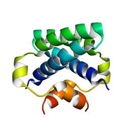
Middle domain of Hsc70-interacting protein, crystal form II
Descriptor: Hsc70-interacting protein
Authors:Li, Z, Bracher, A.
Deposit date:2013-02-14
Release date:2013-07-03
Last modified:2023-09-20
Method:X-RAY DIFFRACTION (2.8 Å)
Cite:Structure and function of Hip, an attenuator of the Hsp70 chaperone cycle.
Nat.Struct.Mol.Biol., 20, 2013
4JA9
DownloadVisualize
BU of 4ja9 by Molmil
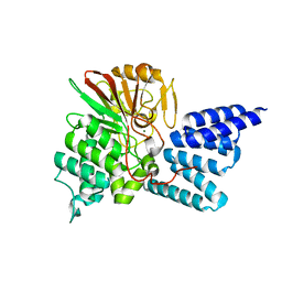
Rat PP5 apo
Descriptor: MAGNESIUM ION, Serine/threonine-protein phosphatase 5
Authors:Haslbeck, V, Helmuth, M, Alte, F, Popowicz, G, Schmidt, W, Weiwad, M, Fischer, G, Gemmecker, G, Sattler, M, Striggow, F, Groll, M, Richter, K.
Deposit date:2013-02-18
Release date:2014-02-19
Last modified:2023-09-20
Method:X-RAY DIFFRACTION (2.3 Å)
Cite:Selective activators of protein phosphatase 5 target the auto-inhibitory mechanism.
Biosci.Rep., 35, 2015
4OPO
DownloadVisualize
BU of 4opo by Molmil
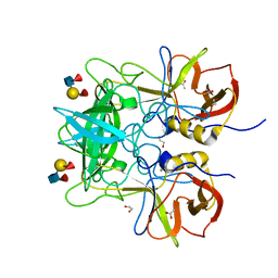
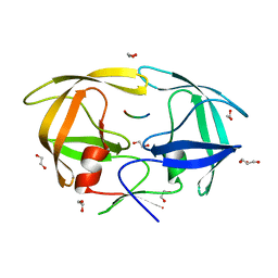
Crystal structure of P domain from norovirus strain Saga4 in complex with HBGA type Leb (tetraglycan)
Descriptor: 1,2-ETHANEDIOL, ACETATE ION, VP1, ...
Authors:Singh, B.K, Hansman, G.S.
Deposit date:2014-02-06
Release date:2014-12-17
Last modified:2024-02-28
Method:X-RAY DIFFRACTION (1.4 Å)
Cite:Human noroviruses' fondness for histo-blood group antigens.
J.Virol., 89, 2015
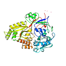
3RE1
DownloadVisualize
BU of 3re1 by Molmil
Crystal structure of uroporphyrinogen III synthase from Pseudomonas syringae pv. tomato DC3000
Descriptor: Uroporphyrinogen-III synthetase
Authors:Chang, W.R, Li, M, Peng, S.X.
Deposit date:2011-04-02
Release date:2011-06-22
Last modified:2024-03-20
Method:X-RAY DIFFRACTION (2.5 Å)
Cite:Crystal structure of uroporphyrinogen III synthase from Pseudomonas syringae pv. tomato DC3000
Biochem.Biophys.Res.Commun., 408, 2011
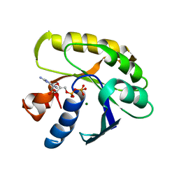
3EZ6
DownloadVisualize
BU of 3ez6 by Molmil
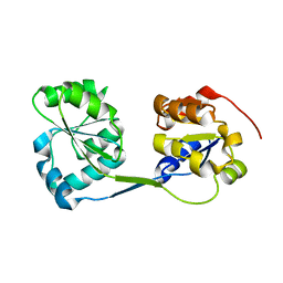
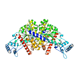
Structure of parA-ADP complex:tetragonal form
Descriptor: ADENOSINE-5'-DIPHOSPHATE, MAGNESIUM ION, Plasmid partition protein A
Authors:Schumacher, M.A.
Deposit date:2008-10-22
Release date:2009-06-02
Last modified:2023-09-06
Method:X-RAY DIFFRACTION (2.58 Å)
Cite:Structural basis for ADP-mediated transcriptional regulation by P1 and P7 ParA.
Embo J., 28, 2009
2XQ5
DownloadVisualize
BU of 2xq5 by Molmil
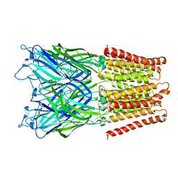
Pentameric ligand gated ion channel GLIC in complex with tetraethylarsonium (TEAs)
Descriptor: ARSENIC, GLR4197 PROTEIN
Authors:Hilf, R.J.C, Bertozzi, C, Zimmermann, I, Reiter, A, Trauner, D, Dutzler, R.
Deposit date:2010-09-01
Release date:2010-11-10
Last modified:2024-05-08
Method:X-RAY DIFFRACTION (3.5 Å)
Cite:Structural Basis of Open Channel Block in a Prokaryotic Pentameric Ligand-Gated Ion Channel
Nat.Struct.Mol.Biol., 17, 2010
4JA7
DownloadVisualize
BU of 4ja7 by Molmil
Rat PP5 co-crystallized with P5SA-2
Descriptor: MAGNESIUM ION, Serine/threonine-protein phosphatase 5
Authors:Haslbeck, V, Helmuth, M, Alte, F, Popowicz, G, Schmidt, W, Weiwad, M, Fischer, G, Gemmecker, G, Sattler, M, Striggow, F, Groll, M, Richter, K.
Deposit date:2013-02-18
Release date:2014-02-19
Last modified:2023-09-20
Method:X-RAY DIFFRACTION (2 Å)
Cite:Selective activators of protein phosphatase 5 target the auto-inhibitory mechanism.
Biosci.Rep., 35, 2015
1DWE
DownloadVisualize
BU of 1dwe by Molmil
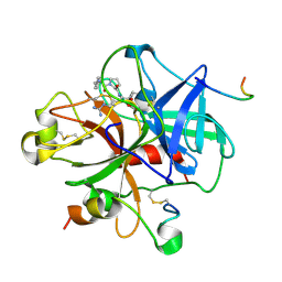
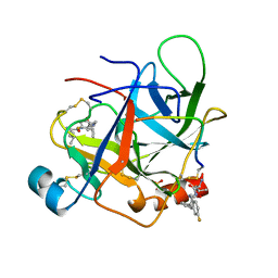
Crystallographic analysis at 3.0-Angstroms resolution of the binding to human thrombin of four active site-directed inhibitors
Descriptor: ALPHA-THROMBIN heavy chain, ALPHA-THROMBIN light chain, D-phenylalanyl-N-[(2S,3S)-6-{[amino(iminio)methyl]amino}-1-chloro-2-hydroxyhexan-3-yl]-L-prolinamide, ...
Authors:Banner, D.W, Hadvary, P.
Deposit date:1992-08-19
Release date:1994-01-31
Last modified:2024-10-16
Method:X-RAY DIFFRACTION (3 Å)
Cite:Crystallographic analysis at 3.0-A resolution of the binding to human thrombin of four active site-directed inhibitors.
J.Biol.Chem., 266, 1991
4GOD
DownloadVisualize
BU of 4god by Molmil
Crystal structure of the SGTA homodimerization domain
Descriptor: (4S)-2-METHYL-2,4-PENTANEDIOL, Small glutamine-rich tetratricopeptide repeat-containing protein alpha
Authors:Chartron, J.W, VanderVelde, D.G, Clemons Jr, W.M.
Deposit date:2012-08-19
Release date:2012-11-21
Last modified:2024-02-28
Method:X-RAY DIFFRACTION (1.4 Å)
Cite:Structures of the Sgt2/SGTA Dimerization Domain with the Get5/UBL4A UBL Domain Reveal an Interaction that Forms a Conserved Dynamic Interface.
Cell Rep, 2, 2012
4C4R
DownloadVisualize
BU of 4c4r by Molmil
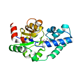
Structure of beta-phosphoglucomutase in complex with a phosphonate analogue of beta-glucose-1-phosphate and magnesium trifluoride
Descriptor: (1R)-1,5-anhydro-1-(phosphonomethyl)-D-glucitol, BETA-PHOSPHOGLUCOMUTASE, MAGNESIUM ION, ...
Authors:Pellegrini, E, Bowler, M.W.
Deposit date:2013-09-09
Release date:2014-07-16
Last modified:2023-12-20
Method:X-RAY DIFFRACTION (1.1 Å)
Cite:Alpha-Fluorophosphonates Reveal How a Phosphomutase Conserves Transition State Conformation Over Hexose Recognition in its Two-Step Reaction.
Proc.Natl.Acad.Sci.USA, 111, 2014
4MXI
DownloadVisualize
BU of 4mxi by Molmil
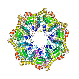
ClpP Ser98dhA
Descriptor: ATP-dependent Clp protease proteolytic subunit
Authors:Gersch, M, Kolb, R, Alte, F, Groll, M, Sieber, S.A.
Deposit date:2013-09-26
Release date:2013-10-23
Last modified:2023-09-20
Method:X-RAY DIFFRACTION (2.3 Å)
Cite:Disruption of Oligomerization and Dehydroalanine Formation as Mechanisms for ClpP Protease Inhibition.
J.Am.Chem.Soc., 136, 2014
1B32
DownloadVisualize
BU of 1b32 by Molmil
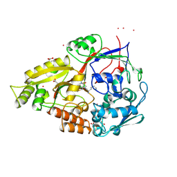
OLIGO-PEPTIDE BINDING PROTEIN (OPPA) COMPLEXED WITH KMK
Descriptor: ACETATE ION, PROTEIN (LYS-MET-LYS), PROTEIN (OLIGO-PEPTIDE BINDING PROTEIN), ...
Authors:Tame, J.R.H, Wilkinson, A.J.
Deposit date:1998-12-15
Release date:1998-12-23
Last modified:2023-08-09
Method:X-RAY DIFFRACTION (1.75 Å)
Cite:Crystallographic and calorimetric analysis of peptide binding to OppA protein.
J.Mol.Biol., 291, 1999
1B3H
DownloadVisualize
BU of 1b3h by Molmil
OLIGO-PEPTIDE BINDING PROTEIN COMPLEXED WITH LYSYL-CYCLOHEXYLALANYL-LYSINE
Descriptor: LYS-ALC-LYS PEPTIDE, PERIPLASMIC OLIGOPEPTIDE-BINDING PROTEIN, URANIUM ATOM
Authors:Davies, T.G, Tame, J.R.H.
Deposit date:1998-11-16
Release date:1998-11-18
Last modified:2023-12-27
Method:X-RAY DIFFRACTION (2 Å)
Cite:Relating structure to thermodynamics: the crystal structures and binding affinity of eight OppA-peptide complexes.
Protein Sci., 8, 1999
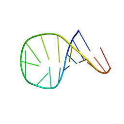
1ATV
DownloadVisualize
BU of 1atv by Molmil
HAIRPIN WITH AGAA TETRALOOP, NMR, 4 STRUCTURES
Descriptor: RNA (5'-R(*GP*GP*GP*AP*CP*CP*AP*GP*AP*AP*GP*GP*UP*CP*CP*CP*G)-3')
Authors:Kang, H.
Deposit date:1997-08-14
Release date:1998-02-25
Last modified:2024-05-22
Method:SOLUTION NMR
Cite:Primary Sequence at the Junction of Stem and Loop in RNA Hairpins Affects the Three-Dimensional Conformation in Solution
To be Published
1EFM
DownloadVisualize
BU of 1efm by Molmil
STRUCTURE OF THE GDP DOMAIN OF EF-TU AND LOCATION OF THE AMINO ACIDS HOMOLOGOUS TO RAS ONCOGENE PROTEINS
Descriptor: ELONGATION FACTOR TU, GUANOSINE-5'-DIPHOSPHATE, MAGNESIUM ION
Authors:Jurnak, F.
Deposit date:1987-05-29
Release date:1987-07-16
Last modified:2024-02-07
Method:X-RAY DIFFRACTION (2.7 Å)
Cite:Structure of the GDP domain of EF-Tu and location of the amino acids homologous to ras oncogene proteins.
Science, 230, 1985
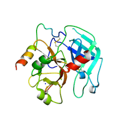
3PMB
DownloadVisualize
BU of 3pmb by Molmil
2.9 Angstrom crystal structure of bovine thrombin in tetragonal spacegroup
Descriptor: SODIUM ION, Thrombin heavy chain, Thrombin light chain
Authors:Wright, H.T, Scarsdale, J.N, Desai, B.J.
Deposit date:2010-11-16
Release date:2011-07-20
Last modified:2024-10-16
Method:X-RAY DIFFRACTION (2.9 Å)
Cite:Interaction of thrombin with sucrose octasulfate.
Biochemistry, 50, 2011
4KM2
DownloadVisualize
BU of 4km2 by Molmil
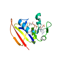
Crystal structure of Dihydrofolate reductase from Mycobacterium tuberculosis in an open conformation in complex with trimethoprim
Descriptor: 2'-MONOPHOSPHOADENOSINE-5'-DIPHOSPHATE, Dihydrofolate reductase, TRIMETHOPRIM
Authors:Dias, M.V.B, Tyrakis, P, Blundell, T.L.
Deposit date:2013-05-07
Release date:2013-12-25
Last modified:2024-02-28
Method:X-RAY DIFFRACTION (1.4 Å)
Cite:Mycobacterium tuberculosis Dihydrofolate Reductase Reveals Two Conformational States and a Possible Low Affinity Mechanism to Antifolate Drugs.
Structure, 22, 2014
4H1P
DownloadVisualize
BU of 4h1p by Molmil
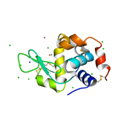
Use of Europium for SAD Phasing at the Cu K alpha wavelength
Descriptor: CHLORIDE ION, EUROPIUM ION, Lysozyme C, ...
Authors:Vijayakumar, B, Velmurugan, D.
Deposit date:2012-09-11
Release date:2012-10-24
Last modified:2020-01-15
Method:X-RAY DIFFRACTION (2.3 Å)
Cite:Use of europium ions for SAD phasing of Lysozyme at the Cu Kalpha wavelength
Acta Crystallogr.,Sect.F, 69, 2013
3Q36
DownloadVisualize
BU of 3q36 by Molmil
Crystal structure of the 4Fe-4S cluster domain of human DNA primase large subunit
Descriptor: DNA primase large subunit, FE (III) ION, IRON/SULFUR CLUSTER
Authors:Agarkar, V.B, Babayeva, N.D, Tahirov, T.H.
Deposit date:2010-12-21
Release date:2011-04-13
Last modified:2023-09-13
Method:X-RAY DIFFRACTION (2.5 Å)
Cite:Crystal structure of the C-terminal domain of human DNA primase large subunit: Implications for the mechanism of the primase - polymerase alpha switch.
Cell Cycle, 10, 2011
1AFQ
DownloadVisualize
BU of 1afq by Molmil
CRYSTAL STRUCTURE OF BOVINE GAMMA-CHYMOTRYPSIN COMPLEXED WITH A SYNTHETIC INHIBITOR
Descriptor: BOVINE GAMMA-CHYMOTRYPSIN, D-leucyl-N-(4-fluorobenzyl)-L-phenylalaninamide, SULFATE ION
Authors:Sugio, S, Kashima, A, Inoue, Y, Maeda, I, Nose, T, Shimohigashi, Y.
Deposit date:1997-03-12
Release date:1997-09-17
Last modified:2024-10-30
Method:X-RAY DIFFRACTION (1.8 Å)
Cite:X-ray crystal structure of a dipeptide-chymotrypsin complex in an inhibitory interaction.
Eur.J.Biochem., 255, 1998
4EQJ
DownloadVisualize
BU of 4eqj by Molmil
Crystal Structure of inactive single chain variant of HIV-1 Protease in Complex with the substrate RT-RH
Descriptor: 1,2-ETHANEDIOL, GLYCEROL, protease, ...
Authors:Schiffer, C.A, Mittal, S.
Deposit date:2012-04-19
Release date:2012-06-06
Last modified:2024-02-28
Method:X-RAY DIFFRACTION (1.8 Å)
Cite:Structural, kinetic, and thermodynamic studies of specificity designed HIV-1 protease.
Protein Sci., 21, 2012
4LOQ
DownloadVisualize
BU of 4loq by Molmil
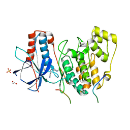
Structural basis of autoactivation of p38 alpha induced by TAB1 (Tetragonal crystal form with bound sulphate)
Descriptor: 1,2-ETHANEDIOL, 4-(4-FLUOROPHENYL)-1-(4-PIPERIDINYL)-5-(2-AMINO-4-PYRIMIDINYL)-IMIDAZOLE, Mitogen-activated protein kinase 14, ...
Authors:Chaikuad, A, DeNicola, G.F, Yue, W.W, von Delft, F, Arrowsmith, C.H, Edwards, A.M, Bountra, C, Marber, M.S, Knapp, S, Structural Genomics Consortium (SGC)
Deposit date:2013-07-13
Release date:2013-08-21
Last modified:2023-09-20
Method:X-RAY DIFFRACTION (2.319 Å)
Cite:Mechanism and consequence of the autoactivation of p38 alpha mitogen-activated protein kinase promoted by TAB1.
Nat.Struct.Mol.Biol., 20, 2013
4CPG
DownloadVisualize
BU of 4cpg by Molmil
Solution structure of the SGTA N-terminal domain
Descriptor: SMALL GLUTAMINE-RICH TETRATRICOPEPTIDE REPEAT-CONTAINING PROTEIN ALPHA
Authors:Darby, J.F, Krysztofinska, E.M, Simpson, P.J, Isaacson, R.L.
Deposit date:2014-02-06
Release date:2014-12-03
Last modified:2024-05-15
Method:SOLUTION NMR
Cite:Solution Structure of the Sgta Dimerisation Domain and Investigation of its Interactions with the Ubiquitin-Like Domains of Bag6 and Ubl4A.
Plos One, 9, 2014

226707

数据于2024-10-30公开中

PDB statisticsPDBj update infoContact PDBjnumon