4ZKH
 
 | |
4ZKX
 | |
4ZMC
 | |
5LHY
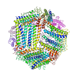
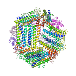
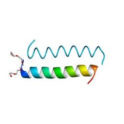
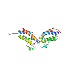
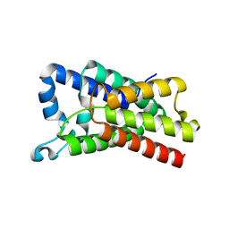
 | | PB3 Domain of Human PLK4 (apo) | | 分子名称: | Serine/threonine-protein kinase PLK4 | | 著者 | Cottee, M.A, Johnson, S, Lea, S.M. | | 登録日 | 2016-07-13 | | 公開日 | 2017-03-01 | | 最終更新日 | 2024-01-10 | | 実験手法 | X-RAY DIFFRACTION (3.31 Å) | | 主引用文献 | A key centriole assembly interaction interface between human PLK4 and STIL appears to not be conserved in flies.
Biol Open, 6, 2017
|
|
5LHZ
 | |
5LHW
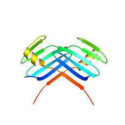
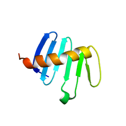
 | | Central Coiled-Coil Domain of Human STIL | | 分子名称: | HEXAETHYLENE GLYCOL, SCL-interrupting locus protein | | 著者 | Cottee, M.A, Lea, S.M. | | 登録日 | 2016-07-13 | | 公開日 | 2017-03-01 | | 最終更新日 | 2024-01-10 | | 実験手法 | X-RAY DIFFRACTION (0.91 Å) | | 主引用文献 | A key centriole assembly interaction interface between human PLK4 and STIL appears to not be conserved in flies.
Biol Open, 6, 2017
|
|
7TO9
 | |
7TO8
 | |
7TOA
 | |
7TO7
 | |
5M9U
 | |
5MLL
 | |
4TMY
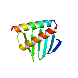
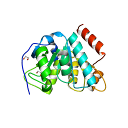
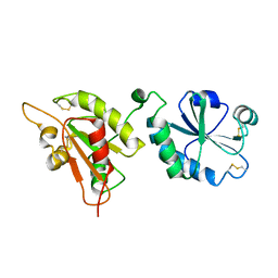
 | | CHEY FROM THERMOTOGA MARITIMA (MG-IV) | | 分子名称: | CHEY PROTEIN, MAGNESIUM ION | | 著者 | Usher, K.C, De La Cruz, A, Dahlquist, F.W, Remington, S.J. | | 登録日 | 1997-06-06 | | 公開日 | 1997-12-03 | | 最終更新日 | 2024-05-22 | | 実験手法 | X-RAY DIFFRACTION (2.8 Å) | | 主引用文献 | Crystal structures of CheY from Thermotoga maritima do not support conventional explanations for the structural basis of enhanced thermostability.
Protein Sci., 7, 1998
|
|
5LHX
 | |
4NJN
 | | Crystal Structure of E.coli GlpG at pH 4.5 | | 分子名称: | Rhomboid protease GlpG | | 著者 | Dickey, S.W, Baker, R.P, Cho, S, Urban, S. | | 登録日 | 2013-11-11 | | 公開日 | 2013-12-25 | | 最終更新日 | 2023-09-20 | | 実験手法 | X-RAY DIFFRACTION (2.4 Å) | | 主引用文献 | Proteolysis inside the Membrane Is a Rate-Governed Reaction Not Driven by Substrate Affinity.
Cell(Cambridge,Mass.), 155, 2013
|
|
4TVE
 | |
4TW5
 | |
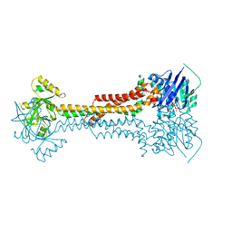
5LJ8
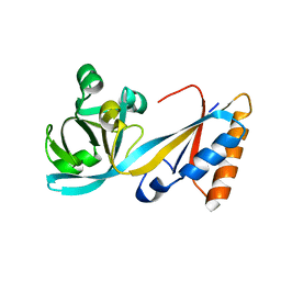 | | Structure of the E. coli MacB periplasmic domain (P21) | | 分子名称: | Macrolide export ATP-binding/permease protein MacB | | 著者 | Crow, A. | | 登録日 | 2016-07-18 | | 公開日 | 2017-11-15 | | 最終更新日 | 2024-01-10 | | 実験手法 | X-RAY DIFFRACTION (1.95 Å) | | 主引用文献 | Structure and mechanotransmission mechanism of the MacB ABC transporter superfamily.
Proc. Natl. Acad. Sci. U.S.A., 114, 2017
|
|
5LJ6
 | |
7Q5G
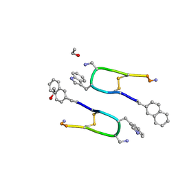 | | LAN-DAP5 DERIVATIVE OF LANREOTIDE: L-DIAMINO PROPIONIC ACID IN POSITION 5 IN PLACE OF L-LYSINE | | 分子名称: | ETHANOL, LAN-DAP5 DERIVATIVE OF LANREOTIDE | | 著者 | Bressanelli, S, Le Du, M.H, Gobeaux, F, Legrand, P, Paternostre, M. | | 登録日 | 2021-11-03 | | 公開日 | 2022-02-02 | | 最終更新日 | 2023-11-15 | | 実験手法 | X-RAY DIFFRACTION (0.83 Å) | | 主引用文献 | Atomic structure of Lanreotide nanotubes revealed by cryo-EM.
Proc.Natl.Acad.Sci.USA, 119, 2022
|
|
5LJ7
 | |
1O45
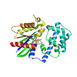
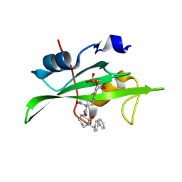 | | CRYSTAL STRUCTURE OF SH2 IN COMPLEX WITH RU84687. | | 分子名称: | N-ACETYL-N-[1-(1,1'-BIPHENYL-4-YLMETHYL)-2-OXOAZEPAN-3-YL]-3-FORMYL-O-PHOSPHONOTYROSINAMIDE, PROTO-ONCOGENE TYROSINE-PROTEIN KINASE SRC | | 著者 | Lange, G, Loenze, P, Liesum, A. | | 登録日 | 2003-06-15 | | 公開日 | 2004-02-17 | | 最終更新日 | 2023-08-16 | | 実験手法 | X-RAY DIFFRACTION (1.8 Å) | | 主引用文献 | Requirements for specific binding of low affinity inhibitor fragments to the SH2 domain of (pp60)Src are identical to those for high affinity binding of full length inhibitors.
J.Med.Chem., 46, 2003
|
|
6ERK
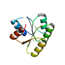
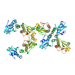
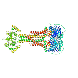
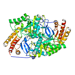 | | Crystal structure of diaminopelargonic acid aminotransferase from Psychrobacter cryohalolentis | | 分子名称: | 1,2-ETHANEDIOL, Aminotransferase, GLYCEROL, ... | | 著者 | Boyko, K.M, Nikolaeva, A.Y, Bezsudnova, E.Y, Stekhanova, T.N, Rakitina, T.V, Popov, V.O. | | 登録日 | 2017-10-18 | | 公開日 | 2018-09-26 | | 最終更新日 | 2024-01-17 | | 実験手法 | X-RAY DIFFRACTION (1.6 Å) | | 主引用文献 | Diaminopelargonic acid transaminase from Psychrobacter cryohalolentis is active towards (S)-(-)-1-phenylethylamine, aldehydes and alpha-diketones.
Appl. Microbiol. Biotechnol., 102, 2018
|
|
5KDL
 | | Crystal structure of the 4 alanine insertion variant of the Gi alpha1 subunit bound to GTPgammaS | | 分子名称: | 5'-GUANOSINE-DIPHOSPHATE-MONOTHIOPHOSPHATE, Guanine nucleotide-binding protein G(i) subunit alpha-1, MAGNESIUM ION | | 著者 | Kaya, A.I, Lokits, A.D, Gilbert, J, Iverson, T.M, Meiler, J, Hamm, H.E. | | 登録日 | 2016-06-08 | | 公開日 | 2016-08-03 | | 最終更新日 | 2023-09-27 | | 実験手法 | X-RAY DIFFRACTION (2.665 Å) | | 主引用文献 | A Conserved Hydrophobic Core in G alpha i1 Regulates G Protein Activation and Release from Activated Receptor.
J.Biol.Chem., 291, 2016
|
|
4WNW
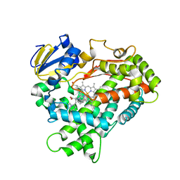 | | Human Cytochrome P450 2D6 Thioridazine Complex | | 分子名称: | 10-{2-[(2R)-1-methylpiperidin-2-yl]ethyl}-2-(methylsulfanyl)-10H-phenothiazine, Cytochrome P450 2D6, NICKEL (II) ION, ... | | 著者 | Wang, A, Stout, C.D, Johnson, E.F. | | 登録日 | 2014-10-14 | | 公開日 | 2015-01-14 | | 最終更新日 | 2023-09-27 | | 実験手法 | X-RAY DIFFRACTION (3.299 Å) | | 主引用文献 | Contributions of Ionic Interactions and Protein Dynamics to Cytochrome P450 2D6 (CYP2D6) Substrate and Inhibitor Binding.
J.Biol.Chem., 290, 2015
|
|


