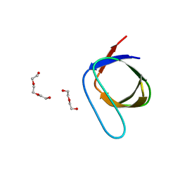
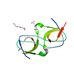
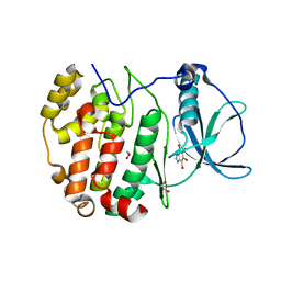
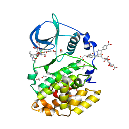
5KDQ
 
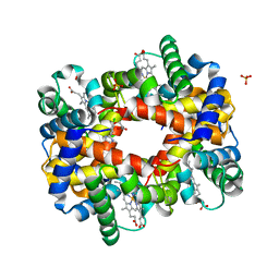 | | Deoxyhemoglobin in Complex with an Aryloxyalkanoic acid | | 分子名称: | 3-[2-chloranyl-4-(1~{H}-imidazol-2-yl)phenoxy]propanoic acid, Hemoglobin subunit alpha, Hemoglobin subunit beta, ... | | 著者 | Ahmed, M.H, Omar, A.M, Safo, M.K. | | 登録日 | 2016-06-08 | | 公開日 | 2016-06-22 | | 最終更新日 | 2023-09-27 | | 実験手法 | X-RAY DIFFRACTION (2.15 Å) | | 主引用文献 | Aryloxyalkanoic Acids as Non-Covalent Modifiers of the Allosteric Properties of Hemoglobin.
Molecules, 21, 2016
|
|
5KDZ
 | |
8C7M
 | |
7PVS
 | |
7PVV
 | |
8C2Z
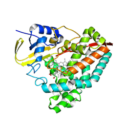
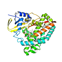
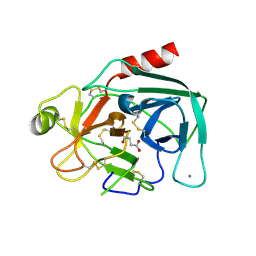
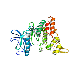 | | Crystal structure of DYRK1B in complex with AZ191 | | 分子名称: | Dual specificity tyrosine-phosphorylation-regulated kinase 1B, MANGANESE (II) ION, N-[2-methoxy-4-(4-methylpiperazin-1-yl)phenyl]-4-(1-methylpyrrolo[2,3-c]pyridin-3-yl)pyrimidin-2-amine | | 著者 | Grygier, P, Pustelny, K, Dubin, G, Czarna, A. | | 登録日 | 2022-12-23 | | 公開日 | 2024-01-17 | | 実験手法 | X-RAY DIFFRACTION (1.91 Å) | | 主引用文献 | Structural perspective on the design of selective DYRK1B inhibitors
To Be Published
|
|
7PVT
 | |
5KGG
 | |
7PVW
 | |
7PW0
 | |
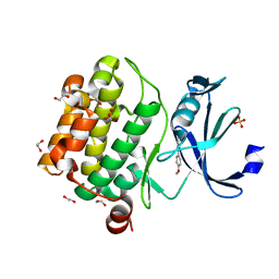
8C5Q
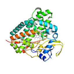
 | | CK2 kinase bound to inhibitor AB668 | | 分子名称: | 2-methylpropyl 5-fluoranyl-3-[1-[[1-[2-[[4-(2-methylpropyl)phenyl]sulfonylamino]ethyl]piperidin-4-yl]methyl]-1,2,3-triazol-4-yl]-1~{H}-indole-2-carboxylate, CHLORIDE ION, Casein kinase II subunit alpha, ... | | 著者 | Krimm, I, Guichou, J.F. | | 登録日 | 2023-01-10 | | 公開日 | 2024-01-24 | | 実験手法 | X-RAY DIFFRACTION (2.5 Å) | | 主引用文献 | CK2 kinase bound to inhibitor AB668
To Be Published
|
|
7PVY
 | |
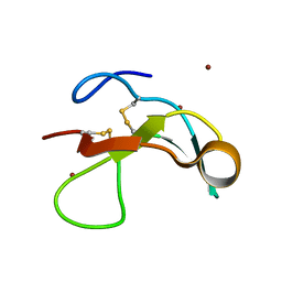
5KAZ
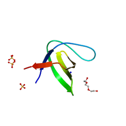
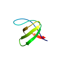
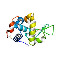
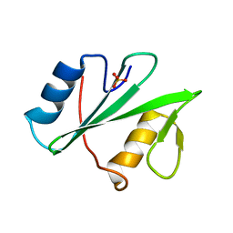 | | Human SH2D1B structure | | 分子名称: | SH2 domain-containing protein 1B, SULFATE ION | | 著者 | Taha, M, Nezerwa, E, Nam, H.-J. | | 登録日 | 2016-06-02 | | 公開日 | 2017-04-12 | | 最終更新日 | 2024-02-28 | | 実験手法 | X-RAY DIFFRACTION (1.7 Å) | | 主引用文献 | The X-ray Crystallographic Structure of Human EAT2 (SH2D1B).
Protein Pept. Lett., 23, 2016
|
|
8QCD
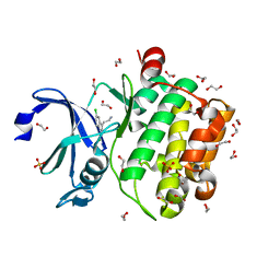
 | | STRUCTURE OF PROTEIN KINASE CK2 CATALYTIC SUBUNIT (ISOFORM CK2ALPHA'; CSNK2A2 GENE PRODUCT) IN COMPLEX WITH THE INHIBITOR 4,5,6,7-TETRABROMOBENZOTRIAZOLE | | 分子名称: | 1,2-ETHANEDIOL, 4,5,6,7-TETRABROMOBENZOTRIAZOLE, Casein kinase II subunit alpha' | | 著者 | Werner, C, Niefind, K. | | 登録日 | 2023-08-25 | | 公開日 | 2023-12-06 | | 実験手法 | X-RAY DIFFRACTION (1.03 Å) | | 主引用文献 | Discovery and Exploration of Protein Kinase CK2 Binding Sites Using CK2alpha Cys336Ser as an Exquisite Crystallographic Tool
Kinases Phosphatases, 2023
|
|
5KDB
 | |
5G5J
 | |
8C9I
 | |
7PSX
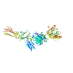
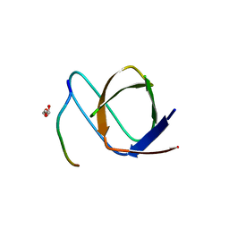
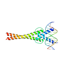
 | | Structure of HOXB13 bound to hydroxymethylated DNA | | 分子名称: | DNA (5'-D(P*GP*GP*AP*CP*CP*TP*5HCP*AP*TP*AP*AP*AP*AP*CP*AP*CP*AP*A)-3'), DNA (5'-D(P*TP*TP*GP*TP*GP*TP*TP*TP*TP*AP*CP*GP*AP*GP*GP*TP*CP*C)-3'), Homeobox protein Hox-B13, ... | | 著者 | Morgunova, E, Popov, A, Yin, Y, Taipale, J. | | 登録日 | 2021-09-24 | | 公開日 | 2022-10-05 | | 最終更新日 | 2024-01-31 | | 実験手法 | X-RAY DIFFRACTION (2 Å) | | 主引用文献 | Structure of HOXB13 bound to hydroxymethylated DNA
To Be Published
|
|
7PRJ
 | |
8QCG
 | |
5KGK
 | |
8Q77
 | | STRUCTURE OF PROTEIN KINASE CK2 CATALYTIC SUBUNIT (ISOFORM CK2ALPHA'; CSNK2A2 GENE PRODUCT) IN COMPLEX WITH THE BISUBSTRATE INHIBITOR ARC-780 | | 分子名称: | (2~{S})-2-[[(2~{S})-2-[[(2~{S})-2-[12-[[4-[[5-(4-carboxyphenyl)-1,3-thiazol-2-yl]amino]-4-oxidanylidene-butanoyl]-(2-hydroxy-2-oxoethyl)amino]dodecanoylamino]-4-oxidanyl-4-oxidanylidene-butanoyl]amino]-4-oxidanyl-4-oxidanylidene-butanoyl]amino]butanedioic acid, 1,2-ETHANEDIOL, CHLORIDE ION, ... | | 著者 | Werner, C, Lindenblatt, D, Niefind, K. | | 登録日 | 2023-08-15 | | 公開日 | 2023-12-06 | | 実験手法 | X-RAY DIFFRACTION (1.255 Å) | | 主引用文献 | Discovery and Exploration of Protein Kinase CK2 Binding Sites Using CK2alpha Cys336Ser as an Exquisite Crystallographic Tool
Kinases Phosphatases, 2023
|
|
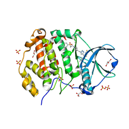
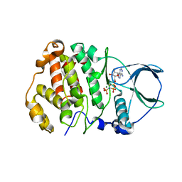
5GIB
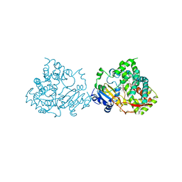
 | | Succinic acid bound trypsin crystallized as dimer | | 分子名称: | CALCIUM ION, Cationic trypsin, SUCCINIC ACID | | 著者 | Kutumbarao, N.H.V, Manohar, R, KarthiK, L, Malathy, P, Gunasekaran, K, Velmurugan, D. | | 登録日 | 2016-06-22 | | 公開日 | 2016-08-10 | | 最終更新日 | 2023-11-08 | | 実験手法 | X-RAY DIFFRACTION (2.697 Å) | | 主引用文献 | Trypsin bound with Succinic acid
To Be Published
|
|
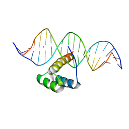
5GNJ
 | | Structure of a transcription factor and DNA complex | | 分子名称: | DNA (5'-D(*AP*GP*GP*AP*AP*CP*AP*CP*GP*TP*GP*AP*CP*CP*C)-3'), DNA (5'-D(*TP*GP*GP*GP*TP*CP*AP*CP*GP*TP*GP*TP*TP*CP*C)-3'), Transcription factor MYC2 | | 著者 | Lian, T, Xu, Y, Su, X. | | 登録日 | 2016-07-21 | | 公開日 | 2017-05-10 | | 最終更新日 | 2024-03-20 | | 実験手法 | X-RAY DIFFRACTION (2.7 Å) | | 主引用文献 | Crystal Structure of Tetrameric Arabidopsis MYC2 Reveals the Mechanism of Enhanced Interaction with DNA.
Cell Rep, 19, 2017
|
|
5K2R
 | |


