3M2M
 
 | |
3EJS
 | | Golgi alpha-Mannosidase II in complex with 5-substituted swainsonine analog: (5S)-5-[2'-(4-tert-butylphenyl)ethyl]-swainsonine | | 分子名称: | (1S,2R,5S,8R,8aR)-5-[2-(4-tert-butylphenyl)ethyl]octahydroindolizine-1,2,8-triol, (4R)-2-METHYLPENTANE-2,4-DIOL, 2-acetamido-2-deoxy-beta-D-glucopyranose, ... | | 著者 | Kuntz, D.A, Shea, K, Rose, D.R. | | 登録日 | 2008-09-18 | | 公開日 | 2009-10-13 | | 最終更新日 | 2024-11-20 | | 実験手法 | X-RAY DIFFRACTION (1.35 Å) | | 主引用文献 | Structural Investigation of the Binding of 5-Substituted Swainsonine Analogues to Golgi alpha-Mannosidase II.
Chembiochem, 11, 2010
|
|
6N2J
 | | Tetrahydropyridopyrimidines as Covalent Inhibitors of KRAS-G12C | | 分子名称: | 1-{4-[7-(naphthalen-1-yl)-5,6,7,8-tetrahydropyrido[3,4-d]pyrimidin-4-yl]piperazin-1-yl}propan-1-one, GTPase KRas, GUANOSINE-5'-DIPHOSPHATE, ... | | 著者 | Vigers, G.P. | | 登録日 | 2018-11-13 | | 公開日 | 2018-12-12 | | 最終更新日 | 2024-11-06 | | 実験手法 | X-RAY DIFFRACTION (1.8 Å) | | 主引用文献 | Discovery of Tetrahydropyridopyrimidines as Irreversible Covalent Inhibitors of KRAS-G12C with In Vivo Activity.
ACS Med Chem Lett, 9, 2018
|
|
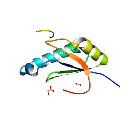
7OES
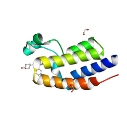
 | | C-TERMINAL BROMODOMAIN OF HUMAN BRD2 WITH 1,5-dimethyl-N-(2-(methylamino)-2-oxoethyl)-6-oxo-N-(2-phenylpropyl)-1,6-dihydropyridine-3-carboxamide | | 分子名称: | 1,2-ETHANEDIOL, 1,5-dimethyl-N-[2-(methylamino)-2-oxidanylidene-ethyl]-6-oxidanylidene-N-[(2S)-2-phenylpropyl]pyridine-3-carboxamide, 2-{2-[2-(2-{2-[2-(2-ETHOXY-ETHOXY)-ETHOXY]-ETHOXY}-ETHOXY)-ETHOXY]-ETHOXY}-ETHANOL, ... | | 著者 | Chung, C. | | 登録日 | 2021-05-03 | | 公開日 | 2021-07-21 | | 最終更新日 | 2024-05-01 | | 実験手法 | X-RAY DIFFRACTION (1.602 Å) | | 主引用文献 | Discovery of a Highly Selective BET BD2 Inhibitor from a DNA-Encoded Library Technology Screening Hit.
J.Med.Chem., 64, 2021
|
|
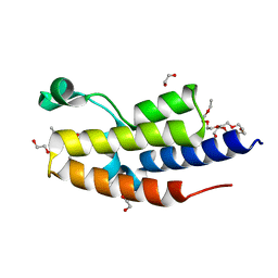
7OE5
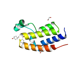
 | | C-TERMINAL BROMODOMAIN OF HUMAN BRD2 WITH N5-hydroxycyclohexyl-N3-methyl-1-phenylethyl-1H-pyrazole-3,5-dicarboxamide | | 分子名称: | 1,2-ETHANEDIOL, 3N-methyl-5N-(4-oxidanylcyclohexyl)-1-[(1S)-1-phenylethyl]pyrazole-3,5-dicarboxamide, Bromodomain-containing protein 2 | | 著者 | Chung, C. | | 登録日 | 2021-05-01 | | 公開日 | 2021-07-28 | | 最終更新日 | 2024-05-01 | | 実験手法 | X-RAY DIFFRACTION (1.602 Å) | | 主引用文献 | Fragment-based Scaffold Hopping: Identification of Potent, Selective, and Highly Soluble Bromo and Extra Terminal Domain (BET) Second Bromodomain (BD2) Inhibitors.
J.Med.Chem., 64, 2021
|
|
6N4K
 | |
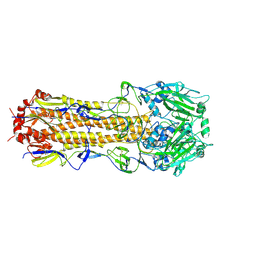
4V4Y
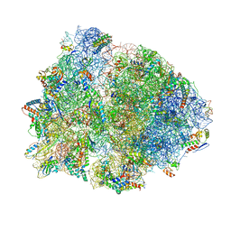 | | Crystal structure of the 70S Thermus thermophilus ribosome with translocated and rotated Shine-Dalgarno Duplex. | | 分子名称: | 16S ribosomal RNA, 23S ribosomal RNA, 30S ribosomal protein S10, ... | | 著者 | Jenner, L, Yusupova, G, Rees, B, Moras, D, Yusupov, M. | | 登録日 | 2006-06-27 | | 公開日 | 2014-07-09 | | 最終更新日 | 2024-11-27 | | 実験手法 | X-RAY DIFFRACTION (5.5 Å) | | 主引用文献 | Structural basis for messenger RNA movement on the ribosome.
Nature, 444, 2006
|
|
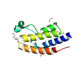
7OE6
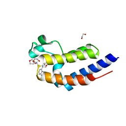
 | | C-TERMINAL BROMODOMAIN OF HUMAN BRD2 WITH N4-hydroxycyclohexyl-N2-methyl-5-phenylethyl-furan-2,4-dicarboxamide | | 分子名称: | 1,2-ETHANEDIOL, 2N-methyl-4N-(4-oxidanylcyclohexyl)-5-[(1S)-1-phenylethyl]furan-2,4-dicarboxamide, Bromodomain-containing protein 2 | | 著者 | Chung, C. | | 登録日 | 2021-05-01 | | 公開日 | 2021-07-28 | | 最終更新日 | 2024-05-01 | | 実験手法 | X-RAY DIFFRACTION (1.762 Å) | | 主引用文献 | Fragment-based Scaffold Hopping: Identification of Potent, Selective, and Highly Soluble Bromo and Extra Terminal Domain (BET) Second Bromodomain (BD2) Inhibitors.
J.Med.Chem., 64, 2021
|
|
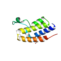
7OE8
 | | C-TERMINAL BROMODOMAIN OF HUMAN BRD2 WITH N5-((1R,5S,6r)-3-oxabicyclo[3.1.0]hexan-6-yl)-3-(1H-indol-4-yl)-N7-methyl-2,3-dihydrobenzofuran-5,7-dicarboxamide | | 分子名称: | (3S)-3-(1H-indol-4-yl)-N7-methyl-N5-[(1R,5S)-3-oxabicyclo[3.1.0]hexan-6-yl]-2,3-dihydro-1-benzofuran-5,7-dicarboxamide, 1,2-ETHANEDIOL, Bromodomain-containing protein 2 | | 著者 | Chung, C. | | 登録日 | 2021-05-02 | | 公開日 | 2021-07-28 | | 最終更新日 | 2024-05-01 | | 実験手法 | X-RAY DIFFRACTION (1.301 Å) | | 主引用文献 | Optimization of a Series of 2,3-Dihydrobenzofurans as Highly Potent, Second Bromodomain (BD2)-Selective, Bromo and Extra-Terminal Domain (BET) Inhibitors.
J.Med.Chem., 64, 2021
|
|
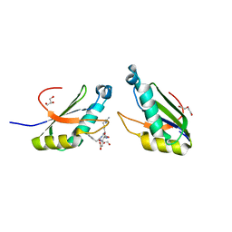
6N3E
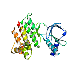
 | | Structure of HIV Tat-specific factor 1 U2AF Homology Motif bound to U2AF ligand motif 4 | | 分子名称: | FORMIC ACID, GLYCEROL, HIV Tat-specific factor 1, ... | | 著者 | Loerch, S, Jenkins, J.L, Kielkopf, C.L. | | 登録日 | 2018-11-15 | | 公開日 | 2019-01-02 | | 最終更新日 | 2024-03-13 | | 実験手法 | X-RAY DIFFRACTION (1.893 Å) | | 主引用文献 | The pre-mRNA splicing and transcription factor Tat-SF1 is a functional partner of the spliceosome SF3b1 subunit via a U2AF homology motif interface.
J. Biol. Chem., 294, 2019
|
|
7OE4
 | | C-TERMINAL BROMODOMAIN OF HUMAN BRD2 WITH N-methyl-4-propionyl-1H-pyrrole-2-carboxamide | | 分子名称: | 1,2-ETHANEDIOL, 2-{2-[2-(2-{2-[2-(2-ETHOXY-ETHOXY)-ETHOXY]-ETHOXY}-ETHOXY)-ETHOXY]-ETHOXY}-ETHANOL, Bromodomain-containing protein 2, ... | | 著者 | Chung, C. | | 登録日 | 2021-05-01 | | 公開日 | 2021-07-28 | | 最終更新日 | 2024-05-01 | | 実験手法 | X-RAY DIFFRACTION (1.653 Å) | | 主引用文献 | Fragment-based Scaffold Hopping: Identification of Potent, Selective, and Highly Soluble Bromo and Extra Terminal Domain (BET) Second Bromodomain (BD2) Inhibitors.
J.Med.Chem., 64, 2021
|
|
7OGY
 | | C-TERMINAL BROMODOMAIN OF HUMAN BRD2 WITH 1-benzyl-N5-cyclopropyl-N3-methyl-1H-pyrazole-3,5-dicarboxamide | | 分子名称: | 1,2-ETHANEDIOL, Bromodomain-containing protein 2, DI(HYDROXYETHYL)ETHER, ... | | 著者 | Chung, C. | | 登録日 | 2021-05-07 | | 公開日 | 2021-07-28 | | 最終更新日 | 2024-05-01 | | 実験手法 | X-RAY DIFFRACTION (1.602 Å) | | 主引用文献 | Fragment-based Scaffold Hopping: Identification of Potent, Selective, and Highly Soluble Bromo and Extra Terminal Domain (BET) Second Bromodomain (BD2) Inhibitors.
J.Med.Chem., 64, 2021
|
|
7OER
 | | C-TERMINAL BROMODOMAIN OF HUMAN BRD2 WITH N-(2,2-diphenylethyl)-1,5-dimethyl-N-(2-(methylamino)-2-oxoethyl)-6-oxo-1,6-dihydropyridine-3-carboxamide | | 分子名称: | 1,2-ETHANEDIOL, Bromodomain-containing protein 2, N-(2,2-diphenylethyl)-1,5-dimethyl-N-[2-(methylamino)-2-oxidanylidene-ethyl]-6-oxidanylidene-pyridine-3-carboxamide | | 著者 | Chung, C. | | 登録日 | 2021-05-03 | | 公開日 | 2021-07-21 | | 最終更新日 | 2024-05-01 | | 実験手法 | X-RAY DIFFRACTION (1.602 Å) | | 主引用文献 | Discovery of a Highly Selective BET BD2 Inhibitor from a DNA-Encoded Library Technology Screening Hit.
J.Med.Chem., 64, 2021
|
|
7O2W
 | | Structure of the C9orf72-SMCR8 complex | | 分子名称: | Guanine nucleotide exchange protein SMCR8,Guanine nucleotide exchange protein SMCR8,Maltose/maltodextrin-binding periplasmic protein, Ubiquitin-like protein SMT3,Guanine nucleotide exchange C9orf72 | | 著者 | Noerpel, J, Cavadini, S, Schenk, A.D, Graff-Meyer, A, Chao, J, Bhaskar, V. | | 登録日 | 2021-03-31 | | 公開日 | 2021-07-21 | | 最終更新日 | 2025-07-02 | | 実験手法 | ELECTRON MICROSCOPY (3.8 Å) | | 主引用文献 | Structure of the human C9orf72-SMCR8 complex reveals a multivalent protein interaction architecture.
Plos Biol., 19, 2021
|
|
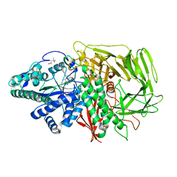
3MOR
 | | Crystal structure of Cathepsin B from Trypanosoma Brucei | | 分子名称: | 2-acetamido-2-deoxy-beta-D-glucopyranose-(1-4)-2-acetamido-2-deoxy-beta-D-glucopyranose, CHLORIDE ION, Cathepsin B-like cysteine protease, ... | | 著者 | Cupelli, K, Stehle, T. | | 登録日 | 2010-04-23 | | 公開日 | 2011-11-02 | | 最終更新日 | 2024-11-06 | | 実験手法 | X-RAY DIFFRACTION (2.55 Å) | | 主引用文献 | In vivo protein crystallization opens new routes in structural biology.
Nat.Methods, 9, 2012
|
|
7OE9
 | | C-TERMINAL BROMODOMAIN OF HUMAN BRD2 WITH rac-N5-((1R,5S)-3-oxabicyclo[3.1.0]hexan-6-yl)-N7,3-dimethyl-3-phenyl-2,3-dihydrobenzofuran-5,7-dicarboxamide | | 分子名称: | (3S)-N7,3-dimethyl-N5-[(1R,5S)-3-oxabicyclo[3.1.0]hexan-6-yl]-3-phenyl-2H-1-benzofuran-5,7-dicarboxamide, 1,2-ETHANEDIOL, Bromodomain-containing protein 2 | | 著者 | Chung, C. | | 登録日 | 2021-05-02 | | 公開日 | 2021-07-28 | | 最終更新日 | 2024-05-01 | | 実験手法 | X-RAY DIFFRACTION (1.602 Å) | | 主引用文献 | Optimization of a Series of 2,3-Dihydrobenzofurans as Highly Potent, Second Bromodomain (BD2)-Selective, Bromo and Extra-Terminal Domain (BET) Inhibitors.
J.Med.Chem., 64, 2021
|
|
7OGT
 | | Folded elbow of cohesin | | 分子名称: | Structural maintenance of chromosomes protein 1, Structural maintenance of chromosomes protein 3 | | 著者 | Lee, B.-G, Gonzalez Llamazares, A, Collier, J, Patele, N.J, Nasmyth, K.A, Lowe, J. | | 登録日 | 2021-05-07 | | 公開日 | 2021-07-28 | | 最終更新日 | 2024-07-10 | | 実験手法 | ELECTRON MICROSCOPY (5.5 Å) | | 主引用文献 | Folding of cohesin's coiled coil is important for Scc2/4-induced association with chromosomes.
Elife, 10, 2021
|
|
3MP6
 | | Complex Structure of Sgf29 and dimethylated H3K4 | | 分子名称: | H3K4me2 peptide, Maltose-binding periplasmic protein,LINKER,SAGA-associated factor 29, alpha-D-glucopyranose-(1-4)-alpha-D-glucopyranose | | 著者 | Li, J, Wu, M, Ruan, J, Zang, J. | | 登録日 | 2010-04-25 | | 公開日 | 2011-05-04 | | 最終更新日 | 2023-11-01 | | 実験手法 | X-RAY DIFFRACTION (1.48 Å) | | 主引用文献 | Sgf29 binds histone H3K4me2/3 and is required for SAGA complex recruitment and histone H3 acetylation
Embo J., 30, 2011
|
|
6MXU
 | |
7OBW
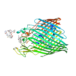 | | Crystal structure of the ferric enterobactin receptor (PfeA) from Pseudomonas aeruginosa in complex with TCV-L6 | | 分子名称: | FE (III) ION, Ferric enterobactin receptor, [4-[[[(2~{S})-2-[[2,3-bis(oxidanyl)phenyl]carbonylamino]-3-[[(2~{S})-2-[[2,3-bis(oxidanyl)phenyl]carbonylamino]-3-[2-[[2,3-bis(oxidanyl)phenyl]carbonylamino]ethanoylamino]propanoyl]amino]propanoyl]amino]methyl]-1,2,3-triazol-1-yl]methyl 4-[4-[(5~{S})-5-(acetamidomethyl)-2-oxidanylidene-1,3-oxazolidin-3-yl]-2-fluoranyl-phenyl]piperazine-1-carboxylate | | 著者 | Moynie, L, Naismith, J.H. | | 登録日 | 2021-04-23 | | 公開日 | 2021-08-04 | | 最終更新日 | 2024-10-23 | | 実験手法 | X-RAY DIFFRACTION (2.66 Å) | | 主引用文献 | Hijacking of the Enterobactin Pathway by a Synthetic Catechol Vector Designed for Oxazolidinone Antibiotic Delivery in Pseudomonas aeruginosa .
Acs Infect Dis., 8, 2022
|
|
3EJR
 | | Golgi alpha-Mannosidase II in complex with 5-substitued swainsonine analog: (5R)-5-[2'-oxo-2'-(4-tert-butylphenyl)ethyl]-swainsonine | | 分子名称: | (4R)-2-METHYLPENTANE-2,4-DIOL, 1-(4-tert-butylphenyl)-2-[(1S,2R,5R,8R,8aR)-1,2,8-trihydroxyoctahydroindolizin-5-yl]ethanone, 2-acetamido-2-deoxy-beta-D-glucopyranose, ... | | 著者 | Kuntz, D.A, Shea, K, Rose, D.R. | | 登録日 | 2008-09-18 | | 公開日 | 2009-10-13 | | 最終更新日 | 2024-10-30 | | 実験手法 | X-RAY DIFFRACTION (1.27 Å) | | 主引用文献 | Structural Investigation of the Binding of 5-Substituted Swainsonine Analogues to Golgi alpha-Mannosidase II.
Chembiochem, 11, 2010
|
|
6N3F
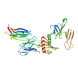
 | | Structure of HIV Tat-specific factor 1 U2AF Homology Motif bound to SF3b1 ULM5 | | 分子名称: | DI(HYDROXYETHYL)ETHER, GLYCEROL, HIV Tat-specific factor 1, ... | | 著者 | Leach, J.R, Jenkins, J.L, Kielkopf, C.L. | | 登録日 | 2018-11-15 | | 公開日 | 2019-01-02 | | 最終更新日 | 2024-03-13 | | 実験手法 | X-RAY DIFFRACTION (2.099 Å) | | 主引用文献 | The pre-mRNA splicing and transcription factor Tat-SF1 is a functional partner of the spliceosome SF3b1 subunit via a U2AF homology motif interface.
J. Biol. Chem., 294, 2019
|
|
7AB0
 | | Apo crystal structure of the MerTK kinase domain | | 分子名称: | CHLORIDE ION, Tyrosine-protein kinase Mer | | 著者 | Pflug, A, Schimpl, M, McCoull, W, Nissink, J.W.M, Overman, R.C, Rawlins, P.B, Truman, C, Underwood, E, Warwicker, J, Winter-Holt, J. | | 登録日 | 2020-09-05 | | 公開日 | 2020-10-28 | | 最終更新日 | 2024-01-31 | | 実験手法 | X-RAY DIFFRACTION (1.74 Å) | | 主引用文献 | A-loop interactions in Mer tyrosine kinase give rise to inhibitors with two-step mechanism and long residence time of binding.
Biochem.J., 477, 2020
|
|
7OHF
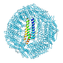 | | Cryo-EM structure of pyrococcus furiosus apoferritin in nanofluidic channels | | 分子名称: | Ferritin | | 著者 | Huber, S.T, Sarajlic, E, Huijink, R, Evers, W.H, Jakobi, A.J. | | 登録日 | 2021-05-10 | | 公開日 | 2021-08-11 | | 最終更新日 | 2024-07-10 | | 実験手法 | ELECTRON MICROSCOPY (3 Å) | | 主引用文献 | Nanofluidic chips for cryo-EM structure determination from picoliter sample volumes.
Elife, 11, 2022
|
|
7OM8
 | |


