5UM6
 
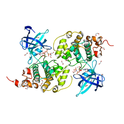
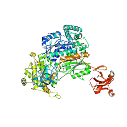 | | Crystal Structure of S. pombe Uba1 in a closed conformation | | 分子名称: | 2-(decylamino)ethane-1-thiol, N-(2-{[(4-chlorophenyl)methyl]disulfanyl}ethyl)decan-1-amine, SULFATE ION, ... | | 著者 | Lv, Z, Yuan, L, Aldana-Masangkay, G, Atkison, J.H, Chen, Y, Olsen, S.K. | | 登録日 | 2017-01-26 | | 公開日 | 2017-06-14 | | 最終更新日 | 2023-10-04 | | 実験手法 | X-RAY DIFFRACTION (2.794 Å) | | 主引用文献 | Domain alternation and active site remodeling are conserved structural features of ubiquitin E1.
J. Biol. Chem., 292, 2017
|
|
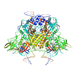
1ZJW
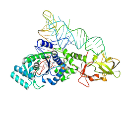 | | Glutaminyl-tRNA synthetase complexed to glutamine and 2'deoxy A76 glutamine tRNA | | 分子名称: | ADENOSINE MONOPHOSPHATE, GLUTAMINE, Glutaminyl-tRNA, ... | | 著者 | Gruic-Sovulj, I, Uter, N, Bullock, T, Perona, J.J. | | 登録日 | 2005-05-01 | | 公開日 | 2005-06-07 | | 最終更新日 | 2024-02-14 | | 実験手法 | X-RAY DIFFRACTION (2.5 Å) | | 主引用文献 | tRNA-dependent Aminoacyl-adenylate Hydrolysis by a Nonediting Class I Aminoacyl-tRNA Synthetase.
J.Biol.Chem., 280, 2005
|
|
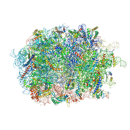
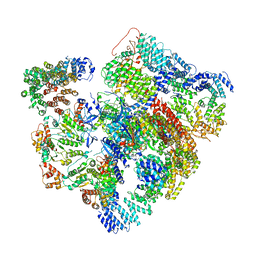
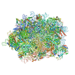
8CVT
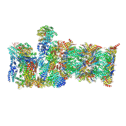 | | Human 19S-20S proteasome, state SD2 | | 分子名称: | 26S protease regulatory subunit 10B, 26S protease regulatory subunit 8, 26S proteasome complex subunit SEM1, ... | | 著者 | Zhao, J. | | 登録日 | 2022-05-18 | | 公開日 | 2022-11-02 | | 実験手法 | ELECTRON MICROSCOPY (3 Å) | | 主引用文献 | Structural insights into the human PA28-20S proteasome enabled by efficient tagging and purification of endogenous proteins.
Proc.Natl.Acad.Sci.USA, 119, 2022
|
|
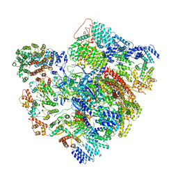
3JBX
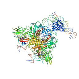
 | | Cryo-electron microscopy structure of RAG Signal End Complex (C2 symmetry) | | 分子名称: | 5'-D(*CP*AP*CP*AP*GP*TP*GP*CP*TP*AP*CP*AP*GP*AP*C)-3', 5'-D(*GP*CP*GP*AP*TP*GP*GP*TP*TP*AP*AP*CP*CP*A)-3', 5'-D(P*GP*TP*CP*TP*GP*TP*AP*GP*CP*AP*CP*TP*GP*TP*G)-3', ... | | 著者 | Ru, H, Chambers, M.G, Fu, T.-M, Tong, A.B, Liao, M, Wu, H. | | 登録日 | 2015-10-22 | | 公開日 | 2015-12-09 | | 最終更新日 | 2024-02-21 | | 実験手法 | ELECTRON MICROSCOPY (3.4 Å) | | 主引用文献 | Molecular Mechanism of V(D)J Recombination from Synaptic RAG1-RAG2 Complex Structures.
Cell(Cambridge,Mass.), 163, 2015
|
|
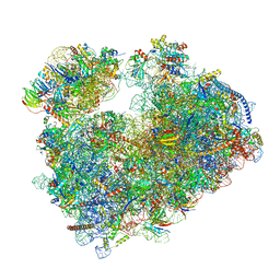
7BHP
 | | Cryo-EM structure of the human Ebp1 - 80S ribosome | | 分子名称: | 28S ribosomal RNA, 5.8S ribosomal RNA, 5S ribosomal RNA, ... | | 著者 | Desogus, J, Bhaskar, V, Chao, J.A. | | 登録日 | 2021-01-11 | | 公開日 | 2021-02-03 | | 最終更新日 | 2021-03-31 | | 実験手法 | ELECTRON MICROSCOPY (3.3 Å) | | 主引用文献 | Dynamic association of human Ebp1 with the ribosome.
Rna, 27, 2021
|
|
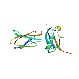
2LAS
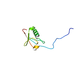
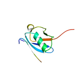 | | Molecular Determinants of Paralogue-Specific SUMO-SIM Recognition | | 分子名称: | M-IR2_peptide, Small ubiquitin-related modifier 1 | | 著者 | Namanja, A, Li, Y, Su, Y, Wong, S, Lu, J, Colson, L, Wu, C, Li, S, Chen, Y. | | 登録日 | 2011-03-20 | | 公開日 | 2011-12-14 | | 最終更新日 | 2024-05-15 | | 実験手法 | SOLUTION NMR | | 主引用文献 | Insights into High Affinity Small Ubiquitin-like Modifier (SUMO) Recognition by SUMO-interacting Motifs (SIMs) Revealed by a Combination of NMR and Peptide Array Analysis.
J.Biol.Chem., 287, 2012
|
|
2N1V
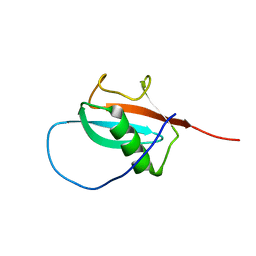 | |
2N1A
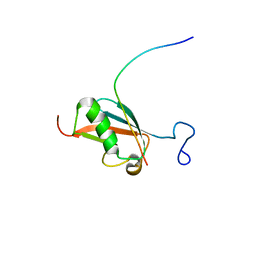
 | | Docked structure between SUMO1 and ZZ-domain from CBP | | 分子名称: | CREB-binding protein, Small ubiquitin-related modifier 1, ZINC ION | | 著者 | Diehl, C. | | 登録日 | 2015-03-26 | | 公開日 | 2016-05-04 | | 最終更新日 | 2024-05-01 | | 実験手法 | SOLUTION NMR | | 主引用文献 | Structural Analysis of a Complex between Small Ubiquitin-like Modifier 1 (SUMO1) and the ZZ Domain of CREB-binding Protein (CBP/p300) Reveals a New Interaction Surface on SUMO.
J.Biol.Chem., 291, 2016
|
|
2MW5
 | |
2BYT
 | | Thermus thermophilus Leucyl-tRNA synthetase complexed with a tRNAleu transcript in the post-editing conformation | | 分子名称: | LEUCINE, LEUCYL-TRNA SYNTHETASE, MERCURY (II) ION, ... | | 著者 | Cusack, S, Tukalo, M, Yaremchuk, A, Fukunaga, R, Yokoyama, S. | | 登録日 | 2005-08-04 | | 公開日 | 2005-09-15 | | 最終更新日 | 2023-12-13 | | 実験手法 | X-RAY DIFFRACTION (3.3 Å) | | 主引用文献 | The Crystal Structure of Leucyl-tRNA Synthetase Complexed with tRNA(Leu) in the Post-Transfer-Editing Conformation.
Nat.Struct.Mol.Biol., 12, 2005
|
|
2KQS
 | |
3JBW
 | | Cryo-electron microscopy structure of RAG Paired Complex (with NBD, no symmetry) | | 分子名称: | 12-RSS signal end forward strand, 5'-D(P*GP*AP*TP*CP*TP*GP*GP*CP*CP*TP*GP*TP*CP*TP*TP*A)-3', Nicked 12-RSS intermediate reverse strand, ... | | 著者 | Ru, H, Chambers, M.G, Fu, T.-M, Tong, A.B, Liao, M, Wu, H. | | 登録日 | 2015-10-21 | | 公開日 | 2015-12-09 | | 最終更新日 | 2024-02-21 | | 実験手法 | ELECTRON MICROSCOPY (4.6 Å) | | 主引用文献 | Molecular Mechanism of V(D)J Recombination from Synaptic RAG1-RAG2 Complex Structures.
Cell(Cambridge,Mass.), 163, 2015
|
|
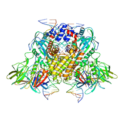
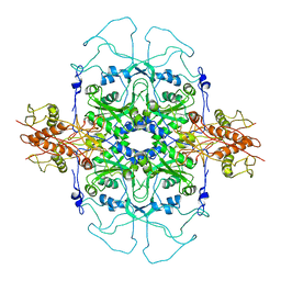
8XN6
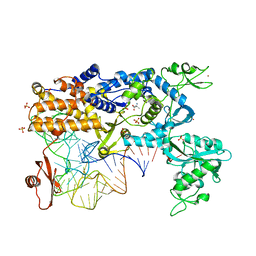
 | | The Crystal Structure of GSK3b from Biortus. | | 分子名称: | 1,2-ETHANEDIOL, DI(HYDROXYETHYL)ETHER, Glycogen synthase kinase-3 beta, ... | | 著者 | Wang, F, Cheng, W, Yuan, Z, Qi, J, Wu, B. | | 登録日 | 2023-12-29 | | 公開日 | 2024-01-24 | | 実験手法 | X-RAY DIFFRACTION (2.4 Å) | | 主引用文献 | The Crystal Structure of GSK3b from Biortus.
To Be Published
|
|
5VHH
 | | Conformational Landscape of the p28-Bound Human Proteasome Regulatory Particle | | 分子名称: | 26S proteasome complex subunit SEM1, 26S proteasome non-ATPase regulatory subunit 1, 26S proteasome non-ATPase regulatory subunit 10, ... | | 著者 | Lu, Y, Wu, J, Dong, Y, Chen, S, Sun, S, Ma, Y.B, Ouyang, Q, Finley, D, Kirschner, M.W, Mao, Y. | | 登録日 | 2017-04-13 | | 公開日 | 2017-08-23 | | 最終更新日 | 2023-08-16 | | 実験手法 | ELECTRON MICROSCOPY (6.1 Å) | | 主引用文献 | Conformational Landscape of the p28-Bound Human Proteasome Regulatory Particle.
Mol. Cell, 67, 2017
|
|
5VHI
 | | Conformational Landscape of the p28-Bound Human Proteasome Regulatory Particle | | 分子名称: | 26S proteasome complex subunit SEM1, 26S proteasome non-ATPase regulatory subunit 1, 26S proteasome non-ATPase regulatory subunit 10, ... | | 著者 | Lu, Y, Wu, J, Dong, Y, Chen, S, Sun, S, Ma, Y.B, Ouyang, Q, Finley, D, Kirschner, M.W, Mao, Y. | | 登録日 | 2017-04-13 | | 公開日 | 2017-08-23 | | 最終更新日 | 2023-08-16 | | 実験手法 | ELECTRON MICROSCOPY (6.8 Å) | | 主引用文献 | Conformational Landscape of the p28-Bound Human Proteasome Regulatory Particle.
Mol. Cell, 67, 2017
|
|
3RZW
 | |
8BPO
 | | Structure of rabbit 80S ribosome translating beta-tubulin in complex with tetratricopeptide protein 5 (TTC5) and S-phase Cyclin A Associated Protein residing in the ER (SCAPER) | | 分子名称: | 28S ribosomal RNA, 5.8S ribosomal RNA, 5S ribosomal RNA, ... | | 著者 | Hopfler, M, Absmeier, E, Passmore, L.A, Hegde, R.S. | | 登録日 | 2022-11-17 | | 公開日 | 2023-07-05 | | 最終更新日 | 2023-07-19 | | 実験手法 | ELECTRON MICROSCOPY (2.8 Å) | | 主引用文献 | Mechanism of ribosome-associated mRNA degradation during tubulin autoregulation.
Mol.Cell, 83, 2023
|
|
3JAG
 | | Structure of a mammalian ribosomal termination complex with ABCE1, eRF1(AAQ), and the UAA stop codon | | 分子名称: | 18S ribosomal RNA, 28S ribosomal RNA, 5.8S ribosomal RNA, ... | | 著者 | Brown, A, Shao, S, Murray, J, Hegde, R.S, Ramakrishnan, V. | | 登録日 | 2015-06-10 | | 公開日 | 2015-08-12 | | 最終更新日 | 2018-07-18 | | 実験手法 | ELECTRON MICROSCOPY (3.65 Å) | | 主引用文献 | Structural basis for stop codon recognition in eukaryotes.
Nature, 524, 2015
|
|
3JAH
 | | Structure of a mammalian ribosomal termination complex with ABCE1, eRF1(AAQ), and the UAG stop codon | | 分子名称: | 18S ribosomal RNA, 28S ribosomal RNA, 5.8S ribosomal RNA, ... | | 著者 | Brown, A, Shao, S, Murray, J, Hegde, R.S, Ramakrishnan, V. | | 登録日 | 2015-06-10 | | 公開日 | 2015-08-12 | | 最終更新日 | 2018-07-18 | | 実験手法 | ELECTRON MICROSCOPY (3.45 Å) | | 主引用文献 | Structural basis for stop codon recognition in eukaryotes.
Nature, 524, 2015
|
|
3JAI
 | | Structure of a mammalian ribosomal termination complex with ABCE1, eRF1(AAQ), and the UGA stop codon | | 分子名称: | 18S ribosomal RNA, 28S ribosomal RNA, 5.8S ribosomal RNA, ... | | 著者 | Brown, A, Shao, S, Murray, J, Hegde, R.S, Ramakrishnan, V. | | 登録日 | 2015-06-10 | | 公開日 | 2015-08-12 | | 最終更新日 | 2018-07-18 | | 実験手法 | ELECTRON MICROSCOPY (3.65 Å) | | 主引用文献 | Structural basis for stop codon recognition in eukaryotes.
Nature, 524, 2015
|
|
2DU7
 | |
1YVL
 | | Structure of Unphosphorylated STAT1 | | 分子名称: | 5-residue peptide, GOLD ION, Signal transducer and activator of transcription 1-alpha/beta | | 著者 | Mao, X, Ren, Z, Parker, G.N, Sondermann, H, Pastorello, M.A, Wang, W, McMurray, J.S, Demeler, B, Darnell Jr, J.E, Chen, X. | | 登録日 | 2005-02-16 | | 公開日 | 2005-03-22 | | 最終更新日 | 2011-07-13 | | 実験手法 | X-RAY DIFFRACTION (3 Å) | | 主引用文献 | Structural bases of unphosphorylated STAT1 association and receptor binding.
Mol.Cell, 17, 2005
|
|
3JBY
 | | Cryo-electron microscopy structure of RAG Paired Complex (C2 symmetry) | | 分子名称: | '-D(P*GP*AP*TP*CP*TP*GP*GP*CP*CP*TP*GP*TP*CP*TP*TP*A)-3', 5'-D(P*CP*AP*CP*AP*GP*TP*GP*CP*TP*AP*CP*AP*GP*AP*C)-3', CALCIUM ION, ... | | 著者 | Ru, H, Chambers, M.G, Fu, T.-M, Tong, A.B, Liao, M, Wu, H. | | 登録日 | 2015-10-22 | | 公開日 | 2015-12-09 | | 最終更新日 | 2024-02-21 | | 実験手法 | ELECTRON MICROSCOPY (3.7 Å) | | 主引用文献 | Molecular Mechanism of V(D)J Recombination from Synaptic RAG1-RAG2 Complex Structures.
Cell(Cambridge,Mass.), 163, 2015
|
|
7PZY
 | | Structure of the vacant Candida albicans 80S ribosome | | 分子名称: | 1,4-DIAMINOBUTANE, 18S ribosomal RNA, 25S ribosomal RNA, ... | | 著者 | Zgadzay, Y, Kolosova, O, Stetsenko, A, Jenner, L, Guskov, A, Yusupova, G, Yusupov, M. | | 登録日 | 2021-10-13 | | 公開日 | 2022-05-18 | | 最終更新日 | 2022-06-08 | | 実験手法 | ELECTRON MICROSCOPY (2.32 Å) | | 主引用文献 | E-site drug specificity of the human pathogen Candida albicans ribosome.
Sci Adv, 8, 2022
|
|
7Q0P
 | | Structure of the Candida albicans 80S ribosome in complex with anisomycin | | 分子名称: | 18S ribosomal RNA, 25S ribosomal RNA, 40S ribosomal protein S0, ... | | 著者 | Kolosova, O, Zgadzay, Y, Stetsenko, A, Jenner, L, Guskov, A, Yusupova, G, Yusupov, M. | | 登録日 | 2021-10-15 | | 公開日 | 2022-05-18 | | 最終更新日 | 2022-06-08 | | 実験手法 | ELECTRON MICROSCOPY (2.77 Å) | | 主引用文献 | E-site drug specificity of the human pathogen Candida albicans ribosome.
Sci Adv, 8, 2022
|
|


