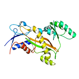
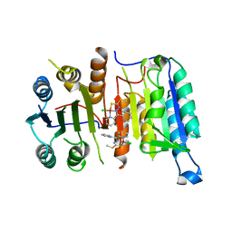
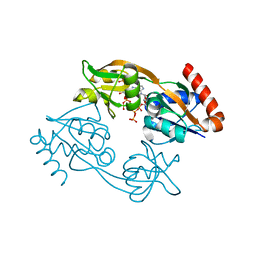
7XV9
 
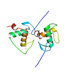 | | Crystal structure of the Human TR4 DNA-Binding Domain | | 分子名称: | Nuclear receptor subfamily 2 group C member 2, ZINC ION | | 著者 | Liu, Y, Chen, Z. | | 登録日 | 2022-05-21 | | 公開日 | 2022-12-28 | | 最終更新日 | 2023-11-29 | | 実験手法 | X-RAY DIFFRACTION (1.599 Å) | | 主引用文献 | Structures of human TR4LBD-JAZF1 and TR4DBD-DNA complexes reveal the molecular basis of transcriptional regulation.
Nucleic Acids Res., 51, 2023
|
|
7XV8
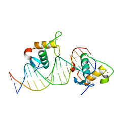 | |
7XV6
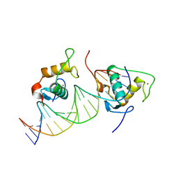 | |
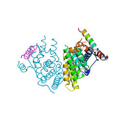
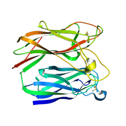
3NG4
 | | Ternary complex of peptidoglycan recognition protein (PGRP-S) with Maltose and N-Acetylglucosamine at 1.7 A Resolution | | 分子名称: | 2-acetamido-2-deoxy-beta-D-glucopyranose, GLYCEROL, Peptidoglycan recognition protein 1, ... | | 著者 | Sharma, P, Dube, D, Kaur, P, Sharma, S, Singh, T.P. | | 登録日 | 2010-06-10 | | 公開日 | 2010-07-14 | | 最終更新日 | 2023-11-01 | | 実験手法 | X-RAY DIFFRACTION (1.73 Å) | | 主引用文献 | Multiligand specificity of pathogen-associated molecular pattern-binding site in peptidoglycan recognition protein
J.Biol.Chem., 286, 2011
|
|
7XVA
 | |
7XZJ
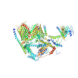 | | Cryo-EM structure of TOC complex from Chlamydomonas reinhardtii. | | 分子名称: | Ctap3, INOSITOL HEXAKISPHOSPHATE, Tic100, ... | | 著者 | Liu, H, Li, A.J, Liu, Z.F. | | 登録日 | 2022-06-02 | | 公開日 | 2023-01-11 | | 最終更新日 | 2024-07-03 | | 実験手法 | ELECTRON MICROSCOPY (2.97 Å) | | 主引用文献 | Architecture of chloroplast TOC-TIC translocon supercomplex.
Nature, 615, 2023
|
|
7XZI
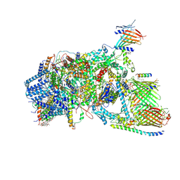 | | Cryo-EM structure of TOC-TIC supercomplex from Chlamydomonas reinhardtii | | 分子名称: | 1,2-DIPALMITOYL-PHOSPHATIDYL-GLYCEROLE, 1,2-DISTEAROYL-MONOGALACTOSYL-DIGLYCERIDE, Ctap3, ... | | 著者 | Liu, H, Li, A.J, Liu, Z.F. | | 登録日 | 2022-06-02 | | 公開日 | 2023-01-11 | | 最終更新日 | 2023-03-22 | | 実験手法 | ELECTRON MICROSCOPY (2.77 Å) | | 主引用文献 | Architecture of chloroplast TOC-TIC translocon supercomplex.
Nature, 615, 2023
|
|
2F55
 | | Two hepatitis c virus ns3 helicase domains complexed with the same strand of dna | | 分子名称: | 5'-D(P*(DU)P*(DU)P*(DU))-3', 5'-D(P*(DU)P*(DU)P*(DU)P*(DU)P*(DU)P*(DU)P*(DU)P*(DU)P*(DU)P*(DU)P*(DU)P*(DU)P*(DU))-3', SULFATE ION, ... | | 著者 | Lu, J.Z, Jordan, J.B, Sakon, J. | | 登録日 | 2005-11-25 | | 公開日 | 2005-12-06 | | 最終更新日 | 2023-08-23 | | 実験手法 | X-RAY DIFFRACTION (3.3 Å) | | 主引用文献 | Structural and biological identification of residues on the surface of NS3 helicase required for optimal replication of the hepatitis C virus
J.Biol.Chem., 281, 2006
|
|
4ZO1
 | | Crystal Structure of the T3-bound TR-beta Ligand-binding Domain in complex with RXR-alpha | | 分子名称: | 3,5,3'TRIIODOTHYRONINE, Nuclear receptor coactivator 2, Retinoic acid receptor RXR-alpha, ... | | 著者 | Bruning, J.B, Kojetin, D.J, Matta-Camacho, E, Hughes, T.S, Srinivasan, S, Nwachukwu, J.C, Cavett, V, Nowak, J, Chalmers, M.J, Marciano, D.P, Kamenecka, T.M, Rance, M, Shulman, A.I, Mangelsdorf, D.J, Griffin, P.R, Nettles, K.W. | | 登録日 | 2015-05-05 | | 公開日 | 2015-09-02 | | 最終更新日 | 2023-11-15 | | 実験手法 | X-RAY DIFFRACTION (3.221 Å) | | 主引用文献 | Structural mechanism for signal transduction in RXR nuclear receptor heterodimers.
Nat Commun, 6, 2015
|
|
4ZHV
 | | Crystal structure of a bacterial signalling protein | | 分子名称: | SULFATE ION, YfiB | | 著者 | Li, S, Li, T, Wang, Y, Bartlam, M. | | 登録日 | 2015-04-27 | | 公開日 | 2016-04-27 | | 最終更新日 | 2024-03-20 | | 実験手法 | X-RAY DIFFRACTION (1.585 Å) | | 主引用文献 | Structural insights into YfiR sequestering by YfiB in Pseudomonas aeruginosa PAO1
Sci Rep, 5, 2015
|
|
3TIG
 | | Tubulin tyrosine ligase | | 分子名称: | MAGNESIUM ION, Ttl protein | | 著者 | Roll-Mecak, A, Szyk, A, Deaconescu, A, Piszczek, G. | | 登録日 | 2011-08-20 | | 公開日 | 2011-10-26 | | 最終更新日 | 2024-02-28 | | 実験手法 | X-RAY DIFFRACTION (2.5 Å) | | 主引用文献 | Tubulin tyrosine ligase structure reveals adaptation of an ancient fold to bind and modify tubulin.
Nat.Struct.Mol.Biol., 18, 2011
|
|
7X32
 | |
1PNG
 | |
2H24
 | | Crystal structure of human IL-10 | | 分子名称: | Interleukin-10 | | 著者 | Yoon, S.I, Walter, M.R. | | 登録日 | 2006-05-18 | | 公開日 | 2006-10-17 | | 最終更新日 | 2023-08-30 | | 実験手法 | X-RAY DIFFRACTION (2 Å) | | 主引用文献 | Conformational changes mediate interleukin-10 receptor 2 (IL-10R2) binding to IL-10 and assembly of the signaling complex.
J.Biol.Chem., 281, 2006
|
|
4FEA
 | |
2LGF
 | |
3TCQ
 | | Crystal Structure of matrix protein VP40 from Ebola virus Sudan | | 分子名称: | Matrix protein VP40 | | 著者 | Clifton, M.C, Edwards, T.E, Anderson, V, Atkins, K, Raymond, A, Saphire, E.O, Seattle Structural Genomics Center for Infectious Disease, Seattle Structural Genomics Center for Infectious Disease (SSGCID) | | 登録日 | 2011-08-09 | | 公開日 | 2012-10-03 | | 最終更新日 | 2023-09-13 | | 実験手法 | X-RAY DIFFRACTION (1.6 Å) | | 主引用文献 | High-resolution Crystal Structure of Dimeric VP40 From Sudan ebolavirus.
J Infect Dis, 212 Suppl 2, 2015
|
|
7Z6R
 | | Psychrobacter arcticus ATPPRT (HisGZ) R56A mutant bound to ATP and PRPP | | 分子名称: | 1-O-pyrophosphono-5-O-phosphono-alpha-D-ribofuranose, ADENOSINE-5'-TRIPHOSPHATE, ATP phosphoribosyltransferase, ... | | 著者 | Alphey, M.S, Fisher, G, da Silva, R.G. | | 登録日 | 2022-03-14 | | 公開日 | 2022-03-23 | | 最終更新日 | 2024-02-07 | | 実験手法 | X-RAY DIFFRACTION (2.55 Å) | | 主引用文献 | Allosteric rescue of catalytically impaired ATP phosphoribosyltransferase variants links protein dynamics to active-site electrostatic preorganisation.
Nat Commun, 13, 2022
|
|
7Z8U
 | | Catalytic subunit HisG R56A mutant from Psychrobacter arcticus ATPPRT (HisGZ) in complex with ATP and PRPP | | 分子名称: | 1-O-pyrophosphono-5-O-phosphono-alpha-D-ribofuranose, ADENOSINE-5'-TRIPHOSPHATE, ATP phosphoribosyltransferase, ... | | 著者 | Alphey, M.S, Fisher, G, da Silva, R.G. | | 登録日 | 2022-03-18 | | 公開日 | 2022-03-30 | | 最終更新日 | 2024-02-07 | | 実験手法 | X-RAY DIFFRACTION (2 Å) | | 主引用文献 | Allosteric rescue of catalytically impaired ATP phosphoribosyltransferase variants links protein dynamics to active-site electrostatic preorganisation.
Nat Commun, 13, 2022
|
|
2FCA
 | | The structure of BsTrmB | | 分子名称: | POTASSIUM ION, tRNA (guanine-N(7)-)-methyltransferase | | 著者 | Zegers, I, Van Vliet, F, Bujnicki, J, Kosinski, J, Gigot, D, Droogmans, L. | | 登録日 | 2005-12-12 | | 公開日 | 2006-08-15 | | 最終更新日 | 2024-02-14 | | 実験手法 | X-RAY DIFFRACTION (2.1 Å) | | 主引用文献 | Crystal structure of Bacillus subtilis TrmB, the tRNA (m7G46) methyltransferase.
Nucleic Acids Res., 34, 2006
|
|
3TII
 | | Tubulin tyrosine ligase | | 分子名称: | MAGNESIUM ION, PHOSPHOAMINOPHOSPHONIC ACID-ADENYLATE ESTER, Ttl protein | | 著者 | Roll-Mecak, A, Szyk, A, Deaconescu, A, Piszczek, G. | | 登録日 | 2011-08-20 | | 公開日 | 2011-10-26 | | 最終更新日 | 2024-02-28 | | 実験手法 | X-RAY DIFFRACTION (2.5 Å) | | 主引用文献 | Tubulin tyrosine ligase structure reveals adaptation of an ancient fold to bind and modify tubulin.
Nat.Struct.Mol.Biol., 18, 2011
|
|
3TXO
 | | PKC eta kinase in complex with a naphthyridine | | 分子名称: | 2-methyl-N~1~-[3-(pyridin-4-yl)-2,6-naphthyridin-1-yl]propane-1,2-diamine, Protein kinase C eta type | | 著者 | Stark, W, Rummel, G, Cowan-Jacob, S.W. | | 登録日 | 2011-09-23 | | 公開日 | 2011-11-30 | | 最終更新日 | 2011-12-07 | | 実験手法 | X-RAY DIFFRACTION (2.05 Å) | | 主引用文献 | 2,6-Naphthyridines as potent and selective inhibitors of the novel protein kinase C isozymes.
Bioorg.Med.Chem.Lett., 21, 2011
|
|
7Z18
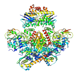 | | E. coli C-P lyase bound to a PhnK ABC dimer and ATP | | 分子名称: | ADENOSINE-5'-TRIPHOSPHATE, Alpha-D-ribose 1-methylphosphonate 5-phosphate C-P lyase, Alpha-D-ribose 1-methylphosphonate 5-triphosphate synthase subunit PhnG, ... | | 著者 | Amstrup, S.K, Sofos, N, Karlsen, J.L, Skjerning, R.B, Boesen, T, Enghild, J.J, Hove-Jensen, B, Brodersen, D.E. | | 登録日 | 2022-02-24 | | 公開日 | 2022-05-25 | | 最終更新日 | 2024-07-17 | | 実験手法 | ELECTRON MICROSCOPY (1.98 Å) | | 主引用文献 | Structural remodelling of the carbon-phosphorus lyase machinery by a dual ABC ATPase.
Nat Commun, 14, 2023
|
|
7Z19
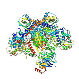 | | E. coli C-P lyase bound to a single PhnK ABC domain | | 分子名称: | Alpha-D-ribose 1-methylphosphonate 5-phosphate C-P lyase, Alpha-D-ribose 1-methylphosphonate 5-triphosphate synthase subunit PhnG, Alpha-D-ribose 1-methylphosphonate 5-triphosphate synthase subunit PhnH, ... | | 著者 | Amstrup, S.K, Sofos, N, Karlsen, J.L, Skjerning, R.B, Boesen, T, Enghild, J.J, Hove-Jensen, B, Brodersen, D.E. | | 登録日 | 2022-02-24 | | 公開日 | 2022-05-25 | | 最終更新日 | 2024-07-17 | | 実験手法 | ELECTRON MICROSCOPY (2.57 Å) | | 主引用文献 | Structural remodelling of the carbon-phosphorus lyase machinery by a dual ABC ATPase.
Nat Commun, 14, 2023
|
|
7Z17
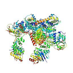 | | E. coli C-P lyase bound to a PhnK ABC dimer in an open conformation | | 分子名称: | Alpha-D-ribose 1-methylphosphonate 5-phosphate C-P lyase, Alpha-D-ribose 1-methylphosphonate 5-triphosphate synthase subunit PhnG, Alpha-D-ribose 1-methylphosphonate 5-triphosphate synthase subunit PhnH, ... | | 著者 | Amstrup, S.K, Sofos, N, Karlsen, J.L, Skjerning, R.B, Boesen, T, Enghild, J.J, Hove-Jensen, B, Brodersen, D.E. | | 登録日 | 2022-02-24 | | 公開日 | 2022-05-25 | | 最終更新日 | 2024-07-17 | | 実験手法 | ELECTRON MICROSCOPY (2.57 Å) | | 主引用文献 | Structural remodelling of the carbon-phosphorus lyase machinery by a dual ABC ATPase.
Nat Commun, 14, 2023
|
|


