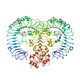
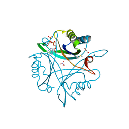
5OAB
 
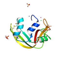 | | A novel crystal form of human RNase6 at atomic resolution | | 分子名称: | CHLORIDE ION, PHOSPHATE ION, POTASSIUM ION, ... | | 著者 | Prats-Ejarque, G, Moussaoui, M, Boix, E. | | 登録日 | 2017-06-21 | | 公開日 | 2018-08-01 | | 最終更新日 | 2024-01-17 | | 実験手法 | X-RAY DIFFRACTION (1.111 Å) | | 主引用文献 | Characterization of an RNase with two catalytic centers. Human RNase6 catalytic and phosphate-binding site arrangement favors the endonuclease cleavage of polymeric substrates.
Biochim Biophys Acta Gen Subj, 1863, 2019
|
|
7TKZ
 | |
7TRN
 | |
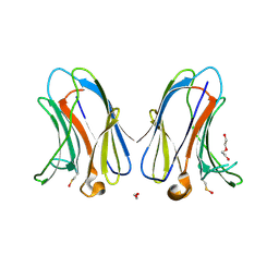
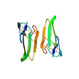
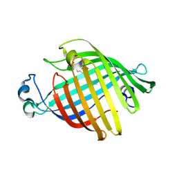
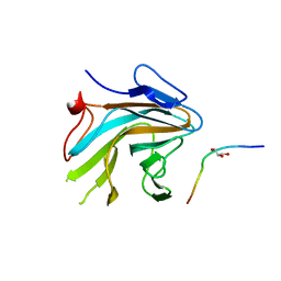
7TRO
 | | Crystal structure of R14A-R20A human Galectin-7 mutant in presence of lactose | | 分子名称: | 1,2-ETHANEDIOL, 2-AMINO-2-HYDROXYMETHYL-PROPANE-1,3-DIOL, Galectin-7, ... | | 著者 | Pham, N.T.H, Calmettes, C, Doucet, N. | | 登録日 | 2022-01-29 | | 公開日 | 2023-02-01 | | 最終更新日 | 2023-10-25 | | 実験手法 | X-RAY DIFFRACTION (1.8 Å) | | 主引用文献 | Crystal structure of R14A-R20A human Galectin-7 mutant in presence of lactose
To Be Published
|
|
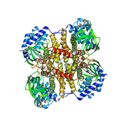
4CY9
 | | DpsA14 from Streptomyces coelicolor | | 分子名称: | DPSA, GLYCEROL | | 著者 | Townsend, P.D, Hitchings, M.D, Del Sol, R, Pohl, E. | | 登録日 | 2014-04-10 | | 公開日 | 2014-06-25 | | 最終更新日 | 2024-05-08 | | 実験手法 | X-RAY DIFFRACTION (1.78 Å) | | 主引用文献 | A Tale of Tails: Deciphering the Contribution of Terminal Tails to the Biochemical Properties of Two Dps Proteins from Streptomyces Coelicolor
Cell.Mol.Life Sci., 71, 2014
|
|
6Z2L
 | |
6PNW
 | |
7QI3
 | | Structure of Fusarium verticillioides NAT1 (FDB2) N-malonyltransferase | | 分子名称: | 1,2-ETHANEDIOL, Arylamine N-acetyltransferase, DI(HYDROXYETHYL)ETHER, ... | | 著者 | Lowe, E.D, Kotomina, E, Karagianni, E, Boukouvala, S. | | 登録日 | 2021-12-14 | | 公開日 | 2022-11-23 | | 最終更新日 | 2024-02-07 | | 実験手法 | X-RAY DIFFRACTION (1.8 Å) | | 主引用文献 | Fusarium verticillioides NAT1 (FDB2) N-malonyltransferase is structurally, functionally and phylogenetically distinct from its N-acetyltransferase (NAT) homologues.
Febs J., 290, 2023
|
|
1FW3
 | | OUTER MEMBRANE PHOSPHOLIPASE A FROM ESCHERICHIA COLI | | 分子名称: | OUTER MEMBRANE PHOSPHOLIPASE A | | 著者 | Snijder, H.J, Kingma, R.L, Kalk, K.H, Dekker, N, Egmond, M.R, Dijkstra, B.W. | | 登録日 | 2000-09-21 | | 公開日 | 2001-06-01 | | 最終更新日 | 2022-12-21 | | 実験手法 | X-RAY DIFFRACTION (2.8 Å) | | 主引用文献 | Structural investigations of calcium binding and its role in activity and activation of outer membrane phospholipase A from Escherichia coli.
J.Mol.Biol., 309, 2001
|
|
6PK1
 | |
8K5C
 | |
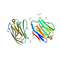
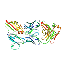
3MBX
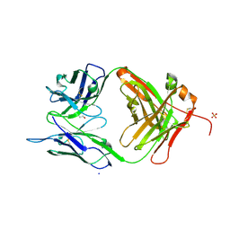 | | Crystal structure of chimeric antibody X836 | | 分子名称: | PHOSPHATE ION, SODIUM ION, X836 HEAVY CHAIN, ... | | 著者 | Teplyakov, A, Obmolova, G, Gilliland, G.L. | | 登録日 | 2010-03-26 | | 公開日 | 2010-06-30 | | 最終更新日 | 2023-09-06 | | 実験手法 | X-RAY DIFFRACTION (1.6 Å) | | 主引用文献 | On the domain pairing in chimeric antibodies.
Mol.Immunol., 47, 2010
|
|
4R07
 | | Crystal structure of human TLR8 in complex with ORN06 | | 分子名称: | 2-acetamido-2-deoxy-beta-D-glucopyranose, 2-acetamido-2-deoxy-beta-D-glucopyranose-(1-4)-2-acetamido-2-deoxy-beta-D-glucopyranose, 3'-O-[(R)-{[(2R,3aR,4R,6R,6aR)-6-(2-amino-6-oxo-1,6-dihydro-9H-purin-9-yl)-2-hydroxy-2-oxidotetrahydrofuro[3,4-d][1,3,2]dioxaphosphol-4-yl]methoxy}(hydroxy)phosphoryl]uridine 5'-(dihydrogen phosphate), ... | | 著者 | Tanji, H, Ohto, U, Shibata, T, Taoka, M, Yamauchi, Y, Isobe, T, Miyake, K, Shimizu, T. | | 登録日 | 2014-07-30 | | 公開日 | 2015-01-14 | | 最終更新日 | 2020-07-29 | | 実験手法 | X-RAY DIFFRACTION (2 Å) | | 主引用文献 | Toll-like receptor 8 senses degradation products of single-stranded RNA
Nat.Struct.Mol.Biol., 22, 2015
|
|
8K5D
 | |
8K5B
 | |
7XND
 | |
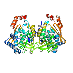
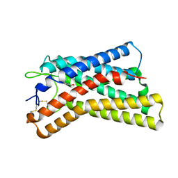
4X28
 | | Crystal structure of the ChsE4-ChsE5 complex from Mycobacterium tuberculosis | | 分子名称: | Acyl-CoA dehydrogenase, DIHYDROFLAVINE-ADENINE DINUCLEOTIDE | | 著者 | Guja, K.E, Yang, M, Sampson, N, Garcia-Diaz, M. | | 登録日 | 2014-11-26 | | 公開日 | 2015-02-18 | | 最終更新日 | 2019-12-18 | | 実験手法 | X-RAY DIFFRACTION (1.99 Å) | | 主引用文献 | Unraveling Cholesterol Catabolism in Mycobacterium tuberculosis: ChsE4-ChsE5 alpha 2 beta 2 Acyl-CoA Dehydrogenase Initiates beta-Oxidation of 3-Oxo-cholest-4-en-26-oyl CoA.
Acs Infect Dis., 1, 2015
|
|
4X8P
 | | Crystal structure of Ash2L SPRY domain in complex with RbBP5 | | 分子名称: | GLYCEROL, Retinoblastoma-binding protein 5, Set1/Ash2 histone methyltransferase complex subunit ASH2,Set1/Ash2 histone methyltransferase complex subunit ASH2 | | 著者 | Zhang, P, Chaturvedi, C.P, Brunzelle, J.S, Skiniotis, G, Brand, M, Shilatifard, A, Couture, J.-F. | | 登録日 | 2014-12-10 | | 公開日 | 2015-01-28 | | 最終更新日 | 2024-02-28 | | 実験手法 | X-RAY DIFFRACTION (2.2 Å) | | 主引用文献 | A phosphorylation switch on RbBP5 regulates histone H3 Lys4 methylation.
Genes Dev., 29, 2015
|
|
7XNP
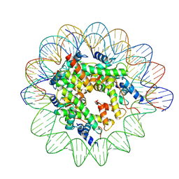 | | Structure of nucleosome-AAG complex (A-55I, post-catalytic state) | | 分子名称: | DNA (152-MER), Histone H2A type 1, Histone H2B 1.1, ... | | 著者 | Zheng, L, Tsai, B, Gao, N. | | 登録日 | 2022-04-29 | | 公開日 | 2023-05-03 | | 最終更新日 | 2023-11-08 | | 実験手法 | ELECTRON MICROSCOPY (2.9 Å) | | 主引用文献 | Structural and mechanistic insights into the DNA glycosylase AAG-mediated base excision in nucleosome.
Cell Discov, 9, 2023
|
|
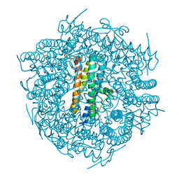
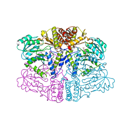
1OHH
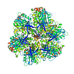 | | BOVINE MITOCHONDRIAL F1-ATPASE complexed with the inhibitor protein IF1 | | 分子名称: | ATP synthase subunit alpha, mitochondrial, ATP synthase subunit beta, ... | | 著者 | Cabezon, E, Montgomery, M.G, Leslie, A.G.W, Walker, J.E. | | 登録日 | 2003-05-27 | | 公開日 | 2003-06-09 | | 最終更新日 | 2023-12-13 | | 実験手法 | X-RAY DIFFRACTION (2.8 Å) | | 主引用文献 | The Structure of Bovine F1-ATPase in Complex with its Regulatory Protein If1
Nat.Struct.Biol., 10, 2003
|
|
7P52
 | |
4CYB
 | | DpsC from Streptomyces coelicolor | | 分子名称: | FE (III) ION, PUTATIVE DNA PROTECTION PROTEIN, SODIUM ION | | 著者 | Townsend, P.D, Hitchings, M.D, Del Sol, R, Pohl, E. | | 登録日 | 2014-04-10 | | 公開日 | 2014-06-25 | | 最終更新日 | 2024-05-08 | | 実験手法 | X-RAY DIFFRACTION (1.78 Å) | | 主引用文献 | A Tale of Tails: Deciphering the Contribution of Terminal Tails to the Biochemical Properties of Two Dps Proteins from Streptomyces Coelicolor
Cell.Mol.Life Sci., 71, 2014
|
|
6PRK
 | | X-ray Crystal Structure of Bacillus subtilis RicA in complex with RicF | | 分子名称: | RicA, RicF | | 著者 | Khaja, F.T, Jeffrey, P.D, Neiditch, M.B, Dubnau, D. | | 登録日 | 2019-07-10 | | 公開日 | 2019-10-02 | | 最終更新日 | 2023-10-11 | | 実験手法 | X-RAY DIFFRACTION (3.2 Å) | | 主引用文献 | Structure-Function Studies of the Bacillus subtilis Ric Proteins Identify the Fe-S Cluster-Ligating Residues and Their Roles in Development and RNA Processing.
Mbio, 10, 2019
|
|
6PRH
 | |
7TV2
 | |


