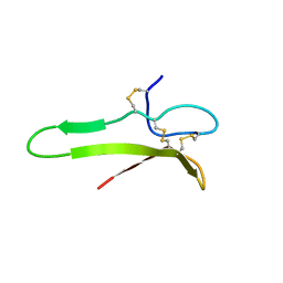
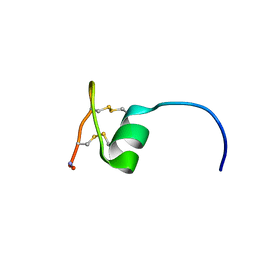
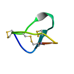
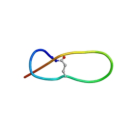
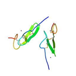
7C13
 
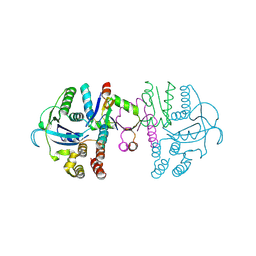 | | beta1 domain-swapped structure of monothiol cGrx1(C16S) | | 分子名称: | Glutaredoxin, Peptide methionine sulfoxide reductase MsrA | | 著者 | Lee, K, Hwang, K.Y. | | 登録日 | 2020-05-02 | | 公開日 | 2020-11-18 | | 最終更新日 | 2024-10-09 | | 実験手法 | X-RAY DIFFRACTION (2.799 Å) | | 主引用文献 | Monothiol and dithiol glutaredoxin-1 from clostridium oremlandii: identification of domain-swapped structures by NMR, X-ray crystallography and HDX mass spectrometry.
Iucrj, 7, 2020
|
|
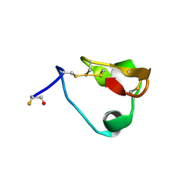
7C12
 | |
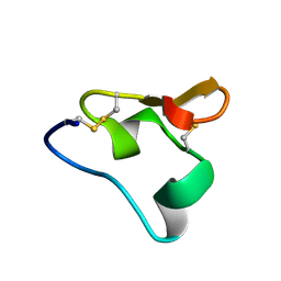
7E51
 | |
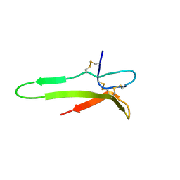
7E4X
 | |
1XI7
 | | NMR structure of the carboxyl-terminal cysteine domain of the VHv1.1 polydnaviral gene product | | 分子名称: | cysteine-rich omega-conotoxin homolog VHv1.1 | | 著者 | Einerwold, J, Jaseja, M, Hapner, K, Webb, B, Copie, V. | | 登録日 | 2004-09-21 | | 公開日 | 2004-10-05 | | 最終更新日 | 2011-08-10 | | 実験手法 | SOLUTION NMR | | 主引用文献 | Solution structure of the carboxyl-terminal cysteine-rich domain of the VHv1.1 polydnaviral gene product: comparison with other cystine knot structural folds
Biochemistry, 40, 2001
|
|
2N8C
 | |
2N8B
 | |
1XJ1
 | | 3D solution structure of the C-terminal cysteine-rich domain of the VHv1.1 polydnaviral gene product | | 分子名称: | cysteine-rich omega-conotoxin homolog VHv1.1 | | 著者 | Einerwold, J, Jaseja, J, Hapner, K, Webb, B, Copie, V. | | 登録日 | 2004-09-22 | | 公開日 | 2004-10-05 | | 最終更新日 | 2024-10-09 | | 実験手法 | SOLUTION NMR | | 主引用文献 | Solution structure of the carboxyl-terminal cysteine-rich domain of the VHv1.1 polydnaviral gene product: comparison with other cystine knot structural folds
Biochemistry, 40, 2001
|
|
2NAV
 | | NMR solution structure of Ex-4[1-16]/pl14a | | 分子名称: | Exendin-4, Alpha/kappa-conotoxin pl14a chimera | | 著者 | Schroeder, C.I, Swedberg, J.E, Craik, D.J. | | 登録日 | 2016-01-11 | | 公開日 | 2016-05-04 | | 最終更新日 | 2016-08-17 | | 実験手法 | SOLUTION NMR | | 主引用文献 | Truncated Glucagon-like Peptide-1 and Exendin-4 alpha-Conotoxin pl14a Peptide Chimeras Maintain Potency and alpha-Helicity and Reveal Interactions Vital for cAMP Signaling in Vitro.
J.Biol.Chem., 291, 2016
|
|
3KFC
 | |
1FYG
 | |
1MVI
 | | N-TYPE CALCIUM CHANNEL BLOCKER, OMEGA-CONOTOXIN MVIIA, NMR, 15 STRUCTURES | | 分子名称: | MVIIA | | 著者 | Nielsen, K.J, Thomas, L, Lewis, R.J, Alewood, P.F, Craik, D.J. | | 登録日 | 1996-08-02 | | 公開日 | 1997-08-12 | | 最終更新日 | 2017-11-29 | | 実験手法 | SOLUTION NMR | | 主引用文献 | A consensus structure for omega-conotoxins with different selectivities for voltage-sensitive calcium channel subtypes: comparison of MVIIA, SVIB and SNX-202.
J.Mol.Biol., 263, 1996
|
|
1MVJ
 | | N-TYPE CALCIUM CHANNEL BLOCKER, OMEGA-CONOTOXIN MVIIA NMR, 15 STRUCTURES | | 分子名称: | SVIB | | 著者 | Nielsen, K.J, Thomas, L, Lewis, R.J, Alewood, P.F, Craik, D.J. | | 登録日 | 1996-08-02 | | 公開日 | 1997-08-12 | | 最終更新日 | 2017-11-29 | | 実験手法 | SOLUTION NMR | | 主引用文献 | A consensus structure for omega-conotoxins with different selectivities for voltage-sensitive calcium channel subtypes: comparison of MVIIA, SVIB and SNX-202.
J.Mol.Biol., 263, 1996
|
|
3KV0
 | | Crystal structure of HET-C2: A FUNGAL GLYCOLIPID TRANSFER PROTEIN (GLTP) | | 分子名称: | HET-C2 | | 著者 | Simanshu, D.K, Kenoth, R, Brown, R.E, Patel, D.J. | | 登録日 | 2009-11-28 | | 公開日 | 2010-02-23 | | 最終更新日 | 2023-09-06 | | 実験手法 | X-RAY DIFFRACTION (1.9 Å) | | 主引用文献 | Structural determination and tryptophan fluorescence of heterokaryon incompatibility C2 protein (HET-C2), a fungal glycolipid transfer protein (GLTP), provide novel insights into glycolipid specificity and membrane interaction by the GLTP fold.
J.Biol.Chem., 285, 2010
|
|
1F27
 | | CRYSTAL STRUCTURE OF A BIOTIN-BINDING RNA PSEUDOKNOT | | 分子名称: | BIOTIN, MAGNESIUM ION, RNA (5'-R(*AP*AP*AP*AP*AP*GP*UP*CP*CP*UP*C)-3'), ... | | 著者 | Nix, J, Sussman, D, Wilson, C. | | 登録日 | 2000-05-23 | | 公開日 | 2000-06-12 | | 最終更新日 | 2024-02-07 | | 実験手法 | X-RAY DIFFRACTION (1.3 Å) | | 主引用文献 | The 1.3 A crystal structure of a biotin-binding pseudoknot and the basis for RNA molecular recognition.
J.Mol.Biol., 296, 2000
|
|
3GSZ
 | |
1PP5
 | | Structure of Antibacterial Peptide Microcin J25: a 21-Residue Lariat Protoknot | | 分子名称: | microcin J25 | | 著者 | Bayro, M.J, Swapna, G.V.T, Huang, J.Y, Ma, L.-C, Mukhopadhyay, J, Ebright, R.H, Montelione, G.T, Northeast Structural Genomics Consortium (NESG) | | 登録日 | 2003-06-16 | | 公開日 | 2003-10-28 | | 最終更新日 | 2012-12-12 | | 実験手法 | SOLUTION NMR | | 主引用文献 | Structure of Antibacterial Peptide Microcin J25: A 21-Residue Lariat Protoknot.
J.Am.Chem.Soc., 125, 2003
|
|
2M8K
 | | A pyrimidine motif triple helix in the Kluyveromyces lactis telomerase RNA pseudoknot is essential for function in vivo | | 分子名称: | RNA (48-MER) | | 著者 | Cash, D.D, Cohen, O, Kim, N, Shefer, K, Brown, Y, Ulyanov, N.B, Tzfati, Y, Feigon, J. | | 登録日 | 2013-05-22 | | 公開日 | 2013-06-19 | | 最終更新日 | 2024-05-15 | | 実験手法 | SOLUTION NMR | | 主引用文献 | Pyrimidine motif triple helix in the Kluyveromyces lactis telomerase RNA pseudoknot is essential for function in vivo.
Proc.Natl.Acad.Sci.USA, 110, 2013
|
|
3E07
 | | Crystal structure of spatzle cystine knot | | 分子名称: | GLYCEROL, Protein spaetzle | | 著者 | Hoffmann, A, Funkner, A, Neumann, P, Juhnke, S, Walther, M, Schierhorn, A, Weininger, U, Balbach, J, Reuter, G, Stubbs, M.T. | | 登録日 | 2008-07-31 | | 公開日 | 2008-09-23 | | 最終更新日 | 2023-11-01 | | 実験手法 | X-RAY DIFFRACTION (2.4 Å) | | 主引用文献 | Biophysical Characterization of Refolded Drosophila Spatzle, a Cystine Knot Protein, Reveals Distinct Properties of Three Isoforms
J.Biol.Chem., 283, 2008
|
|
2G1W
 | |
1EDM
 | | EPIDERMAL GROWTH FACTOR-LIKE DOMAIN FROM HUMAN FACTOR IX | | 分子名称: | CALCIUM ION, FACTOR IX | | 著者 | Rao, Z, Handford, P, Mayhew, M, Knott, V, Brownlee, G.G, Stuart, D. | | 登録日 | 1996-03-21 | | 公開日 | 1996-10-14 | | 最終更新日 | 2024-10-09 | | 実験手法 | X-RAY DIFFRACTION (1.5 Å) | | 主引用文献 | The structure of a Ca(2+)-binding epidermal growth factor-like domain: its role in protein-protein interactions.
Cell(Cambridge,Mass.), 82, 1995
|
|
2GJD
 | | Distinct functional domains of Ubc9 dictate cell survival and resistance to genotoxic stress | | 分子名称: | Ubiquitin-conjugating enzyme E2-18 kDa | | 著者 | van Waardenburg, R.C, Duda, D.M, Lancaster, C.S, Schulman, B.A, Bjornsti, M.A. | | 登録日 | 2006-03-30 | | 公開日 | 2006-07-04 | | 最終更新日 | 2023-08-30 | | 実験手法 | X-RAY DIFFRACTION (1.75 Å) | | 主引用文献 | Distinct functional domains of ubc9 dictate cell survival and resistance to genotoxic stress.
Mol.Cell.Biol., 26, 2006
|
|
387D
 | | RNA Pseudoknot with 3D Domain Swapping | | 分子名称: | RNA Pseudoknot | | 著者 | Lietzke, S.E, Kundrot, C.E, Barnes, C.L. | | 登録日 | 1998-04-14 | | 公開日 | 2003-08-26 | | 最終更新日 | 2023-12-27 | | 実験手法 | X-RAY DIFFRACTION (3.1 Å) | | 主引用文献 | The Structure of an RNA Pseudoknot Shows 3D Domain Swapping
Structure, Motion, Interaction and Expression of Biological Macromolecules, The Proceedings of the Tenth Conversation held at The University-SUNY, Albany NY, June 17-21, 1997, 10, 1998
|
|
3GBA
 | | X-ray structure of iGluR5 ligand-binding core (S1S2) in complex with dysiherbaine at 1.35A resolution | | 分子名称: | (2R,3aR,6S,7R,7aR)-2-[(2S)-2-amino-2-carboxyethyl]-6-hydroxy-7-(methylamino)hexahydro-2H-furo[3,2-b]pyran-2-carboxylic acid, CHLORIDE ION, GLYCEROL, ... | | 著者 | Frydenvang, K, Naur, P, Gajhede, M, Kastrup, J.S. | | 登録日 | 2009-02-19 | | 公開日 | 2009-03-17 | | 最終更新日 | 2023-11-01 | | 実験手法 | X-RAY DIFFRACTION (1.35 Å) | | 主引用文献 | Full Domain Closure of the Ligand-binding Core of the Ionotropic Glutamate Receptor iGluR5 Induced by the High Affinity Agonist Dysiherbaine and the Functional Antagonist 8,9-Dideoxyneodysiherbaine
J.Biol.Chem., 284, 2009
|
|
3GBB
 | | X-ray structure of iGluR5 ligand-binding core (S1S2) in complex with MSVIII-19 at 2.10A resolution | | 分子名称: | (2R,3aR,7aR)-2-[(2S)-2-amino-3-hydroxy-3-oxo-propyl]-3,3a,5,6,7,7a-hexahydrofuro[4,5-b]pyran-2-carboxylic acid, Glutamate receptor, ionotropic kainate 1 | | 著者 | Frydenvang, K, Naur, P, Gajhede, M, Kastrup, J.S. | | 登録日 | 2009-02-19 | | 公開日 | 2009-03-17 | | 最終更新日 | 2023-11-01 | | 実験手法 | X-RAY DIFFRACTION (2.1 Å) | | 主引用文献 | Full Domain Closure of the Ligand-binding Core of the Ionotropic Glutamate Receptor iGluR5 Induced by the High Affinity Agonist Dysiherbaine and the Functional Antagonist 8,9-Dideoxyneodysiherbaine
J.Biol.Chem., 284, 2009
|
|


