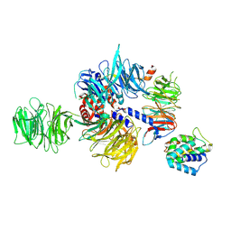
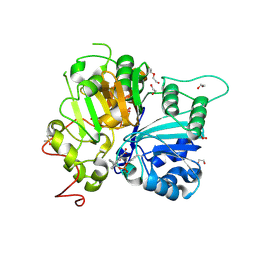
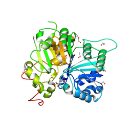
7KIB
 
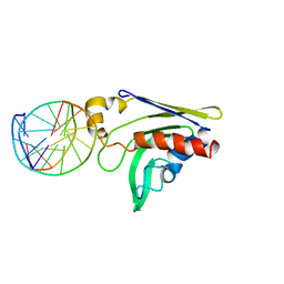
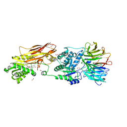 | | PRMT5:MEP50 Complexed with 5,5-Bicyclic Inhibitor Compound 4 | | 分子名称: | (2R,3R,3aS,6S,6aR)-6-[(2-amino-3-bromoquinolin-7-yl)oxy]-2-(4-amino-7H-pyrrolo[2,3-d]pyrimidin-7-yl)hexahydro-3aH-cyclopenta[b]furan-3,3a-diol, 1,2-ETHANEDIOL, CHLORIDE ION, ... | | 著者 | Palte, R.L, Hayes, R.P. | | 登録日 | 2020-10-23 | | 公開日 | 2021-04-21 | | 最終更新日 | 2023-10-18 | | 実験手法 | X-RAY DIFFRACTION (2.52 Å) | | 主引用文献 | The Discovery of Two Novel Classes of 5,5-Bicyclic Nucleoside-Derived PRMT5 Inhibitors for the Treatment of Cancer.
J.Med.Chem., 64, 2021
|
|
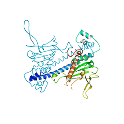
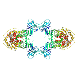
7BV7
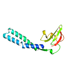
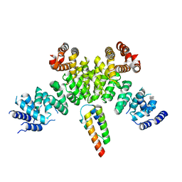 | | INTS3 complexed with INTS6 | | 分子名称: | Integrator complex subunit 3, Integrator complex subunit 6 | | 著者 | Jia, Y, Bharath, S.R, Song, H. | | 登録日 | 2020-04-09 | | 公開日 | 2021-07-14 | | 最終更新日 | 2024-05-29 | | 実験手法 | X-RAY DIFFRACTION (2.4 Å) | | 主引用文献 | Crystal structure of the INTS3/INTS6 complex reveals the functional importance of INTS3 dimerization in DSB repair.
Cell Discov, 7, 2021
|
|
6Y6X
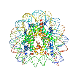
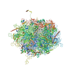 | | Tetracenomycin X bound to the human ribosome | | 分子名称: | 28S ribosomal RNA, 5.8S ribosomal RNA, 5S ribosomal RNA, ... | | 著者 | Buschauer, R, Cheng, J, Berninghausen, O, Beckmann, R, Wilson, D.N. | | 登録日 | 2020-02-27 | | 公開日 | 2020-07-08 | | 最終更新日 | 2023-11-08 | | 実験手法 | ELECTRON MICROSCOPY (2.8 Å) | | 主引用文献 | Tetracenomycin X inhibits translation by binding within the ribosomal exit tunnel.
Nat.Chem.Biol., 16, 2020
|
|
6NT5
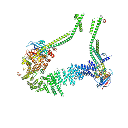
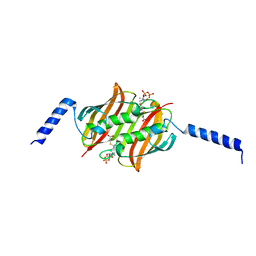
 | | Cryo-EM structure of full-length human STING in the apo state | | 分子名称: | Stimulator of interferon protein | | 著者 | Shang, G, Zhang, C, Chen, Z.J, Bai, X, Zhang, X. | | 登録日 | 2019-01-28 | | 公開日 | 2019-03-06 | | 最終更新日 | 2024-03-20 | | 実験手法 | ELECTRON MICROSCOPY (4.1 Å) | | 主引用文献 | Cryo-EM structures of STING reveal its mechanism of activation by cyclic GMP-AMP.
Nature, 567, 2019
|
|
6YVV
 | | Condensin complex from S.cerevisiae ATP-free apo bridged state | | 分子名称: | Condensin complex subunit 1,Ycs4, Condensin complex subunit 2,Brn1, Structural maintenance of chromosomes protein 2,Structural maintenance of chromosomes protein 2, ... | | 著者 | Lee, B.-G, Cawood, C, Gutierrez-Escribano, P, Nakane, T, Merkel, F, Hassler, M, Haering, C.H, Aragon, L, Lowe, J. | | 登録日 | 2020-04-28 | | 公開日 | 2020-07-15 | | 最終更新日 | 2024-05-22 | | 実験手法 | ELECTRON MICROSCOPY (7.5 Å) | | 主引用文献 | Cryo-EM structures of holo condensin reveal a subunit flip-flop mechanism.
Nat.Struct.Mol.Biol., 27, 2020
|
|
8QH5
 | |
6N2G
 | |
2E1N
 | |
1H56
 | | Structural and biochemical characterization of a new magnesium ion binding site near Tyr94 in the restriction endonuclease PvuII | | 分子名称: | MAGNESIUM ION, TYPE II RESTRICTION ENZYME PVUII | | 著者 | Spyrida, A, Matzen, C, Lanio, T, Jeltsch, A, Simoncsits, A, Athanasiadis, A, Scheuring-Vanamee, E, Kokkinidis, M, Pingoud, A. | | 登録日 | 2001-05-20 | | 公開日 | 2003-08-07 | | 最終更新日 | 2023-12-13 | | 実験手法 | X-RAY DIFFRACTION (3 Å) | | 主引用文献 | Structural and Biochemical Characterization of a New Mg(2+) Binding Site Near Tyr94 in the Restriction Endonuclease PvuII.
J.Mol.Biol., 331, 2003
|
|
2GKD
 | |
1GRJ
 | |
2F3X
 | |
4X23
 | |
9J1S
 | |
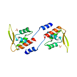
4XLG
 | | C. glabrata Slx1 in complex with Slx4CCD. | | 分子名称: | CHLORIDE ION, Structure-specific endonuclease subunit SLX1, Structure-specific endonuclease subunit SLX4, ... | | 著者 | Gaur, V, Wyatt, H.D.M, Komorowska, W, Szczepanowski, R.H, de Sanctis, D, Gorecka, K.M, West, S.C, Nowotny, M. | | 登録日 | 2015-01-13 | | 公開日 | 2015-03-25 | | 最終更新日 | 2024-01-10 | | 実験手法 | X-RAY DIFFRACTION (1.78 Å) | | 主引用文献 | Structural and Mechanistic Analysis of the Slx1-Slx4 Endonuclease.
Cell Rep, 10, 2015
|
|
8UN9
 | |
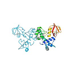
2HTJ
 | | NMR structure of E.coli PapI | | 分子名称: | P fimbrial regulatory protein KS71A | | 著者 | Kawamura, T, Zhou, H, Le, L.U.K, Dahlquist, F.W. | | 登録日 | 2006-07-25 | | 公開日 | 2007-01-30 | | 最終更新日 | 2024-05-29 | | 実験手法 | SOLUTION NMR | | 主引用文献 | Solution Structure of Escherichia coli PapI, a Key Regulator of the Pap Pili Phase Variation.
J.Mol.Biol., 365, 2007
|
|
8TK0
 | |
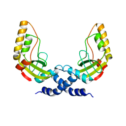
4O5V
 | | Crystal structure of T. acidophilum IdeR | | 分子名称: | FE (II) ION, Iron-dependent transcription repressor related protein | | 著者 | Lee, J.Y, Yeo, H.K. | | 登録日 | 2013-12-20 | | 公開日 | 2014-11-05 | | 実験手法 | X-RAY DIFFRACTION (2.1 Å) | | 主引用文献 | Structural analysis and insight into metal-ion activation of the iron-dependent regulator from Thermoplasma acidophilum.
Acta Crystallogr.,Sect.D, 70, 2014
|
|
8UV1
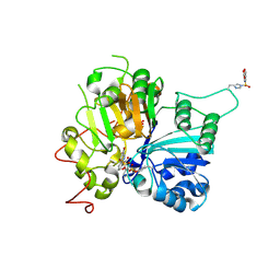 | | Structure of TDP1 complexed with compound IB01 | | 分子名称: | 1,2-ETHANEDIOL, 8-(fluorosulfonyl)-4-oxo-1,4-dihydroquinoline-3-carboxylic acid, Tyrosyl-DNA phosphodiesterase 1 | | 著者 | Lountos, G.T, Zhao, X.Z, Barakat, I, Wang, W, Agama, K, Al Mahmud, M.R, Pommier, Y, Burke, T.R. | | 登録日 | 2023-11-02 | | 公開日 | 2024-09-25 | | 実験手法 | X-RAY DIFFRACTION (1.83 Å) | | 主引用文献 | Structure of TDP1 complexed with compound IB01
To Be Published
|
|
8UZV
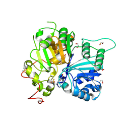 | | Structure of TDP1 catalytic domain complexed with compound IB02 | | 分子名称: | 1,2-ETHANEDIOL, 8-{[2-(fluorosulfonyl)ethyl]amino}-4-oxo-1,4-dihydroquinoline-3-carboxylic acid, DI(HYDROXYETHYL)ETHER, ... | | 著者 | Lountos, G.T, Zhao, X.Z, Barakat, I, Wang, W, Agama, K, Al Mahmud, M.R, Pommier, Y, Burke Jr, T.R. | | 登録日 | 2023-11-16 | | 公開日 | 2024-09-25 | | 実験手法 | X-RAY DIFFRACTION (1.846 Å) | | 主引用文献 | Structure of TDP1 catalytic domain complexed with compound IB02
To Be Published
|
|
8UZZ
 | | Structure of TDP1 catalytic domain complexed with compound IB03 | | 分子名称: | (8M)-8-{2-[(fluorosulfonyl)oxy]phenyl}-4-oxo-1,4-dihydroquinoline-3-carboxylic acid, 1,2-ETHANEDIOL, DI(HYDROXYETHYL)ETHER, ... | | 著者 | Lountos, G.T, Zhao, X.Z, Barakat, I, Wang, W, Agama, K, Al Mahmud, M.R, Pommier, Y, Burke Jr, T.R. | | 登録日 | 2023-11-16 | | 公開日 | 2024-09-25 | | 実験手法 | X-RAY DIFFRACTION (1.93 Å) | | 主引用文献 | Structureal analysis of TDP1 in complex with inhbitors
To Be Published
|
|
8V0C
 | | Structure of TDP1 catalytic domain complexed with compound IB06 | | 分子名称: | (8M)-8-(2-{[2-(fluorosulfonyl)ethyl]amino}phenyl)-4-oxo-1,4-dihydroquinoline-3-carboxylic acid, 1,2-ETHANEDIOL, DI(HYDROXYETHYL)ETHER, ... | | 著者 | Lountos, G.T, Zhao, X.Z, Barakat, I, Wang, W, Agama, K, Al Mahmud, M.R, Pommier, Y, Burke Jr, T.R. | | 登録日 | 2023-11-17 | | 公開日 | 2024-09-25 | | 実験手法 | X-RAY DIFFRACTION (1.62 Å) | | 主引用文献 | Structures of TDP1 complexed with inhibitors
To Be Published
|
|
8V0B
 | | Structure of TDP1 catalytic domain complexed with compound IB05 | | 分子名称: | 1,2-ETHANEDIOL, 8-{4-[(fluorosulfonyl)oxy]phenyl}-4-oxo-1,4-dihydroquinoline-3-carboxylic acid, DI(HYDROXYETHYL)ETHER, ... | | 著者 | Lountos, G.T, Zhao, X.Z, Barakat, I, Wang, W, Agama, K, Al Mahmud, M.R, Pommier, Y, Burke Jr, T.R. | | 登録日 | 2023-11-17 | | 公開日 | 2024-09-25 | | 実験手法 | X-RAY DIFFRACTION (1.65 Å) | | 主引用文献 | Structures of TDP1 complexed with inhibitors
To Be Published
|
|
8TWH
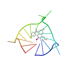 | | Crystal structure of (GGGTT)3GGG G-quadruplex in complex with small molecule ligand RHPS4 | | 分子名称: | (GGGTT)3GGG DNA, 3,11-DIFLUORO-6,8,13-TRIMETHYL-8H-QUINO[4,3,2-KL]ACRIDIN-13-IUM, POTASSIUM ION | | 著者 | Yatsunyk, L.A, Martin, K.N, Lam, G. | | 登録日 | 2023-08-21 | | 公開日 | 2024-09-04 | | 実験手法 | X-RAY DIFFRACTION (2.47 Å) | | 主引用文献 | Crystal structure of (GGGTT)3GGG G-quadruplex in complex with small molecule ligand RHPS4
To be published
|
|


