9AVG
 
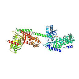
 | | Structure of human calcium-sensing receptor in complex with chimeric Gs (miniGis) protein in nanodiscs | | 分子名称: | (19R,22S,25R)-22,25,26-trihydroxy-16,22-dioxo-17,21,23-trioxa-22lambda~5~-phosphahexacosan-19-yl (9Z)-octadec-9-enoate, 2-acetamido-2-deoxy-beta-D-glucopyranose, 2-acetamido-2-deoxy-beta-D-glucopyranose-(1-4)-2-acetamido-2-deoxy-beta-D-glucopyranose, ... | | 著者 | Zuo, H, Park, J, Frangaj, A, Ye, J, Lu, G, Manning, J.J, Asher, W.B, Lu, Z, Hu, G, Wang, L, Mendez, J, Eng, E, Zhang, Z, Lin, X, Grasucci, R, Hendrickson, W.A, Clarke, O.B, Javitch, J.A, Conigrave, A.D, Fan, Q.R. | | 登録日 | 2024-03-02 | | 公開日 | 2024-04-17 | | 最終更新日 | 2024-05-22 | | 実験手法 | ELECTRON MICROSCOPY (3.6 Å) | | 主引用文献 | Promiscuous G-protein activation by the calcium-sensing receptor.
Nature, 629, 2024
|
|
1GWW
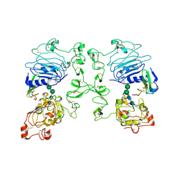
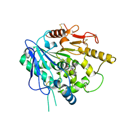
 | | ALPHA-,1,3 GALACTOSYLTRANSFERASE - ALPHA-D-GLUCOSE COMPLEX | | 分子名称: | MANGANESE (II) ION, N-ACETYLLACTOSAMINIDE ALPHA-1,3-GALACTOSYLTRANSFERASE, URIDINE-5'-DIPHOSPHATE, ... | | 著者 | Boix, E, Zhang, Y, Swaminathan, G.J, Brew, K, Acharya, K.R. | | 登録日 | 2002-03-26 | | 公開日 | 2003-03-20 | | 最終更新日 | 2023-12-13 | | 実験手法 | X-RAY DIFFRACTION (1.8 Å) | | 主引用文献 | Structural Basis of Ordered Binding of Donor and Acceptor Substrates to the Retaining Glycosyltransferase, Alpha -1,3 Galactosyltransferase
J.Biol.Chem., 277, 2002
|
|
1HF6
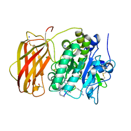
 | | ENDOGLUCANASE CEL5A FROM BACILLUS AGARADHAERENS IN THE ORTHORHOMBIC CRYSTAL FORM IN COMPLEX WITH CELLOTRIOSE | | 分子名称: | ACETIC ACID, ENDOGLUCANASE B, GLYCEROL, ... | | 著者 | Varrot, A, Withers, S, Vasella, A, Schulein, M, Davies, G.J. | | 登録日 | 2000-11-29 | | 公開日 | 2001-11-29 | | 最終更新日 | 2023-12-13 | | 実験手法 | X-RAY DIFFRACTION (1.15 Å) | | 主引用文献 | Direct Experimental Observation of the Hydrogen-Bonding Network of a Glycosidase Along its Reaction Coordinate Revealed by Atomic Resolution Analyses of Endoglucanase Cel5A
Acta Crystallogr.,Sect.D, 59, 2003
|
|
1H11
 | |
1HKC
 | |
1HKB
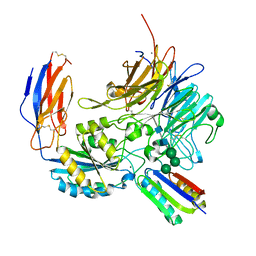
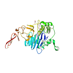
 | | CRYSTAL STRUCTURE OF RECOMBINANT HUMAN BRAIN HEXOKINASE TYPE I COMPLEXED WITH GLUCOSE AND GLUCOSE-6-PHOSPHATE | | 分子名称: | 6-O-phosphono-alpha-D-glucopyranose, CALCIUM ION, D-GLUCOSE 6-PHOSPHOTRANSFERASE, ... | | 著者 | Aleshin, A.E, Zeng, C, Burenkov, G.P, Bartunik, H.D, Fromm, H.J, Honzatko, R.B. | | 登録日 | 1997-12-01 | | 公開日 | 1998-06-03 | | 最終更新日 | 2024-04-03 | | 実験手法 | X-RAY DIFFRACTION (2.8 Å) | | 主引用文献 | The mechanism of regulation of hexokinase: new insights from the crystal structure of recombinant human brain hexokinase complexed with glucose and glucose-6-phosphate.
Structure, 6, 1998
|
|
8TCF
 | | Integrin alpha-v beta-8 in complex with minibinder B8_BP_dsulf | | 分子名称: | 2-acetamido-2-deoxy-beta-D-glucopyranose, 2-acetamido-2-deoxy-beta-D-glucopyranose-(1-4)-2-acetamido-2-deoxy-beta-D-glucopyranose, CALCIUM ION, ... | | 著者 | Campbell, M.G, Fernandez, A, Roy, A, Kraft, J, Baker, D. | | 登録日 | 2023-06-30 | | 公開日 | 2023-09-27 | | 実験手法 | ELECTRON MICROSCOPY (2.9 Å) | | 主引用文献 | De novo design of highly selective miniprotein inhibitors of integrins alpha v beta 6 and alpha v beta 8.
Nat Commun, 14, 2023
|
|
8P7W
 | | Structure of 5D3-Fab and nanobody(Nb8)-bound ABCG2 | | 分子名称: | 5D3(Fab) heavy chain variable domain, 5D3(Fab) light chain variable domain, ATP-binding cassette sub-family G member 2, ... | | 著者 | Irobalieva, R.N, Manolaridis, I, Jackson, S.M, Ni, D, Pardon, E, Stahlberg, H, Steyaert, J, Locher, K.P. | | 登録日 | 2023-05-31 | | 公開日 | 2023-08-30 | | 最終更新日 | 2023-09-13 | | 実験手法 | ELECTRON MICROSCOPY (3.04 Å) | | 主引用文献 | Structural Basis of the Allosteric Inhibition of Human ABCG2 by Nanobodies.
J.Mol.Biol., 435, 2023
|
|
5WB7
 | | Crystal structure of the epidermal growth factor receptor extracellular region in complex with epiregulin | | 分子名称: | 2-acetamido-2-deoxy-beta-D-glucopyranose, 2-acetamido-2-deoxy-beta-D-glucopyranose-(1-4)-2-acetamido-2-deoxy-beta-D-glucopyranose, Epidermal growth factor receptor, ... | | 著者 | Freed, D.M, Bessman, N.J, Ferguson, K.M, Lemmon, M.A. | | 登録日 | 2017-06-28 | | 公開日 | 2017-10-18 | | 最終更新日 | 2023-10-04 | | 実験手法 | X-RAY DIFFRACTION (2.941 Å) | | 主引用文献 | EGFR Ligands Differentially Stabilize Receptor Dimers to Specify Signaling Kinetics.
Cell, 171, 2017
|
|
6ZYF
 | | Notum_Ghrelin complex | | 分子名称: | 1,2-ETHANEDIOL, 2-acetamido-2-deoxy-beta-D-glucopyranose, Palmitoleoyl-protein carboxylesterase NOTUM, ... | | 著者 | Zhao, Y, Jones, E.Y. | | 登録日 | 2020-07-31 | | 公開日 | 2021-03-10 | | 最終更新日 | 2024-01-31 | | 実験手法 | X-RAY DIFFRACTION (2.19 Å) | | 主引用文献 | Notum deacylates octanoylated ghrelin.
Mol Metab, 49, 2021
|
|
8OYH
 | | X-ray structure of furin (PCSK3) in complex with Guanidinomethyl-Phac-Can-Tle-Can-6-(aminomethyl)-3-amino-isoindol | | 分子名称: | CALCIUM ION, CHLORIDE ION, DIMETHYL SULFOXIDE, ... | | 著者 | Dahms, S.O, Brandstetter, H. | | 登録日 | 2023-05-04 | | 公開日 | 2024-03-13 | | 最終更新日 | 2024-05-15 | | 実験手法 | X-RAY DIFFRACTION (1.8 Å) | | 主引用文献 | Fragment-Based Design, Synthesis, and Characterization of Aminoisoindole-Derived Furin Inhibitors.
Chemmedchem, 19, 2024
|
|
8P4M
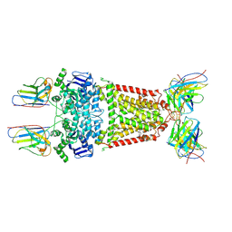
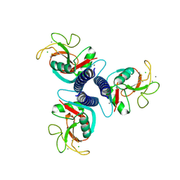
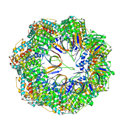 | | CryoEM structure of a C7-symmetrical GroEL7-GroES7 cage in presence of ADP-BeFx | | 分子名称: | ADENOSINE-5'-DIPHOSPHATE, BERYLLIUM TRIFLUORIDE ION, Chaperonin GroEL, ... | | 著者 | Wagner, J, Beck, F, Bracher, A, Caravajal, A.I, Wan, W, Bohn, S, Koerner, R, Baumeister, W, Fernandez-Busnadiego, R, Hartl, F.U. | | 登録日 | 2023-05-23 | | 公開日 | 2024-07-03 | | 実験手法 | ELECTRON MICROSCOPY (2.5 Å) | | 主引用文献 | Visualizing chaperonin function in situ by cryo-electron tomography
Nature, 2024
|
|
8P4O
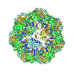 | | CryoEM structure of a GroEL7-GroES7 cage with encapsulated ordered substrate MetK in the presence of ADP-BeFx | | 分子名称: | ADENOSINE-5'-DIPHOSPHATE, BERYLLIUM TRIFLUORIDE ION, Chaperonin GroEL, ... | | 著者 | Wagner, J, Beck, F, Bracher, A, Caravajal, A.I, Wan, W, Bohn, S, Koerner, R, Baumeister, W, Fernandez-Busnadiego, R, Hartl, F.U. | | 登録日 | 2023-05-23 | | 公開日 | 2024-07-03 | | 実験手法 | ELECTRON MICROSCOPY (3.04 Å) | | 主引用文献 | Visualizing chaperonin function in situ by cryo-electron tomography
Nature, 2024
|
|
7PRL
 | | MUC2 D1 with Cu(II) | | 分子名称: | 2-acetamido-2-deoxy-beta-D-glucopyranose, COPPER (II) ION, GLYCEROL, ... | | 著者 | Reznik, N, Fass, D. | | 登録日 | 2021-09-22 | | 公開日 | 2022-10-26 | | 最終更新日 | 2024-01-31 | | 実験手法 | X-RAY DIFFRACTION (2.48 Å) | | 主引用文献 | Intestinal mucin is a chaperone of multivalent copper.
Cell, 185, 2022
|
|
1FIH
 | | N-ACETYLGALACTOSAMINE BINDING MUTANT OF MANNOSE-BINDING PROTEIN A (QPDWG-HDRPY), COMPLEX WITH N-ACETYLGALACTOSAMINE | | 分子名称: | 2-acetamido-2-deoxy-beta-D-galactopyranose, CALCIUM ION, CHLORIDE ION, ... | | 著者 | Feinberg, H, Torgerson, D, Drickamer, K, Weis, W.I. | | 登録日 | 2000-08-03 | | 公開日 | 2000-08-23 | | 最終更新日 | 2021-11-03 | | 実験手法 | X-RAY DIFFRACTION (1.95 Å) | | 主引用文献 | Mechanism of pH-dependent N-acetylgalactosamine binding by a functional mimic of the hepatocyte asialoglycoprotein receptor.
J.Biol.Chem., 275, 2000
|
|
8Q2W
 | |
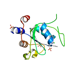
8Q2Q
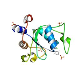
 | | Crystal structure of YTHDC1 in complex with Compound 2b (YL_32) | | 分子名称: | 2-chloranyl-~{N},9-dimethyl-purin-6-amine, SULFATE ION, YTH domain-containing protein 1 | | 著者 | Bedi, R.K, Li, Y, Caflisch, A. | | 登録日 | 2023-08-03 | | 公開日 | 2023-12-06 | | 最終更新日 | 2024-06-19 | | 実験手法 | X-RAY DIFFRACTION (1.3 Å) | | 主引用文献 | Structure-Based Design of a Potent and Selective YTHDC1 Ligand.
J.Med.Chem., 67, 2024
|
|
8Q32
 | |
8Q4U
 | |
8Q38
 | |
8Q4Q
 | |
8Q2T
 | |
8Q33
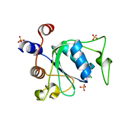
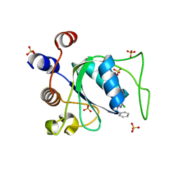 | | Crystal structure of YTHDC1 in complex with Compound 15 (ZA_343) | | 分子名称: | SULFATE ION, YTH domain-containing protein 1, ~{N}-[2-[[2-chloranyl-6-(methylamino)purin-9-yl]methyl]phenyl]-2,2,2-tris(fluoranyl)ethanamide | | 著者 | Bedi, R.K, Zalesak, F, Caflisch, A. | | 登録日 | 2023-08-03 | | 公開日 | 2023-12-06 | | 最終更新日 | 2024-06-19 | | 実験手法 | X-RAY DIFFRACTION (1.43 Å) | | 主引用文献 | Structure-Based Design of a Potent and Selective YTHDC1 Ligand.
J.Med.Chem., 67, 2024
|
|
8Q4V
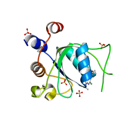 | | Crystal structure of YTHDC1 in complex with Compound 37 (ZA_356) | | 分子名称: | 2-chloranyl-9-[(3-chlorophenyl)methyl]-~{N}-cyclopropyl-7,8-dihydropurin-6-amine, SULFATE ION, YTH domain-containing protein 1 | | 著者 | Bedi, R.K, Zalesak, F, Caflisch, A. | | 登録日 | 2023-08-07 | | 公開日 | 2023-12-06 | | 最終更新日 | 2024-06-19 | | 実験手法 | X-RAY DIFFRACTION (1.36 Å) | | 主引用文献 | Structure-Based Design of a Potent and Selective YTHDC1 Ligand.
J.Med.Chem., 67, 2024
|
|
8Q4R
 | |


