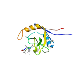
2OGN
 
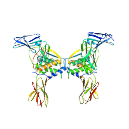
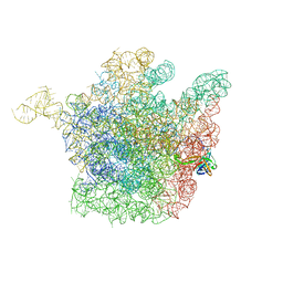 | | The crystal structure of the large ribosomal subunit from Deinococcus radiodurans complexed with the pleuromutilin derivative SB-280080 | | 分子名称: | (3AS,4R,5S,6S,8R,9R,9AR,10R)-5-HYDROXY-4,6,9,10-TETRAMETHYL-1-OXO-6-VINYLDECAHYDRO-3A,9-PROPANOCYCLOPENTA[8]ANNULEN-8-YL (PIPERIDIN-4-YLTHIO)ACETATE, 23S ribosomal RNA, 50S ribosomal protein L3 | | 著者 | Davidovich, C, Bashan, A, Auerbach-Nevo, T, Yonath, A. | | 登録日 | 2007-01-07 | | 公開日 | 2007-05-01 | | 最終更新日 | 2023-12-27 | | 実験手法 | X-RAY DIFFRACTION (3.56 Å) | | 主引用文献 | Induced-fit tightens pleuromutilins binding to ribosomes and remote interactions enable their selectivity.
Proc.Natl.Acad.Sci.Usa, 104, 2007
|
|
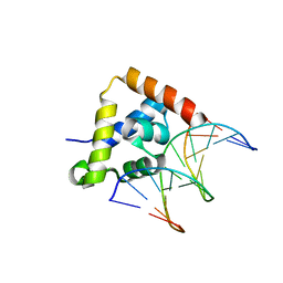
2NSG
 | |
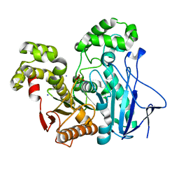
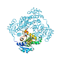
2O7C
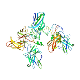
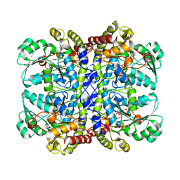 | | Crystal structure of L-methionine-lyase from Pseudomonas | | 分子名称: | Methionine gamma-lyase, SULFATE ION | | 著者 | Misaki, S, Takimoto, A, Takakura, T, Yoshioka, T, Yamashita, M, Tamura, T, Tanaka, H, Inagaki, K. | | 登録日 | 2006-12-10 | | 公開日 | 2007-12-11 | | 最終更新日 | 2023-12-27 | | 実験手法 | X-RAY DIFFRACTION (1.7 Å) | | 主引用文献 | Structure of the antitumour enzyme L-methionine gamma-lyase from Pseudomonas putida at 1.8 A resolution
J.Biochem.(Tokyo), 141, 2007
|
|
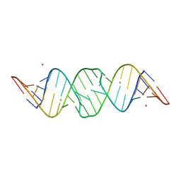
2OGD
 | | T. Brucei Farnesyl Diphosphate Synthase Complexed with Bisphosphonate BPH-527 | | 分子名称: | (4S)-2-METHYL-2,4-PENTANEDIOL, ACETATE ION, BETA-MERCAPTOETHANOL, ... | | 著者 | Cao, R, Gao, Y, Robinson, H, Goddard, A, Oldfield, E. | | 登録日 | 2007-01-05 | | 公開日 | 2007-10-02 | | 最終更新日 | 2023-08-30 | | 実験手法 | X-RAY DIFFRACTION (2 Å) | | 主引用文献 | Bisphosphonates: Teaching Old Drugs with New Tricks
TO BE PUBLISHED
|
|
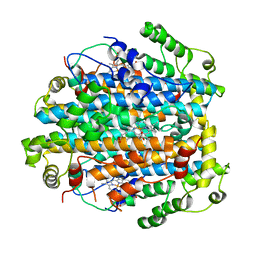
2OGS
 | | Crystal Structure of the GEOBACILLUS STEAROTHERMOPHILUS Carboxylesterase EST55 at pH 6.2 | | 分子名称: | IODIDE ION, Thermostable carboxylesterase Est50 | | 著者 | Liu, P, Ewis, H.E, Tai, P.C, Lu, C.D, Weber, I.T. | | 登録日 | 2007-01-08 | | 公開日 | 2007-02-13 | | 最終更新日 | 2023-12-27 | | 実験手法 | X-RAY DIFFRACTION (2.02 Å) | | 主引用文献 | Crystal Structure of the Geobacillus stearothermophilus Carboxylesterase Est55 and Its Activation of Prodrug CPT-11.
J.Mol.Biol., 367, 2007
|
|
2NW7
 | | Crystal Structure of Tryptophan 2,3-dioxygenase (TDO) from Xanthomonas campestris in complex with ferric heme. Northeast Structural Genomics Target XcR13 | | 分子名称: | PROTOPORPHYRIN IX CONTAINING FE, Tryptophan 2,3-dioxygenase | | 著者 | Forouhar, F, Anderson, J.L.R, Mowat, C.G, Hussain, A, Bruckmann, C, Thackray, S.J, Seetharaman, J, Tucker, T, Ho, C.K, Ma, L.C, Cunningham, K, Janjua, H, Zhao, L, Xiao, R, Liu, J, Baran, M.C, Acton, T.B, Rost, B, Montelione, G.T, Chapman, S.K, Tong, L, Northeast Structural Genomics Consortium (NESG) | | 登録日 | 2006-11-14 | | 公開日 | 2006-12-19 | | 最終更新日 | 2023-08-30 | | 実験手法 | X-RAY DIFFRACTION (2.7 Å) | | 主引用文献 | Molecular insights into substrate recognition and catalysis by tryptophan 2,3-dioxygenase.
Proc.Natl.Acad.Sci.Usa, 104, 2007
|
|
2NUD
 | | The structure of the type III effector AvrB complexed with a high-affinity RIN4 peptide | | 分子名称: | 2-AMINO-2-HYDROXYMETHYL-PROPANE-1,3-DIOL, Avirulence B protein, RPM1-interacting protein 4, ... | | 著者 | Singer, A.U, Desveaux, D, Wu, A.J, McNulty, B, Sondek, J, Dangl, J.L. | | 登録日 | 2006-11-09 | | 公開日 | 2007-05-22 | | 最終更新日 | 2023-08-30 | | 実験手法 | X-RAY DIFFRACTION (2.3 Å) | | 主引用文献 | Type III Effector Activation via Nucleotide Binding, Phosphorylation, and Host Target Interaction.
Plos Pathog., 3, 2007
|
|
2NX1
 | | Structural and mechanistic changes along an engineered path from metallo to non-metallo KDO8P synthase | | 分子名称: | 2-dehydro-3-deoxyphosphooctonate aldolase, 5-O-phosphono-beta-D-ribofuranose, PHOSPHOENOLPYRUVATE | | 著者 | Kona, F, Xu, X, Martin, P, Kuzmic, P, Gatti, D.L. | | 登録日 | 2006-11-16 | | 公開日 | 2007-04-24 | | 最終更新日 | 2023-08-30 | | 実験手法 | X-RAY DIFFRACTION (1.8 Å) | | 主引用文献 | Structural and Mechanistic Changes along an Engineered Path from Metallo to Nonmetallo 3-Deoxy-d-manno-octulosonate 8-Phosphate Synthases.
Biochemistry, 46, 2007
|
|
2O26
 | | Structure of a class III RTK signaling assembly | | 分子名称: | 2-acetamido-2-deoxy-beta-D-glucopyranose-(1-4)-[beta-L-fucopyranose-(1-6)]2-acetamido-2-deoxy-beta-D-glucopyranose, Kit ligand, Mast/stem cell growth factor receptor, ... | | 著者 | Liu, H, Chen, X, Focia, P.J, He, X. | | 登録日 | 2006-11-29 | | 公開日 | 2007-03-13 | | 最終更新日 | 2023-12-27 | | 実験手法 | X-RAY DIFFRACTION (2.5 Å) | | 主引用文献 | Structural basis for stem cell factor-KIT signaling and activation of class III receptor tyrosine kinases.
Embo J., 26, 2007
|
|
2O49
 | |
2O93
 | |
2OIJ
 | |
2OJU
 | |
2NV6
 | |
2OGO
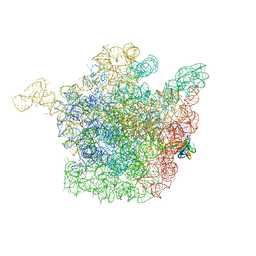 | | The crystal structure of the large ribosomal subunit from Deinococcus radiodurans complexed with the pleuromutilin derivative retapamulin (SB-275833) | | 分子名称: | 23S ribosomal RNA, 50S ribosomal protein L3, Retapamulin | | 著者 | Davidovich, C, Bashan, A, Auerbach-Nevo, T, Yonath, A. | | 登録日 | 2007-01-07 | | 公開日 | 2007-05-01 | | 最終更新日 | 2023-12-27 | | 実験手法 | X-RAY DIFFRACTION (3.66 Å) | | 主引用文献 | Induced-fit tightens pleuromutilins binding to ribosomes and remote interactions enable their selectivity.
Proc.Natl.Acad.Sci.Usa, 104, 2007
|
|
2NW8
 | | Crystal Structure of Tryptophan 2,3-dioxygenase (TDO) from Xanthomonas campestris in complex with ferrous heme and tryptophan. Northeast Structural Genomics Target XcR13. | | 分子名称: | MANGANESE (II) ION, PROTOPORPHYRIN IX CONTAINING FE, TRYPTOPHAN, ... | | 著者 | Forouhar, F, Anderson, J.L.R, Mowat, C.G, Bruckmann, C, Thackray, S.J, Seetharaman, J, Ho, C.K, Ma, L.C, Cunningham, K, Janjua, H, Zhao, L, Xiao, R, Liu, J, Baran, M.C, Acton, T.B, Rost, B, Montelione, G.T, Champman, S.K, Tong, L, Northeast Structural Genomics Consortium (NESG) | | 登録日 | 2006-11-14 | | 公開日 | 2006-12-19 | | 最終更新日 | 2023-08-30 | | 実験手法 | X-RAY DIFFRACTION (1.6 Å) | | 主引用文献 | Molecular insights into substrate recognition and catalysis by tryptophan 2,3-dioxygenase.
Proc.Natl.Acad.Sci.Usa, 104, 2007
|
|
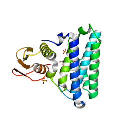
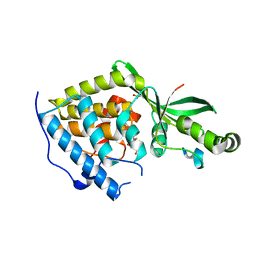
2OIE
 | | Crystal structure of RS21-C6 core segment RSCUT | | 分子名称: | RS21-C6, SULFATE ION | | 著者 | Wu, B, Liu, Y, Zhao, Q, Liao, S, Zhang, J, Bartlam, M, Chen, W, Rao, Z. | | 登録日 | 2007-01-10 | | 公開日 | 2007-03-06 | | 最終更新日 | 2023-12-27 | | 実験手法 | X-RAY DIFFRACTION (2.2 Å) | | 主引用文献 | Crystal Structure of RS21-C6, Involved in Nucleoside Triphosphate Pyrophosphohydrolysis
J.Mol.Biol., 367, 2007
|
|
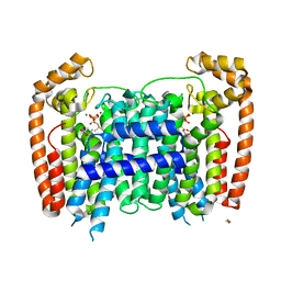
2OAR
 | | Mechanosensitive Channel of Large Conductance (MscL) | | 分子名称: | GOLD ION, Large-conductance mechanosensitive channel | | 著者 | Rees, D.C, Chang, G, Spencer, R.H, Lee, A.T, Steinbacher, S, Strop, P. | | 登録日 | 2006-12-17 | | 公開日 | 2007-01-09 | | 最終更新日 | 2023-12-27 | | 実験手法 | X-RAY DIFFRACTION (3.5 Å) | | 主引用文献 | Structures of the Prokaryotic Mechanosensitive Channels MscL and MscS
Current Topics in Membranes, 58, 2007
|
|
2OG9
 | |
2NRJ
 | | Crystal Structure of Hemolysin binding component from Bacillus cereus | | 分子名称: | Hbl B protein | | 著者 | Madegowda, M, Eswaramoorthy, S, Burley, S.K, Swaminathan, S, New York SGX Research Center for Structural Genomics (NYSGXRC) | | 登録日 | 2006-11-02 | | 公開日 | 2006-11-14 | | 最終更新日 | 2023-12-27 | | 実験手法 | X-RAY DIFFRACTION (2.03 Å) | | 主引用文献 | X-ray crystal structure of the B component of Hemolysin BL from Bacillus cereus
Proteins, 71, 2008
|
|
2NSL
 | | E. coli PurE H45N mutant complexed with CAIR | | 分子名称: | 5-AMINO-1-(5-O-PHOSPHONO-BETA-D-RIBOFURANOSYL)-1H-IMIDAZOLE-4-CARBOXYLIC ACID, Phosphoribosylaminoimidazole carboxylase catalytic subunit | | 著者 | Ealick, S.E, Morar, M. | | 登録日 | 2006-11-04 | | 公開日 | 2007-04-24 | | 最終更新日 | 2023-08-30 | | 実験手法 | X-RAY DIFFRACTION (2 Å) | | 主引用文献 | N(5)-CAIR Mutase: Role of a CO(2) Binding Site and Substrate Movement in Catalysis.
Biochemistry, 46, 2007
|
|
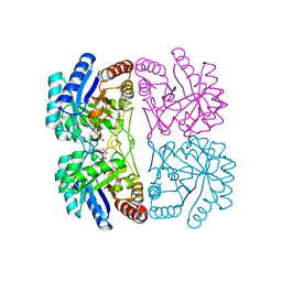
2PH5
 | | Crystal structure of the homospermidine synthase hss from Legionella pneumophila in complex with NAD, Northeast Structural Genomics Target LgR54 | | 分子名称: | Homospermidine synthase, NICOTINAMIDE-ADENINE-DINUCLEOTIDE, SULFATE ION | | 著者 | Forouhar, F, Hussain, M, Seetharaman, J, Fang, Y, Janjua, H, Xiao, R, Cunningham, K, Ma, L.-C, Owens, L, Liu, J, Baran, M.C, Acton, T.B, Montelione, G.T, Hunt, J.F, Tong, L, Northeast Structural Genomics Consortium (NESG) | | 登録日 | 2007-04-10 | | 公開日 | 2007-05-15 | | 最終更新日 | 2018-01-24 | | 実験手法 | X-RAY DIFFRACTION (2.5 Å) | | 主引用文献 | Crystal structure of the homospermidine synthase hss from Legionella pneumophila in complex with NAD.
To be Published
|
|
2PIL
 | | Crystallographic Structure of Phosphorylated Pilin from Neisseria: Phosphoserine Sites Modify Type IV Pilus Surface Chemistry | | 分子名称: | HEPTANE-1,2,3-TRIOL, PLATINUM (II) ION, TYPE 4 PILIN, ... | | 著者 | Forest, K.T, Dunham, S.A, Koomey, M, Tainer, J.A. | | 登録日 | 1998-03-02 | | 公開日 | 1998-05-27 | | 最終更新日 | 2020-07-29 | | 実験手法 | X-RAY DIFFRACTION (2.6 Å) | | 主引用文献 | Crystallographic structure reveals phosphorylated pilin from Neisseria: phosphoserine sites modify type IV pilus surface chemistry and fibre morphology.
Mol.Microbiol., 31, 1999
|
|
2PJC
 | |
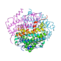
2PHL
 | | THE STRUCTURE OF PHASEOLIN AT 2.2 ANGSTROMS RESOLUTION: IMPLICATIONS FOR A COMMON VICILIN(SLASH)LEGUMIN STRUCTURE AND THE GENETIC ENGINEERING OF SEED STORAGE PROTEINS | | 分子名称: | 2-acetamido-2-deoxy-beta-D-glucopyranose, PHASEOLIN, PHOSPHATE ION | | 著者 | Lawrence, M.C, Izard, T, Beuchat, M, Blagrove, R.J, Colman, P.M. | | 登録日 | 1994-07-07 | | 公開日 | 1994-09-30 | | 最終更新日 | 2020-07-29 | | 実験手法 | X-RAY DIFFRACTION (2.2 Å) | | 主引用文献 | Structure of phaseolin at 2.2 A resolution. Implications for a common vicilin/legumin structure and the genetic engineering of seed storage proteins.
J.Mol.Biol., 238, 1994
|
|


