2FIL
 
 | |
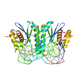
7YOZ
 | | Cryo-EM structure of human subnucleosome (intermediate form) | | 分子名称: | Histone H3.1, Histone H4, Widom601 DNA FW (145-MER), ... | | 著者 | Nozawa, K, Takizawa, Y, Kurumizaka, H. | | 登録日 | 2022-08-02 | | 公開日 | 2022-11-16 | | 最終更新日 | 2024-07-03 | | 実験手法 | ELECTRON MICROSCOPY (4.3 Å) | | 主引用文献 | Cryo-electron microscopy structure of the H3-H4 octasome: A nucleosome-like particle without histones H2A and H2B.
Proc.Natl.Acad.Sci.USA, 119, 2022
|
|
7N9H
 | |
5W7V
 | |
5WKB
 | | MicroED structure of the segment, NFGEFS, from the A315E familial variant of the low complexity domain of TDP-43, residues 312-317 | | 分子名称: | TAR DNA-binding protein 43 | | 著者 | Guenther, E.L, Sawaya, M.R, Cascio, D, Eisenberg, D.S. | | 登録日 | 2017-07-24 | | 公開日 | 2018-05-23 | | 最終更新日 | 2024-03-13 | | 実験手法 | ELECTRON CRYSTALLOGRAPHY (1 Å) | | 主引用文献 | Atomic structures of TDP-43 LCD segments and insights into reversible or pathogenic aggregation.
Nat. Struct. Mol. Biol., 25, 2018
|
|
6JRF
 | | Crystal structure of ZmMoc1-Holliday junction Complex in the presence of Calcium | | 分子名称: | CALCIUM ION, DNA (33-MER), Monokaryotic chloroplast 1, ... | | 著者 | Lin, Z, Lin, H, Zhang, D, Yuan, C. | | 登録日 | 2019-04-03 | | 公開日 | 2019-10-23 | | 最終更新日 | 2023-11-22 | | 実験手法 | X-RAY DIFFRACTION (2.047 Å) | | 主引用文献 | Structural basis of sequence-specific Holliday junction cleavage by MOC1.
Nat.Chem.Biol., 15, 2019
|
|
5WKD
 | | Crystal structure of the segment, GNNQGSN, from the low complexity domain of TDP-43, residues 300-306 | | 分子名称: | TAR DNA-binding protein 43 | | 著者 | Guenther, E.L, Trinh, H, Sawaya, M.R, Cascio, D, Eisenberg, D.S. | | 登録日 | 2017-07-25 | | 公開日 | 2018-04-18 | | 最終更新日 | 2024-04-03 | | 実験手法 | X-RAY DIFFRACTION (1.8 Å) | | 主引用文献 | Atomic structures of TDP-43 LCD segments and insights into reversible or pathogenic aggregation.
Nat. Struct. Mol. Biol., 25, 2018
|
|
5WHP
 | | Crystal structure of the segment, NFGTFS, from the A315T familial variant of the low complexity domain of TDP-43, residues 312-317 | | 分子名称: | Segment of TAR DNA-binding protein 43 | | 著者 | Guenther, E.L, Sawaya, M.R, Eisenberg, D.S. | | 登録日 | 2017-07-17 | | 公開日 | 2018-05-23 | | 最終更新日 | 2024-03-13 | | 実験手法 | X-RAY DIFFRACTION (1 Å) | | 主引用文献 | Atomic structures of TDP-43 LCD segments and insights into reversible or pathogenic aggregation.
Nat. Struct. Mol. Biol., 25, 2018
|
|
5WIQ
 | | Crystal structure of the segment, GFNGGFG, from the low complexity domain of TDP-43, residues 396-402 | | 分子名称: | TAR DNA-binding protein 43 | | 著者 | Guenther, E.L, Sawaya, M.R, Eisenberg, D.S. | | 登録日 | 2017-07-19 | | 公開日 | 2018-04-18 | | 最終更新日 | 2023-10-04 | | 実験手法 | X-RAY DIFFRACTION (1.25 Å) | | 主引用文献 | Atomic structures of TDP-43 LCD segments and insights into reversible or pathogenic aggregation.
Nat. Struct. Mol. Biol., 25, 2018
|
|
7X58
 | | Cryo-EM structure of human subnucleosome (open form) | | 分子名称: | Histone H3.1, Histone H4, Widom601 DNA FW (145-MER), ... | | 著者 | Nozawa, K, Takizawa, Y, Kurumizaka, H. | | 登録日 | 2022-03-04 | | 公開日 | 2022-11-16 | | 最終更新日 | 2024-06-26 | | 実験手法 | ELECTRON MICROSCOPY (3.93 Å) | | 主引用文献 | Cryo-electron microscopy structure of the H3-H4 octasome: A nucleosome-like particle without histones H2A and H2B.
Proc.Natl.Acad.Sci.USA, 119, 2022
|
|
7X57
 | | Cryo-EM structure of human subnucleosome (closed form) | | 分子名称: | Histone H3.1, Histone H4, Widom601 DNA FW (145-MER), ... | | 著者 | Nozawa, K, Takizawa, Y, Kurumizaka, H. | | 登録日 | 2022-03-04 | | 公開日 | 2022-11-16 | | 最終更新日 | 2024-06-26 | | 実験手法 | ELECTRON MICROSCOPY (3.63 Å) | | 主引用文献 | Cryo-electron microscopy structure of the H3-H4 octasome: A nucleosome-like particle without histones H2A and H2B.
Proc.Natl.Acad.Sci.USA, 119, 2022
|
|
5WHN
 | | Crystal structure of the segment, NFGAFS, from the low complexity domain of TDP-43, residues 312-317 | | 分子名称: | Segment of TAR DNA-binding protein 43 | | 著者 | Guenther, E.L, Sawaya, M.R, Eisenberg, D.S. | | 登録日 | 2017-07-17 | | 公開日 | 2018-04-25 | | 最終更新日 | 2023-10-04 | | 実験手法 | X-RAY DIFFRACTION (1.1 Å) | | 主引用文献 | Atomic structures of TDP-43 LCD segments and insights into reversible or pathogenic aggregation.
Nat. Struct. Mol. Biol., 25, 2018
|
|
6E8D
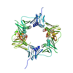 | |
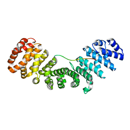
1AY9
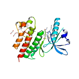
 | | WILD-TYPE UMUD' FROM E. COLI | | 分子名称: | UMUD PROTEIN | | 著者 | Peat, T.S, Frank, E.G, Mcdonald, J.P, Levine, A.S, Woodgate, R, Hendrickson, W.A. | | 登録日 | 1997-11-15 | | 公開日 | 1998-01-28 | | 最終更新日 | 2024-05-22 | | 実験手法 | X-RAY DIFFRACTION (3 Å) | | 主引用文献 | The UmuD' protein filament and its potential role in damage induced mutagenesis.
Structure, 4, 1996
|
|
7AW4
 | | MerTK kinase domain with type 3 inhibitor from a DNA-encoded library | | 分子名称: | 1,2-ETHANEDIOL, 3-methyl-5-(4-methyl-1,2,3-thiadiazol-5-yl)-N-((R)-1-(((R)-3-(methylamino)-3-oxo-1-(4-(trifluoromethyl)phenyl)propyl)amino)-1-oxo-4-phenylbutan-2-yl)isoxazole-4-carboxamide, CHLORIDE ION, ... | | 著者 | Pflug, A, Nissink, J.W.M, Blackett, C, Goldberg, K, Hennessy, E.J, Hardaker, E, McCoull, W, McMurray, L, Collingwood, O, Overman, R, Preston, M, Rawlins, P, Rivers, E, Schimpl, M, Smith, P, Underwood, E, Truman, C, Warwicker, J, Winter, J, Woodcock, S. | | 登録日 | 2020-11-06 | | 公開日 | 2021-03-03 | | 最終更新日 | 2024-05-01 | | 実験手法 | X-RAY DIFFRACTION (1.98 Å) | | 主引用文献 | Generating Selective Leads for Mer Kinase Inhibitors-Example of a Comprehensive Lead-Generation Strategy.
J.Med.Chem., 64, 2021
|
|
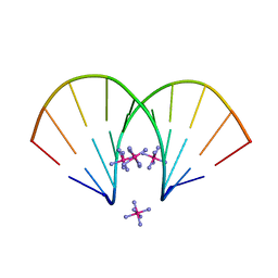
380D
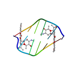 | | BINDING OF THE MODIFIED DAUNORUBICIN WP401 ADJACENT TO A T-G BASE PAIR INDUCES THE REVERSE WATSON-CRICK CONFORMATION: CRYSTAL STRUCTURES OF THE WP401-TGGCCG AND WP401-CGG[BR5C]CG COMPLEXES | | 分子名称: | 2'-BROMO-4'-EPIDAUNORUBICIN, DNA (5'-D(*CP*GP*(G49)P*(CBR)P*CP*G)-3') | | 著者 | Dutta, R, Gao, Y.-G, Priebe, W, Wang, A.H.-J. | | 登録日 | 1998-02-18 | | 公開日 | 1998-07-13 | | 最終更新日 | 2024-02-21 | | 実験手法 | X-RAY DIFFRACTION (2 Å) | | 主引用文献 | Binding of the modified daunorubicin WP401 adjacent to a T-G base pair induces the reverse Watson-Crick conformation: crystal structures of the WP401-TGGCCG and WP401-CGG[br5C]CG complexes.
Nucleic Acids Res., 26, 1998
|
|
4DM0
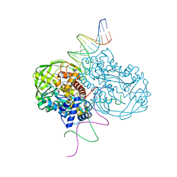 | | TN5 transposase: 20MER OUTSIDE END 2 MN complex | | 分子名称: | 1,2-ETHANEDIOL, DNA NON-TRANSFERRED STRAND, DNA TRANSFERRED STRAND, ... | | 著者 | Klenchin, V.A, Lovell, S, Goryshin, I.Y, Reznikoff, W.R, Rayment, I. | | 登録日 | 2012-02-06 | | 公開日 | 2012-02-22 | | 最終更新日 | 2023-09-13 | | 実験手法 | X-RAY DIFFRACTION (2.5 Å) | | 主引用文献 | Two-metal active site binding of a TN5 transposase synaptic complex
Nat.Struct.Biol., 9, 2002
|
|
8DFA
 | | type I-C Cascade bound to ssDNA target | | 分子名称: | CRISPR-associated protein, CT1133 family, TM1801 family, ... | | 著者 | O'Brien, R.E, Bravo, J.P.K, Ramos, D, Hibshman, G.N, Wright, J.T, Taylor, D.W. | | 登録日 | 2022-06-21 | | 公開日 | 2023-02-22 | | 最終更新日 | 2024-06-12 | | 実験手法 | ELECTRON MICROSCOPY (2.8 Å) | | 主引用文献 | Structural snapshots of R-loop formation by a type I-C CRISPR Cascade.
Mol.Cell, 83, 2023
|
|
6AVK
 | | Streptavidin bound to peptide-like compound KPM-6 | | 分子名称: | 1-hydroxydodecan-4-one, N-[(2H-1,3-benzodioxol-5-yl)methyl]-2-({[(2H-1,3-benzodioxol-5-yl)methyl][2-(chloromethyl)-1,3-oxazole-4-carbonyl]amino}methyl)-N-[(4-carbamoyl-1,3-oxazol-2-yl)methyl]-1,3-oxazole-4-carboxamide, Streptavidin | | 著者 | Park, H, Shamim, R, McEnaney, P, Kodadek, T. | | 登録日 | 2017-09-02 | | 公開日 | 2018-08-08 | | 最終更新日 | 2023-10-04 | | 実験手法 | X-RAY DIFFRACTION (1.4 Å) | | 主引用文献 | Efficient Workflow for Screening DNA-encoded One Bead One Compound Libraries Using a Flow Cytometer
To be Published
|
|
6CFH
 | |
3HO1
 | | Crystal structure of T. thermophilus Argonaute N546 mutant protein complexed with DNA guide strand and 12-nt RNA target strand | | 分子名称: | 5'-D(P*TP*GP*AP*GP*GP*TP*AP*GP*TP*AP*GP*GP*TP*TP*GP*TP*AP*T*AP*GP*T)-3', 5'-R(P*CP*CP*UP*AP*CP*UP*AP*CP*CP*UP*CP*G)-3', MAGNESIUM ION, ... | | 著者 | Wang, Y, Li, H, Sheng, G, Patel, D.J. | | 登録日 | 2009-06-01 | | 公開日 | 2009-10-06 | | 最終更新日 | 2023-09-06 | | 実験手法 | X-RAY DIFFRACTION (2.6 Å) | | 主引用文献 | Nucleation, propagation and cleavage of target RNAs in Ago silencing complexes.
Nature, 461, 2009
|
|
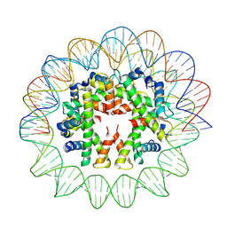
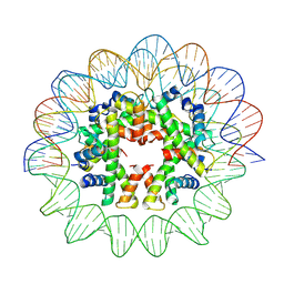
7Y5W
 | | Cryo-EM structure of the left-handed Di-tetrasome | | 分子名称: | Histone H3.1, Histone H4, Widom 601 DNA (147-MER) | | 著者 | Liu, C.P, Yu, Z.Y, Yu, C, Xu, R.M. | | 登録日 | 2022-06-17 | | 公開日 | 2023-08-16 | | 最終更新日 | 2023-09-06 | | 実験手法 | ELECTRON MICROSCOPY (3.5 Å) | | 主引用文献 | Structural insights into histone binding and nucleosome assembly by chromatin assembly factor-1.
Science, 381, 2023
|
|
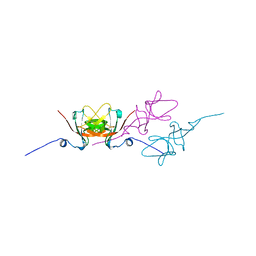
2D1K
 | | Ternary complex of the WH2 domain of mim with actin-dnase I | | 分子名称: | ADENOSINE-5'-TRIPHOSPHATE, Actin, alpha skeletal muscle, ... | | 著者 | Chereau, D, Kerff, F, Dominguez, R. | | 登録日 | 2005-08-26 | | 公開日 | 2006-09-12 | | 最終更新日 | 2023-10-25 | | 実験手法 | X-RAY DIFFRACTION (2.5 Å) | | 主引用文献 | Structural basis for the actin-binding function of missing-in-metastasis
Structure, 15, 2007
|
|
5W7X
 | |
1H15
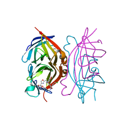
 | | X-ray crystal structure of HLA-DRA1*0101/DRB5*0101 complexed with a peptide from Epstein Barr Virus DNA polymerase | | 分子名称: | 2-acetamido-2-deoxy-beta-D-glucopyranose, 2-acetamido-2-deoxy-beta-D-glucopyranose-(1-4)-2-acetamido-2-deoxy-beta-D-glucopyranose, DNA POLYMERASE, ... | | 著者 | Lang, H, Jacobsen, H, Ikemizu, S, Andersson, C, Harlos, K, Madsen, L, Hjorth, P, Sondergaard, L, Svejgaard, A, Wucherpfennig, K, Stuart, D.I, Bell, J.I, Jones, E.Y, Fugger, L. | | 登録日 | 2002-07-02 | | 公開日 | 2002-10-03 | | 最終更新日 | 2023-12-13 | | 実験手法 | X-RAY DIFFRACTION (3.1 Å) | | 主引用文献 | A Functional and Structural Basis for Tcr Cross-Reactivity in Multiple Sclerosis
Nat.Immunol., 3, 2002
|
|


