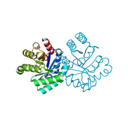
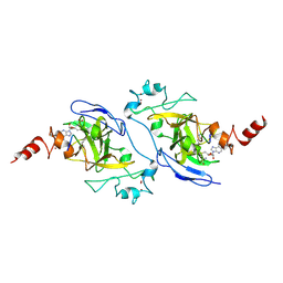
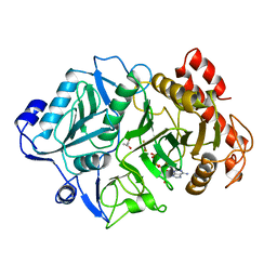
5JWK
 
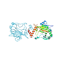 | | Factor Inhibiting HIF D201E in Complex with Fe, and Alpha-Ketoglutarate | | 分子名称: | 2-OXOGLUTARIC ACID, DI(HYDROXYETHYL)ETHER, FE (II) ION, ... | | 著者 | Taabazuing, C.Y, Garman, S.C, Knapp, M.J, Eron, S. | | 登録日 | 2016-05-12 | | 公開日 | 2017-05-17 | | 最終更新日 | 2023-09-27 | | 実験手法 | X-RAY DIFFRACTION (2.3 Å) | | 主引用文献 | Factor Inhibiting HIF D201E in Complex with Fe, and Alpha-Ketoglutarate
To Be Published
|
|
5KER
 | | Deer mouse recombinant hemoglobin from high altitude species | | 分子名称: | Alpha-globin, Beta globin, PROTOPORPHYRIN IX CONTAINING FE | | 著者 | Inoguchi, N, Natarajan, C, Storz, J.F, Moriyama, H. | | 登録日 | 2016-06-10 | | 公開日 | 2017-04-05 | | 最終更新日 | 2023-09-27 | | 実験手法 | X-RAY DIFFRACTION (2.202 Å) | | 主引用文献 | Alteration of the alpha 1 beta 2/ alpha 2 beta 1 subunit interface contributes to the increased hemoglobin-oxygen affinity of high-altitude deer mice.
PLoS ONE, 12, 2017
|
|
1KD1
 | | Co-crystal Structure of Spiramycin bound to the 50S Ribosomal Subunit of Haloarcula marismortui | | 分子名称: | 23S RRNA, 5S RRNA, CADMIUM ION, ... | | 著者 | Hansen, J.L, Ippolito, J.A, Ban, N, Nissen, P, Moore, P.B, Steitz, T.A. | | 登録日 | 2001-11-12 | | 公開日 | 2002-07-19 | | 最終更新日 | 2023-08-16 | | 実験手法 | X-RAY DIFFRACTION (3 Å) | | 主引用文献 | The structures of four macrolide antibiotics bound to the large ribosomal subunit.
Mol.Cell, 10, 2002
|
|
1KQ9
 | | Human methionine aminopeptidase type II in complex with L-methionine | | 分子名称: | METHIONINE, Methionine aminopeptidase 2, TERTIARY-BUTYL ALCOHOL, ... | | 著者 | Nonato, M.C, Widom, J, Clardy, J. | | 登録日 | 2002-01-04 | | 公開日 | 2003-12-09 | | 最終更新日 | 2023-08-16 | | 実験手法 | X-RAY DIFFRACTION (1.9 Å) | | 主引用文献 | Human methionine aminopeptidase type 2 in complex with L- and D-methionine
Bioorg.Med.Chem.Lett., 16, 2006
|
|
1KKH
 | | Crystal Structure of the Methanococcus jannaschii Mevalonate Kinase | | 分子名称: | 1,4-DIETHYLENE DIOXIDE, Mevalonate Kinase | | 著者 | Yang, D, Shipman, L.W, Roessner, C.A, Scott, A.I, Sacchettini, J.C. | | 登録日 | 2001-12-08 | | 公開日 | 2002-03-27 | | 最終更新日 | 2011-07-13 | | 実験手法 | X-RAY DIFFRACTION (2.4 Å) | | 主引用文献 | Structure of the Methanococcus jannaschii mevalonate kinase, a member of the GHMP kinase superfamily.
J.Biol.Chem., 277, 2002
|
|
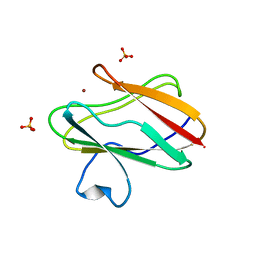
1KM2
 | |
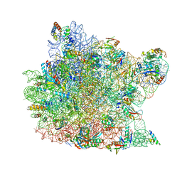
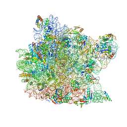
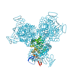
5K36
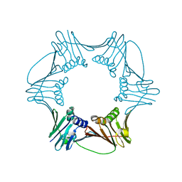
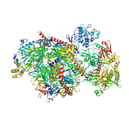 | | Structure of an eleven component nuclear RNA exosome complex bound to RNA | | 分子名称: | Exosome complex component CSL4, Exosome complex component MTR3, Exosome complex component RRP4, ... | | 著者 | Lima, C.D, Zinder, J.C. | | 登録日 | 2016-05-19 | | 公開日 | 2016-11-02 | | 最終更新日 | 2023-09-27 | | 実験手法 | X-RAY DIFFRACTION (3.1 Å) | | 主引用文献 | Nuclear RNA Exosome at 3.1 angstrom Reveals Substrate Specificities, RNA Paths, and Allosteric Inhibition of Rrp44/Dis3.
Mol.Cell, 64, 2016
|
|
1KW2
 | |
1UOZ
 | | Structure of the endoglucanase Cel6 from Mycobacterium tuberculosis in complex with thiocellopentaose at 1.1 angstrom | | 分子名称: | 4-thio-beta-D-glucopyranose, GLYCEROL, PUTATIVE CELLULASE, ... | | 著者 | Varrot, A, Leydier, S, Pell, G, Gilbert, H.J, Davies, G.J. | | 登録日 | 2003-09-26 | | 公開日 | 2004-11-18 | | 最終更新日 | 2023-12-13 | | 実験手法 | X-RAY DIFFRACTION (1.1 Å) | | 主引用文献 | Mycobacterium Tuberculosis Strains Possess Functional Cellulases.
J.Biol.Chem., 280, 2005
|
|
1RWZ
 | | Crystal Structure of Proliferating Cell Nuclear Antigen (PCNA) from A. fulgidus | | 分子名称: | DNA polymerase sliding clamp | | 著者 | Chapados, B.R, Hosfield, D.J, Han, S, Qiu, J, Yelent, B, Shen, B, Tainer, J.A. | | 登録日 | 2003-12-17 | | 公開日 | 2004-01-27 | | 最終更新日 | 2024-04-03 | | 実験手法 | X-RAY DIFFRACTION (1.8 Å) | | 主引用文献 | Structural Basis for FEN-1 Substrate Specificity and PCNA-Mediated Activation in DNA Replication and Repair
Cell(Cambridge,Mass.), 116, 2004
|
|
5J8A
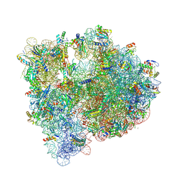 | | Structure of the E coli 70S ribosome with the U1052G mutation in 16S rRNA bound to tigecycline | | 分子名称: | (4S)-2-METHYL-2,4-PENTANEDIOL, 1,2-ETHANEDIOL, 1,4-DIAMINOBUTANE, ... | | 著者 | Cocozaki, A, Ferguson, A. | | 登録日 | 2016-04-07 | | 公開日 | 2016-07-06 | | 最終更新日 | 2016-08-03 | | 実験手法 | X-RAY DIFFRACTION (3.1 Å) | | 主引用文献 | Resistance mutations generate divergent antibiotic susceptibility profiles against translation inhibitors.
Proc.Natl.Acad.Sci.USA, 113, 2016
|
|
1K8U
 | |
1S1Y
 | | Photoactivated chromophore conformation in Photoactive Yellow Protein (E46Q mutant) from 10 microseconds to 3 milliseconds | | 分子名称: | 4'-HYDROXYCINNAMIC ACID, Photoactive yellow protein | | 著者 | Anderson, S, Srajer, V, Pahl, R, Rajagopal, S, Schotte, F, Anfinrud, P, Wulff, M, Moffat, K. | | 登録日 | 2004-01-07 | | 公開日 | 2004-06-15 | | 最終更新日 | 2021-10-27 | | 実験手法 | X-RAY DIFFRACTION (1.6 Å) | | 主引用文献 | Chromophore conformation and the evolution of tertiary structural changes in photoactive yellow protein
Structure, 12, 2004
|
|
1K3V
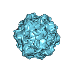 | | Porcine Parvovirus Capsid | | 分子名称: | capsid protein VP2 | | 著者 | Simpson, A.A, Hebert, B, Sullivan, G.M, Parrish, C.R, Zadori, Z, Tijssen, P, Rossmann, M.G. | | 登録日 | 2001-10-04 | | 公開日 | 2001-10-24 | | 最終更新日 | 2024-04-03 | | 実験手法 | X-RAY DIFFRACTION (3.5 Å) | | 主引用文献 | The structure of porcine parvovirus: comparison with related viruses.
J.Mol.Biol., 315, 2002
|
|
5JIY
 | | Structure of G9a SET-domain with Histone H3K9norLeucine mutant peptide and bound S-adenosylmethionine | | 分子名称: | Histone H3.1 mutant peptide with H3K9nor-leucine, Histone-lysine N-methyltransferase EHMT2, S-ADENOSYLMETHIONINE, ... | | 著者 | Jayaram, H, Bellon, S.F, Poy, F. | | 登録日 | 2016-04-22 | | 公開日 | 2016-09-14 | | 最終更新日 | 2023-11-15 | | 実験手法 | X-RAY DIFFRACTION (1.48 Å) | | 主引用文献 | S-adenosyl methionine is necessary for inhibition of the methyltransferase G9a by the lysine 9 to methionine mutation on histone H3.
Proc.Natl.Acad.Sci.USA, 113, 2016
|
|
1S3G
 | |
1K73
 | | Co-crystal Structure of Anisomycin Bound to the 50S Ribosomal Subunit | | 分子名称: | 23S RRNA, 5S RRNA, ANISOMYCIN, ... | | 著者 | Hansen, J, Ban, N, Nissen, P, Moore, P.B, Steitz, T.A. | | 登録日 | 2001-10-18 | | 公開日 | 2003-07-22 | | 最終更新日 | 2023-08-16 | | 実験手法 | X-RAY DIFFRACTION (3.01 Å) | | 主引用文献 | Structures of Five Antibiotics Bound at the Peptidyl Transferase Center of
the Large Ribosomal Subunit
J.Mol.Biol., 330, 2003
|
|
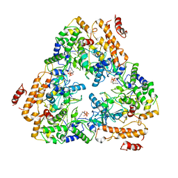
1S5N
 | | Xylose Isomerase in Substrate and Inhibitor Michaelis States: Atomic Resolution Studies of a Metal-Mediated Hydride Shift | | 分子名称: | HYDROXIDE ION, MANGANESE (II) ION, SODIUM ION, ... | | 著者 | Fenn, T.D, Ringe, D, Petsko, G.A. | | 登録日 | 2004-01-21 | | 公開日 | 2004-02-10 | | 最終更新日 | 2023-08-23 | | 実験手法 | X-RAY DIFFRACTION (0.95 Å) | | 主引用文献 | Xylose isomerase in substrate and inhibitor michaelis States: atomic resolution studies of a metal-mediated hydride shift(,).
Biochemistry, 43, 2004
|
|
1K90
 | | Crystal structure of the adenylyl cyclase domain of anthrax edema factor (EF) in complex with calmodulin and 3' deoxy-ATP | | 分子名称: | 3'-DEOXYADENOSINE-5'-TRIPHOSPHATE, CALCIUM ION, CALMODULIN, ... | | 著者 | Drum, C.L, Yan, S.-Z, Bard, J, Shen, Y.-Q, Lu, D, Soelaiman, S, Grabarek, Z, Bohm, A, Tang, W.-J. | | 登録日 | 2001-10-26 | | 公開日 | 2002-01-23 | | 最終更新日 | 2024-02-07 | | 実験手法 | X-RAY DIFFRACTION (2.75 Å) | | 主引用文献 | Structural basis for the activation of anthrax adenylyl cyclase exotoxin by calmodulin.
Nature, 415, 2002
|
|
1K3C
 | | Phosphoenolpyruvate carboxykinase in complex with ADP, AlF3 and Pyruvate | | 分子名称: | ADENOSINE-5'-DIPHOSPHATE, ALUMINUM FLUORIDE, MAGNESIUM ION, ... | | 著者 | Sudom, A.M, Prasad, L, Goldie, H, Delbaere, L.T.J. | | 登録日 | 2001-10-02 | | 公開日 | 2001-12-19 | | 最終更新日 | 2023-11-15 | | 実験手法 | X-RAY DIFFRACTION (2 Å) | | 主引用文献 | The phosphoryl-transfer mechanism of Escherichia coli phosphoenolpyruvate carboxykinase from the use of AlF(3).
J.Mol.Biol., 314, 2001
|
|
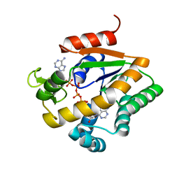
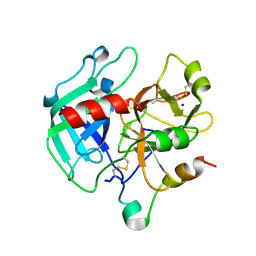
1SF5
 | | Structure of oxidized state of the P94A mutant of amicyanin | | 分子名称: | Amicyanin, COPPER (II) ION | | 著者 | Carrell, C.J, Sun, D, Jiang, S, Davidson, V.L, Mathews, F.S. | | 登録日 | 2004-02-19 | | 公開日 | 2004-07-27 | | 最終更新日 | 2023-08-23 | | 実験手法 | X-RAY DIFFRACTION (1.1 Å) | | 主引用文献 | Structural Studies of Two Mutants of Amicyanin from Paracoccus denitrificans That Stabilize the Reduced State of the Copper.
Biochemistry, 43, 2004
|
|
1SFD
 | | oxidized form of amicyanin mutant P94F | | 分子名称: | Amicyanin, COPPER (II) ION, SULFATE ION | | 著者 | Carrell, C.J, Sun, D, Jiang, S, Davidson, V.L, Mathews, F.S. | | 登録日 | 2004-02-19 | | 公開日 | 2004-07-27 | | 最終更新日 | 2023-08-23 | | 実験手法 | X-RAY DIFFRACTION (0.99 Å) | | 主引用文献 | Structural Studies of Two Mutants of Amicyanin from Paracoccus denitrificans That Stabilize the Reduced State of the Copper.
Biochemistry, 43, 2004
|
|
1SG8
 | | Crystal structure of the procoagulant fast form of thrombin | | 分子名称: | 2-acetamido-2-deoxy-beta-D-glucopyranose, SODIUM ION, thrombin | | 著者 | Pineda, A.O, Carrell, C.J, Bush, L.A, Prasad, S, Caccia, S, Chen, Z.W, Mathews, F.S, Di Cera, E. | | 登録日 | 2004-02-23 | | 公開日 | 2004-06-08 | | 最終更新日 | 2023-08-23 | | 実験手法 | X-RAY DIFFRACTION (2.3 Å) | | 主引用文献 | Molecular dissection of na+ binding to thrombin.
J.Biol.Chem., 279, 2004
|
|
1K6W
 | | The Structure of Escherichia coli Cytosine Deaminase | | 分子名称: | Cytosine Deaminase, FE (III) ION | | 著者 | Ireton, G.C, McDermott, G, Black, M.E, Stoddard, B.L. | | 登録日 | 2001-10-17 | | 公開日 | 2002-02-06 | | 最終更新日 | 2024-02-07 | | 実験手法 | X-RAY DIFFRACTION (1.75 Å) | | 主引用文献 | The structure of Escherichia coli cytosine deaminase.
J.Mol.Biol., 315, 2002
|
|
1S1Z
 | | Photoactivated chromophore conformation in Photoactive Yellow Protein (E46Q mutant) from 10 to 500 nanoseconds | | 分子名称: | 4'-HYDROXYCINNAMIC ACID, Photoactive Yellow Protein | | 著者 | Anderson, S, Srajer, V, Pahl, R, Rajagopal, S, Schotte, F, Anfinrud, P, Wulff, M, Moffat, K. | | 登録日 | 2004-01-07 | | 公開日 | 2004-06-15 | | 最終更新日 | 2021-10-27 | | 実験手法 | X-RAY DIFFRACTION (1.6 Å) | | 主引用文献 | Chromophore conformation and the evolution of tertiary structural changes in photoactive yellow protein
Structure, 12, 2004
|
|


