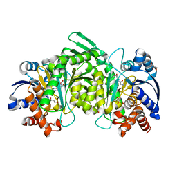
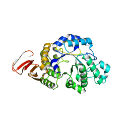
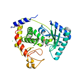
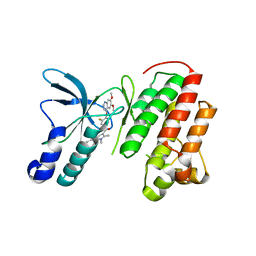
2Q6B
 
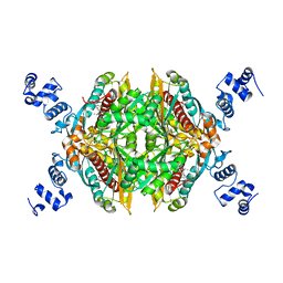 | | Design and synthesis of novel, conformationally restricted HMG-COA reductase inhibitors | | 分子名称: | (3R,5R)-7-[3-(4-FLUOROPHENYL)-1-ISOPROPYL-8-OXO-7-PHENYL-1,4,5,6,7,8-HEXAHYDROPYRROLO[2,3-C]AZEPIN-2-YL]-3,5-DIHYDROXYHEPTANOIC ACID, 3-hydroxy-3-methylglutaryl-coenzyme A reductase, SULFATE ION | | 著者 | Pavlovsky, A, Pfefferkorn, J.A, Harris, M.S, Finzel, B.C. | | 登録日 | 2007-06-04 | | 公開日 | 2007-07-17 | | 最終更新日 | 2024-02-21 | | 実験手法 | X-RAY DIFFRACTION (2 Å) | | 主引用文献 | Design and synthesis of novel, conformationally restricted HMG-CoA reductase inhibitors.
Bioorg.Med.Chem.Lett., 17, 2007
|
|
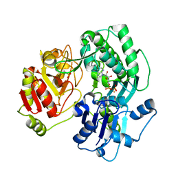
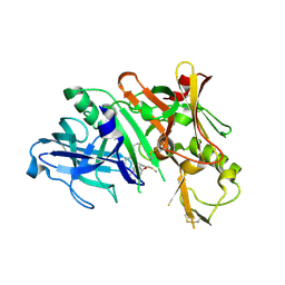
3DEK
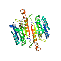 | | Crystal Structures of Caspase-3 with Bound Isoquinoline-1,3,4-trione Derivative Inhibitors | | 分子名称: | Caspase-3, N-[3-(2-fluoroethoxy)phenyl]-N'-(1,3,4-trioxo-1,2,3,4-tetrahydroisoquinolin-6-yl)butanediamide | | 著者 | Wu, J, Du, J, Li, J, Ding, J. | | 登録日 | 2008-06-10 | | 公開日 | 2008-09-02 | | 最終更新日 | 2023-11-15 | | 実験手法 | X-RAY DIFFRACTION (2.4 Å) | | 主引用文献 | Isoquinoline-1,3,4-trione Derivatives Inactivate Caspase-3 by Generation of Reactive Oxygen Species
J.Biol.Chem., 283, 2008
|
|
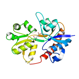
2Q7A
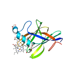 | | Crystal structure of the cell surface heme transfer protein Shp | | 分子名称: | Cell surface heme-binding protein, GLYCEROL, PROTOPORPHYRIN IX CONTAINING FE | | 著者 | Aranda IV, R, Worley, C.E, Bitto, E, Phillips Jr, G.N. | | 登録日 | 2007-06-06 | | 公開日 | 2007-09-18 | | 最終更新日 | 2024-02-21 | | 実験手法 | X-RAY DIFFRACTION (2.1 Å) | | 主引用文献 | Bis-methionyl coordination in the crystal structure of the heme-binding domain of the streptococcal cell surface protein Shp.
J.Mol.Biol., 374, 2007
|
|
3VEE
 | | Rhodococcus jostii RHA1 DypB N246A variant in complex with heme | | 分子名称: | CHLORIDE ION, DypB, FORMIC ACID, ... | | 著者 | Grigg, J.C, Singh, R, Armstrong, Z, Eltis, L.D, Murphy, M.E.P. | | 登録日 | 2012-01-07 | | 公開日 | 2012-01-18 | | 最終更新日 | 2023-09-13 | | 実験手法 | X-RAY DIFFRACTION (2.4 Å) | | 主引用文献 | Distal heme pocket residues of B-type dye-decolorizing peroxidase: arginine but not aspartate is essential for peroxidase activity.
J.Biol.Chem., 287, 2012
|
|
3TY3
 | |
3VEW
 | | Crystal structure of the O-carbamoyltransferase TobZ in complex with ADP | | 分子名称: | ADENOSINE-5'-DIPHOSPHATE, FE (II) ION, O-carbamoyltransferase TobZ, ... | | 著者 | Parthier, C, Stubbs, M.T, Goerlich, S, Jaenecke, F. | | 登録日 | 2012-01-09 | | 公開日 | 2012-01-25 | | 最終更新日 | 2023-09-13 | | 実験手法 | X-RAY DIFFRACTION (2.352 Å) | | 主引用文献 | The O-Carbamoyltransferase TobZ Catalyzes an Ancient Enzymatic Reaction.
Angew.Chem.Int.Ed.Engl., 51, 2012
|
|
3DP4
 | | Crystal structure of the binding domain of the AMPA subunit GluR3 bound to AMPA | | 分子名称: | (S)-ALPHA-AMINO-3-HYDROXY-5-METHYL-4-ISOXAZOLEPROPIONIC ACID, Glutamate receptor 3, ZINC ION | | 著者 | Ahmed, A.H, Wang, Q, Sondermann, H, Oswald, R.E. | | 登録日 | 2008-07-07 | | 公開日 | 2008-11-25 | | 最終更新日 | 2023-08-30 | | 実験手法 | X-RAY DIFFRACTION (2.11 Å) | | 主引用文献 | Structure of the S1S2 glutamate binding domain of GLuR3.
Proteins, 75, 2008
|
|
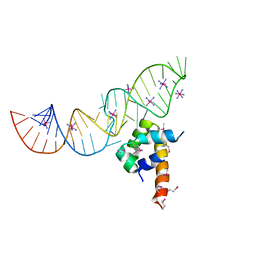
2PXU
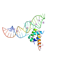 | | Variant 16 of Ribonucleoprotein Core of the E. Coli Signal Recognition Particle | | 分子名称: | 4.5 S RNA, COBALT HEXAMMINE(III), Signal recognition particle protein | | 著者 | Keel, A.Y, Rambo, R.P, Batey, R.T, Kieft, J.S. | | 登録日 | 2007-05-14 | | 公開日 | 2007-08-07 | | 最終更新日 | 2021-10-20 | | 実験手法 | X-RAY DIFFRACTION (2.5 Å) | | 主引用文献 | A General Strategy to Solve the Phase Problem in RNA Crystallography.
Structure, 15, 2007
|
|
3DHU
 | | Crystal structure of an alpha-amylase from Lactobacillus plantarum | | 分子名称: | Alpha-amylase | | 著者 | Bonanno, J.B, Dickey, M, Bain, K.T, Iizuka, M, Ozyurt, S, Smith, D, Wasserman, S, Sauder, J.M, Burley, S.K, Almo, S.C, New York SGX Research Center for Structural Genomics (NYSGXRC) | | 登録日 | 2008-06-18 | | 公開日 | 2008-08-12 | | 最終更新日 | 2024-02-21 | | 実験手法 | X-RAY DIFFRACTION (2 Å) | | 主引用文献 | Crystal structure of an alpha-amylase from Lactobacillus plantarum
To be Published
|
|
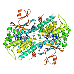
3U39
 | | Crystal Structure of the apo Bacillus Stearothermophilus phosphofructokinase | | 分子名称: | 6-phosphofructokinase, CALCIUM ION | | 著者 | Mosser, R, Reddy, M.C.M, Bruning, J.B, Sacchettini, J.C, Reinhart, G.D. | | 登録日 | 2011-10-05 | | 公開日 | 2012-03-28 | | 最終更新日 | 2023-09-13 | | 実験手法 | X-RAY DIFFRACTION (2.7921 Å) | | 主引用文献 | Structure of the apo form of Bacillus stearothermophilus phosphofructokinase.
Biochemistry, 51, 2012
|
|
3U3R
 | | Crystal structure of D249G mutated Human SULT1A1 bound to PAP and P-NITROPHENOL | | 分子名称: | ADENOSINE-3'-5'-DIPHOSPHATE, P-NITROPHENOL, Sulfotransferase 1A1 | | 著者 | Guttman, C, Berger, I, Aharoni, A, Zarivach, R. | | 登録日 | 2011-10-06 | | 公開日 | 2011-11-16 | | 最終更新日 | 2024-02-28 | | 実験手法 | X-RAY DIFFRACTION (2.36 Å) | | 主引用文献 | The molecular basis for the broad substrate specificity of human sulfotransferase 1A1.
Plos One, 6, 2011
|
|
3DK7
 | |
2Q68
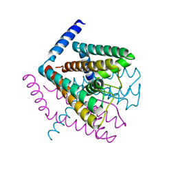 | | Crystal Structure of Nak channel D66A, S70E double mutants | | 分子名称: | CALCIUM ION, Potassium channel protein, SODIUM ION | | 著者 | Alam, A, Shi, N, Jiang, Y. | | 登録日 | 2007-06-04 | | 公開日 | 2007-10-02 | | 最終更新日 | 2023-08-30 | | 実験手法 | X-RAY DIFFRACTION (2.5 Å) | | 主引用文献 | Structural insight into Ca2+ specificity in tetrameric cation channels.
Proc.Natl.Acad.Sci.Usa, 104, 2007
|
|
3U2H
 | | Crystal structure of the C-terminal DUF1608 domain of the Methanosarcina acetivorans S-layer (MA0829) protein | | 分子名称: | GLYCEROL, S-layer protein MA0829 | | 著者 | Chan, S, Phan, T, Ahn, C.J, Shin, A, Rohlin, L, Gunsalus, R.P, Arbing, M.A. | | 登録日 | 2011-10-03 | | 公開日 | 2012-07-04 | | 最終更新日 | 2023-09-13 | | 実験手法 | X-RAY DIFFRACTION (2.36 Å) | | 主引用文献 | Structure of the surface layer of the methanogenic archaean Methanosarcina acetivorans.
Proc.Natl.Acad.Sci.USA, 109, 2012
|
|
3DKL
 | | Crystal structure of phosphorylated mimic form of human NAMPT complexed with benzamide and phosphoribosyl pyrophosphate | | 分子名称: | 1-O-pyrophosphono-5-O-phosphono-alpha-D-ribofuranose, BENZAMIDE, BERYLLIUM TRIFLUORIDE ION, ... | | 著者 | Ho, M, Burgos, E.S, Almo, S.C, Schramm, V.L. | | 登録日 | 2008-06-25 | | 公開日 | 2009-08-18 | | 最終更新日 | 2023-08-30 | | 実験手法 | X-RAY DIFFRACTION (1.89 Å) | | 主引用文献 | A phosphoenzyme mimic, overlapping catalytic sites and reaction coordinate motion for human NAMPT.
Proc.Natl.Acad.Sci.USA, 106, 2009
|
|
3U45
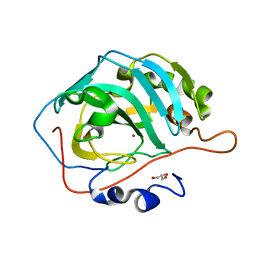 | | Human Carbonic Anhydrase II V143A | | 分子名称: | Carbonic anhydrase 2, GLYCEROL, ZINC ION | | 著者 | West, D, Mckenna, R. | | 登録日 | 2011-10-07 | | 公開日 | 2012-10-10 | | 最終更新日 | 2023-09-13 | | 実験手法 | X-RAY DIFFRACTION (1.699 Å) | | 主引用文献 | Structural Modification of the hydrophobic pocket in Human Carbonic Anhydrase II
To be Published
|
|
2Q6R
 | |
2Q0G
 | |
3VSC
 | | Crystal Structure of the K127A Mutant of O-Phosphoserine Sulfhydrylase Complexed with External Schiff Base of Pyridoxal 5'-Phosphate with O-Phospho-L-Serine | | 分子名称: | (4S)-2-METHYL-2,4-PENTANEDIOL, PHOSPHOSERINE, PYRIDOXAL-5'-PHOSPHATE, ... | | 著者 | Nakamura, T, Kawai, Y, Kataoka, M, Ishikawa, K. | | 登録日 | 2012-04-24 | | 公開日 | 2012-05-16 | | 最終更新日 | 2017-11-22 | | 実験手法 | X-RAY DIFFRACTION (2.07 Å) | | 主引用文献 | Structural analysis of the substrate recognition mechanism in O-phosphoserine sulfhydrylase from the hyperthermophilic archaeon Aeropyrum pernix K1
J.Mol.Biol., 422, 2012
|
|
3U4X
 | | Crystal structure of a lectin from Camptosema pedicellatum seeds in complex with 5-bromo-4-chloro-3-indolyl-alpha-D-mannose | | 分子名称: | 5-bromo-4-chloro-1H-indol-3-yl alpha-D-mannopyranoside, CALCIUM ION, Camptosema pedicellatum lectin (CPL), ... | | 著者 | Rocha, B.A.M, Teixeira, C.S, Moura, T.R, Silva, H.C, Pereira-Junior, F.N, Nagano, C.S, Delatorre, P, Cavada, B.S. | | 登録日 | 2011-10-10 | | 公開日 | 2012-09-12 | | 最終更新日 | 2024-02-28 | | 実験手法 | X-RAY DIFFRACTION (2.16 Å) | | 主引用文献 | Crystal structure of the lectin of Camptosema pedicellatum: implications of a conservative substitution at the hydrophobic subsite.
J.Biochem., 152, 2012
|
|
2Q87
 | |
3DTN
 | | Crystal structure of putative Methyltransferase-MM_2633 from Methanosarcina mazei . | | 分子名称: | ACETATE ION, CALCIUM ION, Putative Methyltransferase MM_2633 | | 著者 | Ramagopal, U.A, Toro, R, Meyer, A.J, Burley, S.K, Almo, S.C, New York SGX Research Center for Structural Genomics (NYSGXRC) | | 登録日 | 2008-07-15 | | 公開日 | 2008-09-09 | | 最終更新日 | 2024-02-21 | | 実験手法 | X-RAY DIFFRACTION (2.09 Å) | | 主引用文献 | Crystal structure of putative Methyltransferase-MM_2633 from Methanosarcina mazei
To be published
|
|
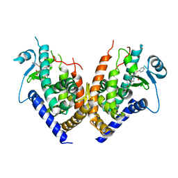
3U6H
 | | Crystal structure of c-Met in complex with pyrazolone inhibitor 26 | | 分子名称: | Hepatocyte growth factor receptor, N-{4-[(6,7-dimethoxyquinolin-4-yl)oxy]-3-fluorophenyl}-1,5-dimethyl-3-oxo-2-phenyl-2,3-dihydro-1H-pyrazole-4-carboxamide | | 著者 | Bellon, S.F, Whittington, D.A, Long, A.L. | | 登録日 | 2011-10-12 | | 公開日 | 2012-02-22 | | 最終更新日 | 2023-09-13 | | 実験手法 | X-RAY DIFFRACTION (2 Å) | | 主引用文献 | Structure-based design of novel class II c-Met inhibitors: 1. Identification of pyrazolone-based derivatives.
J.Med.Chem., 55, 2012
|
|
3DUY
 | | Crystal structure of human beta-secretase in complex with NVP-AFJ144 | | 分子名称: | (2R,4S,5S)-N-butyl-4-hydroxy-2,7-dimethyl-5-{[N-(4-methylpentanoyl)-L-methionyl]amino}octanamide, Beta-secretase 1 | | 著者 | Rondeau, J.-M. | | 登録日 | 2008-07-18 | | 公開日 | 2009-02-24 | | 最終更新日 | 2023-08-30 | | 実験手法 | X-RAY DIFFRACTION (1.97 Å) | | 主引用文献 | Structure-based design and synthesis of macrocyclic peptidomimetic beta-secretase (BACE-1) inhibitors.
Bioorg.Med.Chem.Lett., 19, 2009
|
|
2PXF
 | | Variant 5 of Ribonucleoprotein Core of the E. Coli Signal Recognition Particle | | 分子名称: | 4.5 S RNA, COBALT HEXAMMINE(III), Signal recognition particle protein | | 著者 | Keel, A.Y, Rambo, R.P, Batey, R.T, Kieft, J.S. | | 登録日 | 2007-05-14 | | 公開日 | 2007-08-07 | | 最終更新日 | 2021-10-20 | | 実験手法 | X-RAY DIFFRACTION (2 Å) | | 主引用文献 | A General Strategy to Solve the Phase Problem in RNA Crystallography.
Structure, 15, 2007
|
|


