2WHO
 
 | |
4EM6
 | |
4PEP
 | |
1W3U
 | |
7JXS
 | | Crystal Structure Human Immunodeficiency Virus-1 Matrix protein Mutant Q63R Crystal Form 2 | | 分子名称: | ACETATE ION, DODECAETHYLENE GLYCOL, HEXAETHYLENE GLYCOL, ... | | 著者 | Green, T.J, Eastep, G.N, Ghanam, R.H, Saad, J.S. | | 登録日 | 2020-08-27 | | 公開日 | 2021-04-14 | | 最終更新日 | 2023-10-18 | | 実験手法 | X-RAY DIFFRACTION (2.35 Å) | | 主引用文献 | Structural characterization of HIV-1 matrix mutants implicated in envelope incorporation.
J.Biol.Chem., 296, 2021
|
|
4JLM
 | | Human dCK C4S-S74E mutant in complex with UDP and the F2.3.1 inhibitor (2-[({5-ETHYL-2-[3-(2-FLUOROETHOXY)-4-METHOXYPHENYL]-1,3-THIAZOL-4-YL}METHYL)SULFANYL]PYRIMIDINE-4,6-DIAMINE) | | 分子名称: | 2-[({5-ethyl-2-[3-(2-fluoroethoxy)-4-methoxyphenyl]-1,3-thiazol-4-yl}methyl)sulfanyl]pyrimidine-4,6-diamine, Deoxycytidine kinase, URIDINE-5'-DIPHOSPHATE | | 著者 | Nomme, J, Lavie, A. | | 登録日 | 2013-03-12 | | 公開日 | 2014-01-22 | | 最終更新日 | 2024-02-28 | | 実験手法 | X-RAY DIFFRACTION (2.18 Å) | | 主引用文献 | Structural characterization of new deoxycytidine kinase inhibitors rationalizes the affinity-determining moieties of the molecules.
Acta Crystallogr.,Sect.D, 70, 2014
|
|
3VLB
 | | Crystal structure of xeg-edgp | | 分子名称: | EDGP, Xyloglucan-specific endo-beta-1,4-glucanase A | | 著者 | Yoshizawa, T, Shimizu, T, Hirano, H, Sato, M, Hashimoto, H. | | 登録日 | 2011-11-30 | | 公開日 | 2012-04-18 | | 最終更新日 | 2024-11-20 | | 実験手法 | X-RAY DIFFRACTION (2.7 Å) | | 主引用文献 | Structural basis for inhibition of xyloglucan-specific endo-beta-1,4-glucanase (XEG) by XEG-protein inhibitor
J.Biol.Chem., 287, 2012
|
|
1B57
 | | CLASS II FRUCTOSE-1,6-BISPHOSPHATE ALDOLASE IN COMPLEX WITH PHOSPHOGLYCOLOHYDROXAMATE | | 分子名称: | CHLORIDE ION, PHOSPHOGLYCOLOHYDROXAMIC ACID, PROTEIN (FRUCTOSE-BISPHOSPHATE ALDOLASE II), ... | | 著者 | Hall, D.R, Hunter, W.N. | | 登録日 | 1999-01-12 | | 公開日 | 2000-01-07 | | 最終更新日 | 2023-08-09 | | 実験手法 | X-RAY DIFFRACTION (2 Å) | | 主引用文献 | The crystal structure of Escherichia coli class II fructose-1, 6-bisphosphate aldolase in complex with phosphoglycolohydroxamate reveals details of mechanism and specificity.
J.Mol.Biol., 287, 1999
|
|
2ZB2
 | | Human liver glycogen phosphorylase a complexed with glcose and 5-chloro-N-[4-(1,2-dihydroxyethyl)phenyl]-1H-indole-2-carboxamide | | 分子名称: | (4S)-2-METHYL-2,4-PENTANEDIOL, 2-(N-MORPHOLINO)-ETHANESULFONIC ACID, 5-chloro-N-{4-[(1R)-1,2-dihydroxyethyl]phenyl}-1H-indole-2-carboxamide, ... | | 著者 | Katayama, N, Onda, K. | | 登録日 | 2007-10-15 | | 公開日 | 2008-09-02 | | 最終更新日 | 2023-11-01 | | 実験手法 | X-RAY DIFFRACTION (2.45 Å) | | 主引用文献 | Synthesis of 5-chloro-N-aryl-1H-indole-2-carboxamide derivatives as inhibitors of human liver glycogen phosphorylase a.
Bioorg.Med.Chem., 16, 2008
|
|
5WCZ
 | |
4PP8
 | |
3B7F
 | |
5EPQ
 | | Structure at 1.75 A resolution of a glycosylated, lipid-binding, lipocalin-like protein | | 分子名称: | 1,2-ETHANEDIOL, 2-acetamido-2-deoxy-beta-D-glucopyranose, 2-acetamido-2-deoxy-beta-D-glucopyranose-(1-4)-2-acetamido-2-deoxy-beta-D-glucopyranose, ... | | 著者 | Banerjee, S, Chavas, L.M.G, Ramaswamy, S. | | 登録日 | 2015-11-12 | | 公開日 | 2015-12-02 | | 最終更新日 | 2024-10-16 | | 実験手法 | X-RAY DIFFRACTION (1.752 Å) | | 主引用文献 | Structure of a heterogeneous, glycosylated, lipid-bound, in vivo-grown protein crystal at atomic resolution from the viviparous cockroach Diploptera punctata.
Iucrj, 3, 2016
|
|
4B03
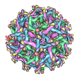 | | 6A Electron cryomicroscopy structure of immature Dengue virus serotype 1 | | 分子名称: | DENGUE VIRUS 1 E PROTEIN, DENGUE VIRUS 1 PRM PROTEIN | | 著者 | Kostyuchenko, V.A, Zhang, Q, Tan, L.C, Ng, T.S, Lok, S.M. | | 登録日 | 2012-06-28 | | 公開日 | 2013-06-05 | | 最終更新日 | 2024-05-08 | | 実験手法 | ELECTRON MICROSCOPY (6 Å) | | 主引用文献 | Immature and Mature Dengue Serotype 1 Virus Structures Provide Insight Into the Maturation Process.
J.Virol., 87, 2013
|
|
1TGJ
 | | HUMAN TRANSFORMING GROWTH FACTOR-BETA 3, CRYSTALLIZED FROM DIOXANE | | 分子名称: | 1,4-DIETHYLENE DIOXIDE, TRANSFORMING GROWTH FACTOR-BETA 3 | | 著者 | Mittl, P.R.E, Priestle, J.P, Gruetter, M.G. | | 登録日 | 1996-07-09 | | 公開日 | 1997-01-11 | | 最終更新日 | 2024-10-23 | | 実験手法 | X-RAY DIFFRACTION (2 Å) | | 主引用文献 | The crystal structure of TGF-beta 3 and comparison to TGF-beta 2: implications for receptor binding.
Protein Sci., 5, 1996
|
|
4OQ8
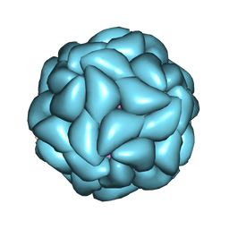 | | Satellite Tobacco Mosaic Virus Refined to 1.4 A Resolution using icosahedral constraints | | 分子名称: | Coat protein, MAGNESIUM ION, PHOSPHATE ION, ... | | 著者 | Larson, S.B, Day, J.S, McPherson, A. | | 登録日 | 2014-02-07 | | 公開日 | 2014-09-10 | | 最終更新日 | 2023-09-20 | | 実験手法 | X-RAY DIFFRACTION (1.45 Å) | | 主引用文献 | Satellite tobacco mosaic virus refined to 1.4 angstrom resolution.
Acta Crystallogr.,Sect.D, 70, 2014
|
|
6B3J
 | | 3.3 angstrom phase-plate cryo-EM structure of a biased agonist-bound human GLP-1 receptor-Gs complex | | 分子名称: | Exendin-P5, Glucagon-like peptide 1 receptor, Guanine nucleotide-binding protein G(I)/G(S)/G(O) subunit gamma-2, ... | | 著者 | Liang, Y.L, Khoshouei, M, Glukhova, A, Furness, S.G.B, Koole, C, Zhao, P, Clydesdale, L, Thal, D.M, Radjainia, M, Danev, R, Baumeister, W, Wang, M.W, Miller, L.J, Christopoulos, A, Sexton, P.M, Wootten, D. | | 登録日 | 2017-09-22 | | 公開日 | 2018-02-21 | | 最終更新日 | 2024-10-23 | | 実験手法 | ELECTRON MICROSCOPY (3.3 Å) | | 主引用文献 | Phase-plate cryo-EM structure of a biased agonist-bound human GLP-1 receptor-Gs complex.
Nature, 555, 2018
|
|
5UUM
 | | Human Mcl-1 in complex with a Bfl-1-specific selected peptide | | 分子名称: | Bfl-1 specific peptide FS2, Induced myeloid leukemia cell differentiation protein Mcl-1, SULFATE ION, ... | | 著者 | Jenson, J.M, Grant, R.A, Keating, A.E. | | 登録日 | 2017-02-17 | | 公開日 | 2017-06-21 | | 最終更新日 | 2024-10-16 | | 実験手法 | X-RAY DIFFRACTION (2.346 Å) | | 主引用文献 | Epistatic mutations in PUMA BH3 drive an alternate binding mode to potently and selectively inhibit anti-apoptotic Bfl-1.
Elife, 6, 2017
|
|
1RQD
 | | deoxyhypusine synthase holoenzyme in its low ionic strength, high pH crystal form with the inhibitor GC7 bound in the active site | | 分子名称: | 1-GUANIDINIUM-7-AMINOHEPTANE, Deoxyhypusine synthase, NICOTINAMIDE-ADENINE-DINUCLEOTIDE | | 著者 | Umland, T.C, Wolff, E.C, Park, M.-H, Davies, D.R. | | 登録日 | 2003-12-04 | | 公開日 | 2004-07-13 | | 最終更新日 | 2023-08-23 | | 実験手法 | X-RAY DIFFRACTION (3 Å) | | 主引用文献 | A New Crystal Structure of Deoxyhypusine Synthase Reveals the Configuration of the Active Enzyme and of an Enzyme-NAD-Inhibitor Ternary Complex
J.Biol.Chem., 279, 2004
|
|
2X76
 | | The crystal structure of PhaZ7 at atomic (1.2 Angstrom) resolution reveals details of the active site and suggests a substrate binding mode | | 分子名称: | CHLORIDE ION, GLYCEROL, IODIDE ION, ... | | 著者 | Wakadkar, S, Hermawan, S, Jendrossek, D, Papageorgiou, A.C. | | 登録日 | 2010-02-24 | | 公開日 | 2010-06-09 | | 最終更新日 | 2024-10-16 | | 実験手法 | X-RAY DIFFRACTION (1.45 Å) | | 主引用文献 | The structure of PhaZ7 at atomic (1.2 A) resolution reveals details of the active site and suggests a substrate-binding mode.
Acta Crystallogr. Sect. F Struct. Biol. Cryst. Commun., 66, 2010
|
|
2OAT
 | | ORNITHINE AMINOTRANSFERASE COMPLEXED WITH 5-FLUOROMETHYLORNITHINE | | 分子名称: | 1-AMINO-7-(2-METHYL-3-OXIDO-5-((PHOSPHONOXY)METHYL)-4-PYRIDOXAL-5-OXO-6-HEPTENATE, ORNITHINE AMINOTRANSFERASE | | 著者 | Storici, P, Schirmer, T. | | 登録日 | 1998-05-07 | | 公開日 | 1998-12-09 | | 最終更新日 | 2024-05-22 | | 実験手法 | X-RAY DIFFRACTION (1.95 Å) | | 主引用文献 | Crystal structure of human ornithine aminotransferase complexed with the highly specific and potent inhibitor 5-fluoromethylornithine.
J.Mol.Biol., 285, 1999
|
|
2WZ1
 | | STRUCTURE OF THE CATALYTIC DOMAIN OF HUMAN SOLUBLE GUANYLATE CYCLASE 1 BETA 3. | | 分子名称: | 1,2-ETHANEDIOL, GUANYLATE CYCLASE SOLUBLE SUBUNIT BETA-1 | | 著者 | Allerston, C.K, Cooper, C.D.O, Muniz, J, Pike, A.C.W, von Delft, F, Arrowsmith, C.H, Weigelt, J, Edwards, A, Bountra, C, Gileadi, O. | | 登録日 | 2009-11-23 | | 公開日 | 2009-12-01 | | 最終更新日 | 2023-12-20 | | 実験手法 | X-RAY DIFFRACTION (1.63 Å) | | 主引用文献 | Crystal Structures of the Catalytic Domain of Human Soluble Guanylate Cyclase.
Plos One, 8, 2013
|
|
7COH
 | | Dimeric Form of Bovine Heart Cytochrome c Oxidase in the Fully Oxidized State | | 分子名称: | (1R)-2-{[{[(2S)-2,3-DIHYDROXYPROPYL]OXY}(HYDROXY)PHOSPHORYL]OXY}-1-[(PALMITOYLOXY)METHYL]ETHYL (11E)-OCTADEC-11-ENOATE, (1S)-2-{[(2-AMINOETHOXY)(HYDROXY)PHOSPHORYL]OXY}-1-[(STEAROYLOXY)METHYL]ETHYL (5E,8E,11E,14E)-ICOSA-5,8,11,14-TETRAENOATE, 1,2-ETHANEDIOL, ... | | 著者 | Shinzawa-Itoh, K, Muramoto, K. | | 登録日 | 2020-08-04 | | 公開日 | 2021-04-07 | | 最終更新日 | 2025-03-12 | | 実験手法 | X-RAY DIFFRACTION (1.3 Å) | | 主引用文献 | The 1.3-A Resolution structure of bovine cytochrome c oxidase suggests a dimerization mechanism
Biochim.Biophys.Acta, 2021
|
|
1RII
 | | Crystal structure of phosphoglycerate mutase from M. Tuberculosis | | 分子名称: | 2,3-bisphosphoglycerate-dependent phosphoglycerate mutase, GLYCEROL | | 著者 | Mueller, P, Sawaya, M.R, Chan, S, Wu, Y, Pashkova, I, Perry, J, Eisenberg, D, TB Structural Genomics Consortium (TBSGC) | | 登録日 | 2003-11-17 | | 公開日 | 2004-10-05 | | 最終更新日 | 2023-08-23 | | 実験手法 | X-RAY DIFFRACTION (1.7 Å) | | 主引用文献 | The 1.70 angstroms X-ray crystal structure of Mycobacterium tuberculosis phosphoglycerate mutase.
Acta Crystallogr.,Sect.D, 61, 2005
|
|
4CP5
 | | ndpK in complex with (Rp)-SPMPApp | | 分子名称: | NUCLEOSIDE DIPHOSPHATE KINASE, CYTOSOLIC, [[(2R)-1-(6-aminopurin-9-yl)propan-2-yl]oxymethyl-sulfanyl-phosphoryl] phosphono hydrogen phosphate | | 著者 | Priet, S, Ferron, F, Alvarez, K, Verron, M, Canard, B. | | 登録日 | 2014-01-31 | | 公開日 | 2015-02-11 | | 最終更新日 | 2023-12-20 | | 実験手法 | X-RAY DIFFRACTION (2.32 Å) | | 主引用文献 | Enzymatic Synthesis of Acyclic Nucleoside Thiophosphonate Diphosphates: Effect of the Alpha-Phosphorus Configuration on HIV-1 RT Activity.
Antiviral Res., 117, 2015
|
|


