181L
 
 | |
188L
 | |
8BF1
 | |
4YIT
 | |
7WJV
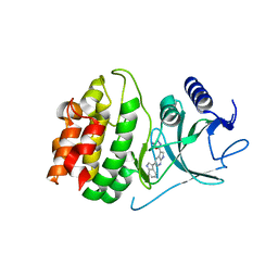
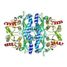 | | Crystal structure of human liver FBPase complexed with an covalent inhibitor | | 分子名称: | 1,2-BENZISOTHIAZOL-3(2H)-ONE 1,1-DIOXIDE, ADENOSINE MONOPHOSPHATE, Fructose-1,6-bisphosphatase 1, ... | | 著者 | Cao, H, Huang, Y, Ren, Y, Wan, J. | | 登録日 | 2022-01-08 | | 公開日 | 2022-07-13 | | 最終更新日 | 2023-11-29 | | 実験手法 | X-RAY DIFFRACTION (1.724 Å) | | 主引用文献 | N -Acylamino Saccharin as an Emerging Cysteine-Directed Covalent Warhead and Its Application in the Identification of Novel FBPase Inhibitors toward Glucose Reduction.
J.Med.Chem., 65, 2022
|
|
8BF2
 | |
6ZOZ
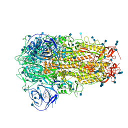 | | Structure of Disulphide-stabilized SARS-CoV-2 Spike Protein Trimer (x1 disulphide-bond mutant, S383C, D985C, K986P, V987P, single Arg S1/S2 cleavage site) in Locked State | | 分子名称: | 2-acetamido-2-deoxy-beta-D-glucopyranose, 2-acetamido-2-deoxy-beta-D-glucopyranose-(1-4)-2-acetamido-2-deoxy-beta-D-glucopyranose, BILIVERDINE IX ALPHA, ... | | 著者 | Xiong, X, Qu, K, Scheres, S.H.W, Briggs, J.A.G. | | 登録日 | 2020-07-08 | | 公開日 | 2020-07-22 | | 最終更新日 | 2022-03-02 | | 実験手法 | ELECTRON MICROSCOPY (3.5 Å) | | 主引用文献 | A thermostable, closed SARS-CoV-2 spike protein trimer.
Nat.Struct.Mol.Biol., 27, 2020
|
|
2XHY
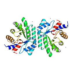
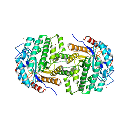 | | Crystal Structure of E.coli BglA | | 分子名称: | 6-PHOSPHO-BETA-GLUCOSIDASE BGLA, BROMIDE ION, SULFATE ION | | 著者 | Totir, M, Zubieta, C, Echols, N, May, A.P, Gee, C.L, nanao, M, alber, T. | | 登録日 | 2010-06-24 | | 公開日 | 2011-07-06 | | 最終更新日 | 2023-12-20 | | 実験手法 | X-RAY DIFFRACTION (2.3 Å) | | 主引用文献 | Macro-to-Micro Structural Proteomics: Native Source Proteins for High-Throughput Crystallization.
Plos One, 7, 2012
|
|
4Y85
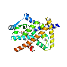
 | | Crystal structure of COT kinase domain in complex with 5-(5-(1H-indol-3-yl)-1H-pyrrolo[2,3-b]pyridin-3-yl)-1,3,4-oxadiazol-2-amine | | 分子名称: | 5-[5-(1H-indol-3-yl)-1H-pyrrolo[2,3-b]pyridin-3-yl]-1,3,4-oxadiazol-2-amine, Mitogen-activated protein kinase kinase kinase 8 | | 著者 | Gutmann, S, Hinniger, A. | | 登録日 | 2015-02-16 | | 公開日 | 2015-05-06 | | 最終更新日 | 2024-05-08 | | 実験手法 | X-RAY DIFFRACTION (2.33 Å) | | 主引用文献 | The Crystal Structure of Cancer Osaka Thyroid Kinase Reveals an Unexpected Kinase Domain Fold.
J.Biol.Chem., 290, 2015
|
|
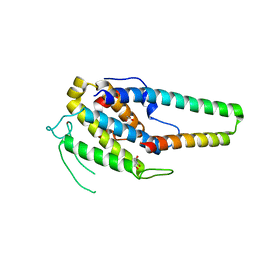
2XTD
 | | Structure of the TBL1 tetramerisation domain | | 分子名称: | TBL1 F-BOX-LIKE/WD REPEAT-CONTAINING PROTEIN TBL1X | | 著者 | Oberoi, J, Fairall, L, Watson, P.J, Greenwood, J.A, Schwabe, J.W.R. | | 登録日 | 2010-10-06 | | 公開日 | 2011-01-19 | | 最終更新日 | 2023-12-20 | | 実験手法 | X-RAY DIFFRACTION (3.2 Å) | | 主引用文献 | Structural Basis for the Assembly of the Smrt/Ncor Core Transcriptional Repression Machinery.
Nat.Struct.Mol.Biol., 18, 2011
|
|
1PMC
 | |
5XQU
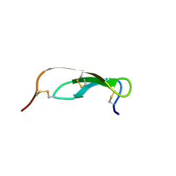
 | | Crystal structure of Notched-fin eelpout type III antifreeze protein A20I mutant (NFE6, AFP), P212121 form | | 分子名称: | Ice-structuring protein | | 著者 | Adachi, M, Shimizu, R, Shibazaki, C, Kondo, H, Tsuda, S. | | 登録日 | 2017-06-07 | | 公開日 | 2018-05-16 | | 最終更新日 | 2023-11-22 | | 実験手法 | X-RAY DIFFRACTION (1 Å) | | 主引用文献 | Polypentagonal ice-like water networks emerge solely in an activity-improved variant of ice-binding protein
Proc. Natl. Acad. Sci. U.S.A., 115, 2018
|
|
6Z9J
 | |
5XQZ
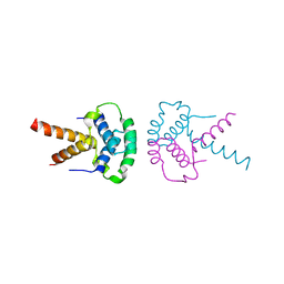
 | | Structure of the MOB1-NDR2 complex | | 分子名称: | GLYCEROL, MOB kinase activator 1A, Serine/threonine-protein kinase 38-like, ... | | 著者 | Wu, G, Lin, K. | | 登録日 | 2017-06-07 | | 公開日 | 2018-08-29 | | 最終更新日 | 2024-03-27 | | 実験手法 | X-RAY DIFFRACTION (2.1 Å) | | 主引用文献 | Stable MOB1 interaction with Hippo/MST is not essential for development and tissue growth control.
Nat Commun, 8, 2017
|
|
4X66
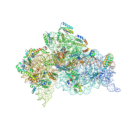 | | Crystal Structure of 30S ribosomal subunit from Thermus thermophilus | | 分子名称: | 16S rRNA, 30S ribosomal protein S10, 30S ribosomal protein S11, ... | | 著者 | Demirci, H, Chen, J, Choi, J, Soltis, M, Puglisi, J.D. | | 登録日 | 2014-12-06 | | 公開日 | 2015-11-18 | | 最終更新日 | 2019-12-25 | | 実験手法 | X-RAY DIFFRACTION (3.446 Å) | | 主引用文献 | N(6)-methyladenosine in mRNA disrupts tRNA selection and translation-elongation dynamics.
Nat.Struct.Mol.Biol., 23, 2016
|
|
5XOU
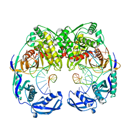 | | Crystal structure of T. thermophilus Argonaute protein complexed with a bulge 7T8 on the guide strand | | 分子名称: | DNA (5'-D(*AP*CP*AP*AP*CP*CP*TP*AP*CP*TP*AP*CP*CP*TP*CP*G)-3'), DNA (5'-D(P*TP*GP*AP*GP*GP*TP*AP*TP*GP*GP*TP*TP*GP*T)-3'), MAGNESIUM ION, ... | | 著者 | Sheng, G, Wang, J, Zhao, H, Wang, Y. | | 登録日 | 2017-05-31 | | 公開日 | 2017-10-04 | | 最終更新日 | 2023-11-22 | | 実験手法 | X-RAY DIFFRACTION (2.63 Å) | | 主引用文献 | Structure/cleavage-based insights into helical perturbations at bulge sites within T. thermophilus Argonaute silencing complexes
Nucleic Acids Res., 45, 2017
|
|
1PTQ
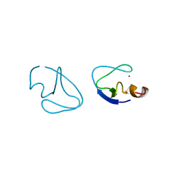 | | PROTEIN KINASE C DELTA CYS2 DOMAIN | | 分子名称: | PROTEIN KINASE C DELTA TYPE, ZINC ION | | 著者 | Zhang, G, Hurley, J.H. | | 登録日 | 1995-05-11 | | 公開日 | 1995-07-31 | | 最終更新日 | 2024-02-14 | | 実験手法 | X-RAY DIFFRACTION (1.95 Å) | | 主引用文献 | Crystal structure of the cys2 activator-binding domain of protein kinase C delta in complex with phorbol ester.
Cell(Cambridge,Mass.), 81, 1995
|
|
1PTR
 | |
4WTX
 | | Crystal structure of the fourth FnIII domain of integrin beta4 | | 分子名称: | Integrin beta-4 | | 著者 | Alonso-Garcia, N, Urien, H, Buey, R.M, de Pereda, J.M. | | 登録日 | 2014-10-30 | | 公開日 | 2015-02-11 | | 最終更新日 | 2024-05-08 | | 実験手法 | X-RAY DIFFRACTION (1.5 Å) | | 主引用文献 | Combination of X-ray crystallography, SAXS and DEER to obtain the structure of the FnIII-3,4 domains of integrin alpha6beta4
Acta Crystallogr.,Sect.D, 71, 2015
|
|
4X0E
 | |
4WV6
 | |
8B6N
 | |
4X2C
 | | Clostridium difficile Fic protein_0569 mutant S31A, E35A | | 分子名称: | 4-(2-HYDROXYETHYL)-1-PIPERAZINE ETHANESULFONIC ACID, Fic family protein putative filamentation induced by cAMP protein, GLYCEROL, ... | | 著者 | Jorgensen, R, Dedic, E. | | 登録日 | 2014-11-26 | | 公開日 | 2015-12-23 | | 最終更新日 | 2024-01-10 | | 実験手法 | X-RAY DIFFRACTION (1.8 Å) | | 主引用文献 | Structure of Clostridium difficile Fic_0569 S31A, E35A mutant at 1.8 Angstroms resolution
To Be Published
|
|
8B6P
 | |
7Y6W
 | | RRGSGG-AtPRT6 UBR box (I222) | | 分子名称: | E3 ubiquitin-protein ligase PRT6, ZINC ION | | 著者 | Kim, L, Song, H.K. | | 登録日 | 2022-06-21 | | 公開日 | 2023-07-05 | | 最終更新日 | 2023-11-29 | | 実験手法 | X-RAY DIFFRACTION (1.95 Å) | | 主引用文献 | Structural analyses of plant PRT6-UBR box for Cys-Arg/N-degron pathway and insights into the plant submergence resistance
To Be Published
|
|


