6M1X
 
 | |
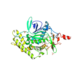
7NQN
 | | Mycobacterium tuberculosis Cytochrome P450 CYP121 in complex with lead compound 14 | | 分子名称: | 2-(N-MORPHOLINO)-ETHANESULFONIC ACID, 4-[4-[2-(1~{H}-indol-3-yl)ethyl]pyrimidin-2-yl]morpholine, CHLORIDE ION, ... | | 著者 | Selvam, I.R. | | 登録日 | 2021-03-01 | | 公開日 | 2022-02-02 | | 最終更新日 | 2024-01-31 | | 実験手法 | X-RAY DIFFRACTION (1.6 Å) | | 主引用文献 | A new strategy for hit generation: Novel in cellulo active inhibitors of CYP121A1 from Mycobacterium tuberculosis via a combined X-ray crystallographic and phenotypic screening approach (XP screen).
Eur.J.Med.Chem., 230, 2022
|
|
2N3H
 | |
2N3F
 | |
4MGN
 | |
2JN5
 | |
4MK7
 | |
2I0H
 | | The structure of p38alpha in complex with an arylpyridazinone | | 分子名称: | 2-(3-{(2-CHLORO-4-FLUOROPHENYL)[1-(2-CHLOROPHENYL)-6-OXO-1,6-DIHYDROPYRIDAZIN-3-YL]AMINO}PROPYL)-1H-ISOINDOLE-1,3(2H)-DIONE, GLYCEROL, Mitogen-activated protein kinase 14 | | 著者 | Natarajan, S.R, Heller, S.T, Nam, K, Singh, S.B, Scapin, G, Patel, S, Thompson, J.E, Fitzgerald, C.E, O'Keefe, S.J. | | 登録日 | 2006-08-10 | | 公開日 | 2006-10-17 | | 最終更新日 | 2023-08-30 | | 実験手法 | X-RAY DIFFRACTION (2 Å) | | 主引用文献 | p38 MAP Kinase Inhibitors Part 6: 2-Arylpyridazin-3-ones as templates for inhibitor design.
Bioorg.Med.Chem.Lett., 16, 2006
|
|
7WIT
 | | Structure of SUR1 in complex with mitiglinide | | 分子名称: | (2S)-4-[(3aR,7aS)-1,3,3a,4,5,6,7,7a-octahydroisoindol-2-yl]-4-oxidanylidene-2-(phenylmethyl)butanoic acid, ADENOSINE-5'-TRIPHOSPHATE, ATP-sensitive inward rectifier potassium channel 11,ATP-binding cassette sub-family C member 8 isoform X1 | | 著者 | Chen, L, Wang, M.M. | | 登録日 | 2022-01-04 | | 公開日 | 2022-11-16 | | 最終更新日 | 2024-06-26 | | 実験手法 | ELECTRON MICROSCOPY (3.21 Å) | | 主引用文献 | Structural Insights Into the High Selectivity of the Anti-Diabetic Drug Mitiglinide
Front Pharmacol, 13, 2022
|
|
8TZZ
 | | SpG Cas9 with NGC PAM DNA target | | 分子名称: | CRISPR-associated endonuclease Cas9/Csn1, DNA (5'-D(P*CP*GP*TP*TP*TP*GP*TP*AP*CP*TP*GP*CP*AP*GP*CP*G)-3'), DNA (5'-D(P*TP*CP*TP*CP*AP*TP*CP*TP*TP*TP*AP*TP*GP*CP*GP*TP*C)-3'), ... | | 著者 | Bravo, J.P.K, Hibshman, G.N, Taylor, D.W. | | 登録日 | 2023-08-28 | | 公開日 | 2024-05-01 | | 最終更新日 | 2024-05-15 | | 実験手法 | ELECTRON MICROSCOPY (3.56 Å) | | 主引用文献 | Unraveling the mechanisms of PAMless DNA interrogation by SpRY-Cas9.
Nat Commun, 15, 2024
|
|
8U3Y
 | | SpG Cas9 with NGG PAM DNA target | | 分子名称: | CRISPR-associated endonuclease Cas9/Csn1, DNA (5'-D(P*CP*GP*TP*TP*TP*GP*TP*AP*CP*TP*CP*CP*AP*GP*CP*G)-3'), DNA (5'-D(P*TP*CP*TP*CP*AP*TP*CP*TP*TP*TP*AP*TP*GP*CP*GP*TP*C)-3'), ... | | 著者 | Bravo, J.P.K, Hibshman, G.N, Taylor, D.W. | | 登録日 | 2023-09-08 | | 公開日 | 2024-05-01 | | 最終更新日 | 2024-05-15 | | 実験手法 | ELECTRON MICROSCOPY (3.1 Å) | | 主引用文献 | Unraveling the mechanisms of PAMless DNA interrogation by SpRY-Cas9.
Nat Commun, 15, 2024
|
|
7N2W
 | | The crystal structure of an FMN-dependent NADH-azoreductase, AzoA in complex with Red 40 | | 分子名称: | 6-hydroxy-5-[(E)-(2-methoxy-5-methyl-4-sulfophenyl)diazenyl]naphthalene-2-sulfonic acid, FLAVIN MONONUCLEOTIDE, FMN dependent NADH:quinone oxidoreductase | | 著者 | Arcinas, A.J, Fedorov, E, Kelly, L, Almo, S.C, Ghosh, A. | | 登録日 | 2021-05-30 | | 公開日 | 2022-06-01 | | 最終更新日 | 2023-10-18 | | 実験手法 | X-RAY DIFFRACTION (2.65 Å) | | 主引用文献 | Uncovering a novel mechanism of enzyme activation in multimeric azoreductases
To Be Published
|
|
4M1R
 | | Structure of a novel cellulase 5 from a sugarcane soil metagenomic library | | 分子名称: | 2-AMINO-2-HYDROXYMETHYL-PROPANE-1,3-DIOL, Cellulase 5 | | 著者 | Paiva, J.H, Alvarez, T.M, Cairo, J.P, Paixao, D.A, Almeida, R.A, Tonoli, C.C.C, Ruiz, D.M, Ruller, R, Santos, C.R, Squina, F.M, Murakami, M.T. | | 登録日 | 2013-08-03 | | 公開日 | 2014-02-05 | | 最終更新日 | 2024-02-28 | | 実験手法 | X-RAY DIFFRACTION (1.8 Å) | | 主引用文献 | Structure and function of a novel cellulase 5 from sugarcane soil metagenome.
Plos One, 8, 2013
|
|
2I53
 | | Crystal structure of Cyclin K | | 分子名称: | ACETATE ION, Cyclin K | | 著者 | Baek, K, Brown, R.S, Birrane, G, Ladias, J.A.A. | | 登録日 | 2006-08-23 | | 公開日 | 2007-01-02 | | 最終更新日 | 2024-02-21 | | 実験手法 | X-RAY DIFFRACTION (1.5 Å) | | 主引用文献 | Crystal Structure of Human Cyclin K, a Positive Regulator of Cyclin-dependent Kinase 9.
J.Mol.Biol., 366, 2007
|
|
7N2X
 | | The crystal structure of an FMN-dependent NADH:quinone oxidoreductase, AzoR from Escherichia coli | | 分子名称: | 1,2-ETHANEDIOL, 1-ETHOXY-2-(2-ETHOXYETHOXY)ETHANE, 2-AMINO-ACRYLIC ACID, ... | | 著者 | Arcinas, A.J, Fedorov, E, Kelly, L, Almo, S.C, Ghosh, A. | | 登録日 | 2021-05-30 | | 公開日 | 2022-08-31 | | 最終更新日 | 2023-10-18 | | 実験手法 | X-RAY DIFFRACTION (1.7 Å) | | 主引用文献 | Uncovering a novel mechanism of enzyme activation in multimeric azoreductases
To Be Published
|
|
4MGM
 | |
8WDG
 | |
2JMW
 | | Structure of DNA-Binding Domain of Arabidopsis GT-1 | | 分子名称: | DNA binding protein GT-1 | | 著者 | Nagata, T, Niyada, E, Noto, K, Ikeda, Y, Yamamoto, Y, Uesugi, S, Murata, J, Hiratsuka, K, Katahira, M. | | 登録日 | 2006-12-11 | | 公開日 | 2007-12-11 | | 最終更新日 | 2023-12-20 | | 実験手法 | SOLUTION NMR | | 主引用文献 | Solution structures of the trihelix DNA-binding domains of the wild-type and a phosphomimetic mutant of Arabidopsis GT-1: mechanism for an increase in DNA-binding affinity through phosphorylation.
Proteins, 78, 2010
|
|
2JNH
 | |
2YPD
 | | Crystal structure of the Jumonji domain of human Jumonji domain containing 1C protein | | 分子名称: | 2-(ETHYLMERCURI-THIO)-BENZOIC ACID, PROBABLE JMJC DOMAIN-CONTAINING HISTONE DEMETHYLATION PROT EIN 2C, SODIUM ION | | 著者 | Vollmar, M, Johansson, C, Krojer, T, Berridge, G, Burgess-Brown, N, Strain-Damerell, C, Froese, S, Williams, E, Goubin, S, Coutandin, D, von Delft, F, Arrowsmith, C.H, Bountra, C, Edwards, A, Oppermann, U. | | 登録日 | 2012-10-30 | | 公開日 | 2012-12-12 | | 最終更新日 | 2024-05-08 | | 実験手法 | X-RAY DIFFRACTION (2.1 Å) | | 主引用文献 | Crystal Structure of the Jumonji Domain of Human Jumonji Domain Containing 1C Protein
To be Published
|
|
3D3X
 | |
8WG5
 | | Cryo-EM structure of USP16 bound to H2AK119Ub nucleosome | | 分子名称: | DNA (147-MER), Histone H2A type 1-B/E, Histone H2B type 1-K, ... | | 著者 | Ai, H.S, He, Z.Z, Deng, Z.H, Liu, L. | | 登録日 | 2023-09-20 | | 公開日 | 2023-12-27 | | 最終更新日 | 2024-07-10 | | 実験手法 | ELECTRON MICROSCOPY (3.05 Å) | | 主引用文献 | Structural and mechanistic basis for nucleosomal H2AK119 deubiquitination by single-subunit deubiquitinase USP16.
Nat.Struct.Mol.Biol., 2024
|
|
2JES
 | | Portal protein (gp6) from bacteriophage SPP1 | | 分子名称: | CALCIUM ION, MERCURY (II) ION, PORTAL PROTEIN, ... | | 著者 | Lebedev, A.A, Krause, M.H, Isidro, A.L, Vagin, A.A, Orlova, E.V, Turner, J, Dodson, E.J, Tavares, P, Antson, A.A. | | 登録日 | 2007-01-21 | | 公開日 | 2007-03-27 | | 最終更新日 | 2024-05-08 | | 実験手法 | X-RAY DIFFRACTION (3.4 Å) | | 主引用文献 | Structural Framework for DNA Translocation Via the Viral Portal Protein
Embo J., 26, 2007
|
|
4MK8
 | | Hepatitis C Virus polymerase NS5B genotype 1b (BK) in complex with inhibitor 4 (N-(4-{2-[3-tert-butyl-2-methoxy-5-(2-oxo-1,2-dihydropyridin-3-yl)phenyl]ethyl}phenyl)methanesulfonamide) | | 分子名称: | DIMETHYL SULFOXIDE, GLYCEROL, N-(4-{2-[3-tert-butyl-2-methoxy-5-(2-oxo-1,2-dihydropyridin-3-yl)phenyl]ethyl}phenyl)methanesulfonamide, ... | | 著者 | Harris, S.F, Wong, A. | | 登録日 | 2013-09-04 | | 公開日 | 2013-10-09 | | 最終更新日 | 2014-01-15 | | 実験手法 | X-RAY DIFFRACTION (2.09 Å) | | 主引用文献 | Discovery of a Novel Series of Potent Non-Nucleoside Inhibitors of Hepatitis C Virus NS5B.
J.Med.Chem., 56, 2013
|
|
7DWA
 | | Structure of a novel beta-mannanase BaMan113A with mannotriose, N236Y mutation | | 分子名称: | Endo-beta-1,4-mannanase, beta-D-mannopyranose-(1-4)-beta-D-mannopyranose-(1-4)-alpha-D-mannopyranose | | 著者 | Liu, W.T, Liu, W.D, Zheng, Y.Y. | | 登録日 | 2021-01-15 | | 公開日 | 2021-06-02 | | 最終更新日 | 2023-11-29 | | 実験手法 | X-RAY DIFFRACTION (1.62 Å) | | 主引用文献 | Functional and structural investigation of a novel beta-mannanase BaMan113A from Bacillus sp. N16-5.
Int.J.Biol.Macromol., 182, 2021
|
|


