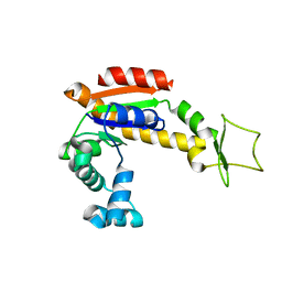
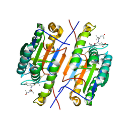
8OVN
 
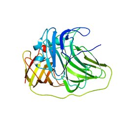
 | | X-ray structure of the SF-iGluSnFR-S72A | | 分子名称: | CITRIC ACID, Putative periplasmic binding transport protein,Green fluorescent protein | | 著者 | Tarnawski, M, Hellweg, L, Bergner, A, Hiblot, J, Leippe, P, Johnsson, K. | | 登録日 | 2023-04-26 | | 公開日 | 2023-05-17 | | 実験手法 | X-RAY DIFFRACTION (2.6 Å) | | 主引用文献 | X-ray structure of the SF-iGluSnFR-S72A
To Be Published
|
|
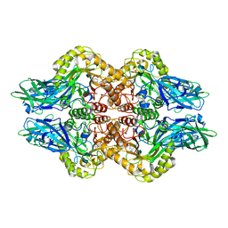
8CV1
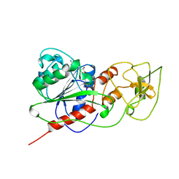
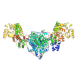 | | ACP1-KS-AT domains of mycobacterial Pks13 | | 分子名称: | Polyketide synthase PKS13, UNKNOWN LIGAND | | 著者 | Kim, S.K, Dickinson, M.S, Finer-Moore, J.S, Rosenberg, O.S, Stroud, R.M. | | 登録日 | 2022-05-17 | | 公開日 | 2023-02-15 | | 最終更新日 | 2023-03-29 | | 実験手法 | ELECTRON MICROSCOPY (2.6 Å) | | 主引用文献 | Structure and dynamics of the essential endogenous mycobacterial polyketide synthase Pks13.
Nat.Struct.Mol.Biol., 30, 2023
|
|
5FGF
 | |
7DIG
 | |
8J7F
 | | ion channel | | 分子名称: | (2S)-3-(hexadecanoyloxy)-2-[(9Z)-octadec-9-enoyloxy]propyl 2-(trimethylammonio)ethyl phosphate, CALCIUM ION, CHOLESTEROL, ... | | 著者 | Chen, H.W, Chen, H.W. | | 登録日 | 2023-04-27 | | 公開日 | 2024-05-15 | | 実験手法 | ELECTRON MICROSCOPY (2.6 Å) | | 主引用文献 | structure of ion channel
To Be Published
|
|
8T4S
 | |
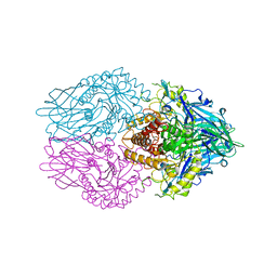
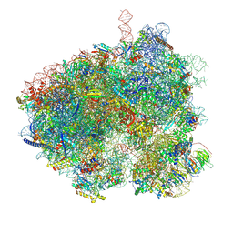
7ZAI
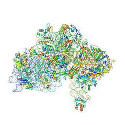 | | Cryo-EM structure of a Pyrococcus abyssi 30S bound to Met-initiator tRNA, mRNA and aIF1A. | | 分子名称: | 16S rRNA, 30S ribosomal protein S10, 30S ribosomal protein S11, ... | | 著者 | Coureux, P.D, Bourgeois, G, Mechulam, Y, Schmitt, E, Kazan, R. | | 登録日 | 2022-03-22 | | 公開日 | 2022-07-13 | | 最終更新日 | 2024-04-24 | | 実験手法 | ELECTRON MICROSCOPY (2.6 Å) | | 主引用文献 | Role of aIF5B in archaeal translation initiation.
Nucleic Acids Res., 50, 2022
|
|
4X8M
 | | Crystal structure of E. coli Adenylate kinase Y171W mutant | | 分子名称: | Adenylate kinase | | 著者 | Sauer-Eriksson, A.E, Kovermann, M, Aden, J, Grundstrom, C, Wolf-Watz, M, Sauer, U.H. | | 登録日 | 2014-12-10 | | 公開日 | 2015-07-15 | | 最終更新日 | 2024-01-10 | | 実験手法 | X-RAY DIFFRACTION (2.6 Å) | | 主引用文献 | Structural basis for catalytically restrictive dynamics of a high-energy enzyme state.
Nat Commun, 6, 2015
|
|
6LOF
 | | Crystal structure of ZsYellow soaked by Cu2+ | | 分子名称: | GFP-like fluorescent chromoprotein FP538 | | 著者 | Nam, K.H. | | 登録日 | 2020-01-05 | | 公開日 | 2020-01-22 | | 最終更新日 | 2023-11-29 | | 実験手法 | X-RAY DIFFRACTION (2.6 Å) | | 主引用文献 | Spectroscopic and Structural Analysis of Cu 2+ -Induced Fluorescence Quenching of ZsYellow.
Biosensors (Basel), 10, 2020
|
|
5C5X
 | | CRYSTAL STRUCTURE OF THE S156E MUTANT OF HUMAN AQUAPORIN 5 | | 分子名称: | Aquaporin-5, O-[(S)-{[(2S)-2-(hexanoyloxy)-3-(tetradecanoyloxy)propyl]oxy}(hydroxy)phosphoryl]-D-serine | | 著者 | Kitchen, P, Oeberg, F, Sjoehamn, J, Hedfalk, K, Bill, R.M, Conner, A.C, Conner, M.T, Toernroth-Horsefield, S. | | 登録日 | 2015-06-22 | | 公開日 | 2015-12-02 | | 最終更新日 | 2024-01-10 | | 実験手法 | X-RAY DIFFRACTION (2.6 Å) | | 主引用文献 | Plasma Membrane Abundance of Human Aquaporin 5 Is Dynamically Regulated by Multiple Pathways.
Plos One, 10, 2015
|
|
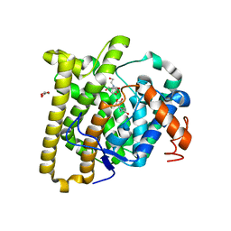
2NTL
 | | Crystal structure of PurO/AICAR from Methanothermobacter thermoautotrophicus | | 分子名称: | AMINOIMIDAZOLE 4-CARBOXAMIDE RIBONUCLEOTIDE, IMP cyclohydrolase | | 著者 | Kang, Y.N, Tran, A, White, R.H, Ealick, S.E. | | 登録日 | 2006-11-07 | | 公開日 | 2007-04-24 | | 最終更新日 | 2023-08-30 | | 実験手法 | X-RAY DIFFRACTION (2.6 Å) | | 主引用文献 | A novel function for the N-terminal nucleophile hydrolase fold demonstrated by the structure of an archaeal inosine monophosphate cyclohydrolase.
Biochemistry, 46, 2007
|
|
2EMO
 | |
2NTM
 | |
4ZVU
 | |
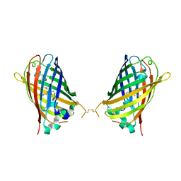
6OFO
 | | Crystal structure of split green fluorescent protein (GFP); s10 circular permutant (194-195) | | 分子名称: | Green fluorescent protein (GFP); s10 circular permutant (194-195) | | 著者 | Lin, C.-Y, Romei, M.G, Deller, M.C, Doukov, T.I, Boxer, S.G. | | 登録日 | 2019-03-31 | | 公開日 | 2019-07-10 | | 最終更新日 | 2023-11-15 | | 実験手法 | X-RAY DIFFRACTION (2.603 Å) | | 主引用文献 | Unified Model for Photophysical and Electro-Optical Properties of Green Fluorescent Proteins.
J.Am.Chem.Soc., 141, 2019
|
|
6LEG
 | | Structure of E. coli beta-glucuronidase complex with uronic isofagomine | | 分子名称: | (3S,4R,5R)-4,5-dihydroxypiperidine-3-carboxylic acid, Beta-D-glucuronidase | | 著者 | Lin, H.-Y, Kuo, Y.-H, Lin, C.-H. | | 登録日 | 2019-11-25 | | 公開日 | 2021-01-27 | | 最終更新日 | 2023-11-22 | | 実験手法 | X-RAY DIFFRACTION (2.603 Å) | | 主引用文献 | Entropy-driven binding of gut bacterial beta-glucuronidase inhibitors ameliorates irinotecan-induced toxicity.
Commun Biol, 4, 2021
|
|
6VPD
 | | Crystal structure of Trgpx in apo form | | 分子名称: | Glutathione peroxidase | | 著者 | Adriani, P.P, De Oliveira, G.S, Paiva, F.C.R, Dias, M.V.B, Chambergo, F.S. | | 登録日 | 2020-02-03 | | 公開日 | 2020-12-16 | | 最終更新日 | 2023-10-11 | | 実験手法 | X-RAY DIFFRACTION (2.603 Å) | | 主引用文献 | Structural and functional characterization of the glutathione peroxidase-like thioredoxin peroxidase from the fungus Trichoderma reesei.
Int.J.Biol.Macromol., 167, 2020
|
|
4PFE
 | | Crystal structure of vsfGFP-0 | | 分子名称: | Green fluorescent protein | | 著者 | Jauch, R, Chen, S.L. | | 登録日 | 2014-04-29 | | 公開日 | 2015-06-24 | | 最終更新日 | 2023-11-15 | | 実験手法 | X-RAY DIFFRACTION (2.603 Å) | | 主引用文献 | Rational Structure-Based Design of Bright GFP-Based Complexes with Tunable Dimerization.
Angew.Chem.Int.Ed.Engl., 54, 2015
|
|
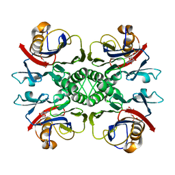
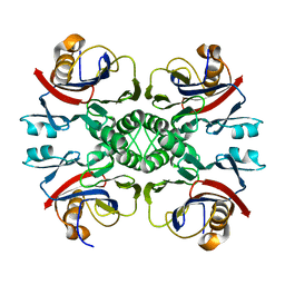
2UXV
 | | SufI Protein from Escherichia Coli | | 分子名称: | PROTEIN SUFI | | 著者 | Tarry, M.J, Roversi, P, Sargent, F, Berks, B.C, Lea, S.M. | | 登録日 | 2007-03-30 | | 公開日 | 2008-05-13 | | 最終更新日 | 2023-12-13 | | 実験手法 | X-RAY DIFFRACTION (2.61 Å) | | 主引用文献 | The Escherichia Coli Cell Division Protein and Model Tat Substrate Sufi (Ftsp) Localizes to the Septal Ring and Has a Multicopper Oxidase-Like Structure.
J.Mol.Biol., 386, 2009
|
|
2HMY
 | | BINARY COMPLEX OF HHAI METHYLTRANSFERASE WITH ADOMET FORMED IN THE PRESENCE OF A SHORT NONPSECIFIC DNA OLIGONUCLEOTIDE | | 分子名称: | PROTEIN (CYTOSINE-SPECIFIC METHYLTRANSFERASE HHAI), S-ADENOSYLMETHIONINE | | 著者 | O'Gara, M, Zhang, X, Roberts, R.J, Cheng, X. | | 登録日 | 1999-02-08 | | 公開日 | 1999-03-19 | | 最終更新日 | 2023-08-30 | | 実験手法 | X-RAY DIFFRACTION (2.61 Å) | | 主引用文献 | Structure of a binary complex of HhaI methyltransferase with S-adenosyl-L-methionine formed in the presence of a short non-specific DNA oligonucleotide.
J.Mol.Biol., 287, 1999
|
|
6LEJ
 | | Structure of E. coli beta-glucuronidase complex with C6-propyl uronic isofagomine | | 分子名称: | (2~{S},3~{S},4~{R},5~{R})-4,5-bis(oxidanyl)-2-propyl-piperidine-3-carboxylic acid, Beta-D-glucuronidase | | 著者 | Lin, H.-Y, Kuo, Y.-H, Lin, C.-H. | | 登録日 | 2019-11-25 | | 公開日 | 2021-01-27 | | 最終更新日 | 2023-11-22 | | 実験手法 | X-RAY DIFFRACTION (2.617 Å) | | 主引用文献 | Entropy-driven binding of gut bacterial beta-glucuronidase inhibitors ameliorates irinotecan-induced toxicity.
Commun Biol, 4, 2021
|
|
8D7F
 | |
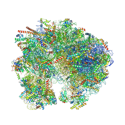
8EVS
 | | Hypopseudouridylated yeast 80S bound with Taura syndrome virus (TSV) internal ribosome entry site (IRES), eEF2 and GDP, Structure II | | 分子名称: | 18S rRNA, 25S rRNA, 40S ribosomal protein S0-A, ... | | 著者 | Zhao, Y, Rai, J, Li, H. | | 登録日 | 2022-10-20 | | 公開日 | 2023-09-06 | | 最終更新日 | 2023-11-01 | | 実験手法 | ELECTRON MICROSCOPY (2.62 Å) | | 主引用文献 | Regulation of translation by ribosomal RNA pseudouridylation.
Sci Adv, 9, 2023
|
|
4W6I
 | |
9BH5
 | | High-resolution C. elegans 80S ribosome structure - class 1 | | 分子名称: | 18S rRNA, 28S rRNA, 4-{(2R)-2-[(1S,3S,5S)-3,5-dimethyl-2-oxocyclohexyl]-2-hydroxyethyl}piperidine-2,6-dione, ... | | 著者 | Sehgal, E, Serrao, V.H.B, Arribere, J. | | 登録日 | 2024-04-19 | | 公開日 | 2024-09-04 | | 最終更新日 | 2024-09-11 | | 実験手法 | ELECTRON MICROSCOPY (2.63 Å) | | 主引用文献 | High-Resolution Reconstruction of a C. elegans Ribosome Sheds Light on Evolutionary Dynamics and Tissue Specificity.
Rna, 2024
|
|


