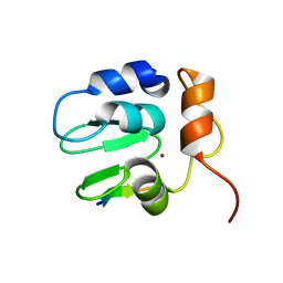
1LFU
 
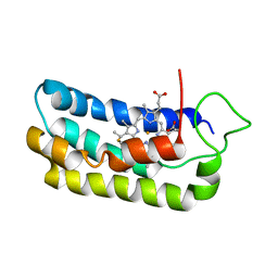
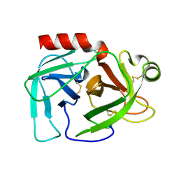
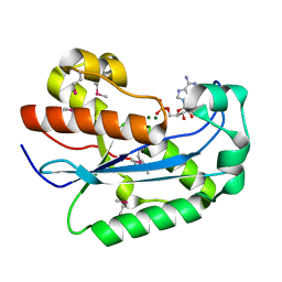
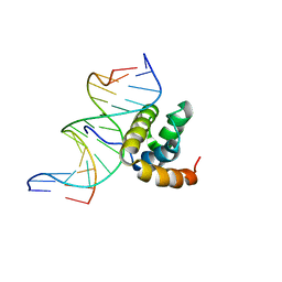 | | NMR Solution Structure of the Extended PBX Homeodomain Bound to DNA | | 分子名称: | 5'-D(*GP*CP*GP*CP*AP*TP*GP*AP*TP*TP*GP*CP*CP*C)-3', 5'-D(*GP*GP*GP*CP*AP*AP*TP*CP*AP*TP*GP*CP*GP*C)-3', homeobox protein PBX1 | | 著者 | Sprules, T, Green, N, Featherstone, M, Gehring, K. | | 登録日 | 2002-04-12 | | 公開日 | 2003-01-14 | | 最終更新日 | 2024-05-22 | | 実験手法 | SOLUTION NMR | | 主引用文献 | Lock and Key Binding of the HOX YPWM Peptide to the PBX Homeodomain
J.Biol.Chem., 278, 2003
|
|
1UWJ
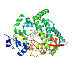
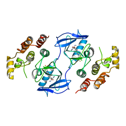 | | The complex of mutant V599E B-RAF and BAY439006. | | 分子名称: | 4-{4-[({[4-CHLORO-3-(TRIFLUOROMETHYL)PHENYL]AMINO}CARBONYL)AMINO]PHENOXY}-N-METHYLPYRIDINE-2-CARBOXAMIDE, B-RAF PROTO-ONCOGENE SERINE/THREONINE-PROTEIN KINASE | | 著者 | Barford, D, Roe, S.M, Wan, P.T.C, Cancer Genome Project | | 登録日 | 2004-02-05 | | 公開日 | 2004-03-19 | | 最終更新日 | 2024-05-01 | | 実験手法 | X-RAY DIFFRACTION (3.5 Å) | | 主引用文献 | Mechanism of Activation of the Raf-Erk Signaling Pathway by Oncogenic Mutations of B-Raf
Cell(Cambridge,Mass.), 116, 2004
|
|
1LPB
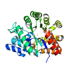
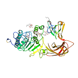 | | THE 2.46 ANGSTROMS RESOLUTION STRUCTURE OF THE PANCREATIC LIPASE COLIPASE COMPLEX INHIBITED BY A C11 ALKYL PHOSPHONATE | | 分子名称: | CALCIUM ION, COLIPASE, LIPASE, ... | | 著者 | Egloff, M.-P, Van Tilbeurgh, H, Cambillau, C. | | 登録日 | 1994-08-19 | | 公開日 | 1994-11-01 | | 最終更新日 | 2020-07-29 | | 実験手法 | X-RAY DIFFRACTION (2.46 Å) | | 主引用文献 | The 2.46 A resolution structure of the pancreatic lipase-colipase complex inhibited by a C11 alkyl phosphonate.
Biochemistry, 34, 1995
|
|
1LT6
 | |
1S05
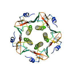
 | | NMR-validated structural model for oxidized R.palustris cytochrome c556 | | 分子名称: | Cytochrome c-556, HEME C | | 著者 | Bertini, I, Faraone-Mennella, J, Gray, H.B, Luchinat, C, Parigi, G, Winkler, J.R. | | 登録日 | 2003-12-30 | | 公開日 | 2004-01-20 | | 最終更新日 | 2021-03-03 | | 実験手法 | SOLUTION NMR | | 主引用文献 | NMR-validated structural model for oxidized Rhodopseudomonas palustris cytochrome c(556).
J.Biol.Inorg.Chem., 9, 2004
|
|
1S4Y
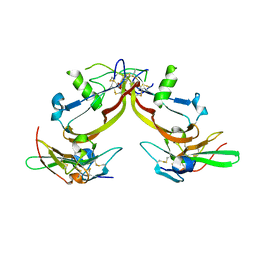 | | Crystal structure of the activin/actrIIb extracellular domain | | 分子名称: | Activin receptor type IIB precursor, Inhibin beta A chain | | 著者 | Greenwald, J, Vega, M.E, Allendorph, G.P, Fischer, W.H, Vale, W, Choe, S, Joint Center for Structural Genomics (JCSG) | | 登録日 | 2004-01-19 | | 公開日 | 2004-08-10 | | 最終更新日 | 2011-07-13 | | 実験手法 | X-RAY DIFFRACTION (2.3 Å) | | 主引用文献 | A Flexible Activin Explains the Membrane-Dependent Cooperative Assembly of TGF-beta Family Receptors.
Mol.Cell, 15, 2004
|
|
1SC3
 | | Crystal structure of the human caspase-1 C285A mutant in complex with malonate | | 分子名称: | Interleukin-1 beta convertase, MALONATE ION | | 著者 | Romanowski, M.J, Scheer, J.M, O'Brien, T, McDowell, R.S. | | 登録日 | 2004-02-11 | | 公開日 | 2004-08-10 | | 最終更新日 | 2023-08-23 | | 実験手法 | X-RAY DIFFRACTION (1.8 Å) | | 主引用文献 | Crystal structures of a ligand-free and malonate-bound human caspase-1: implications for the mechanism of substrate binding.
Structure, 12, 2004
|
|
1LTT
 | |
1LOI
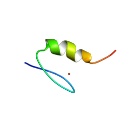
 | | N-TERMINAL SPLICE REGION OF RAT C-AMP PHOSPHODIESTERASE, NMR, 7 STRUCTURES | | 分子名称: | CYCLIC 3',5'-AMP SPECIFIC PHOSPHODIESTERASE RD1 | | 著者 | Smith, K.J, Scotland, G, Beattie, J, Trayer, I.P, Houslay, M.D. | | 登録日 | 1996-05-21 | | 公開日 | 1997-05-15 | | 最終更新日 | 2022-02-23 | | 実験手法 | SOLUTION NMR | | 主引用文献 | Determination of the structure of the N-terminal splice region of the cyclic AMP-specific phosphodiesterase RD1 (RNPDE4A1) by 1H NMR and identification of the membrane association domain using chimeric constructs.
J.Biol.Chem., 271, 1996
|
|
1SH5
 | |
1LFO
 | | LIVER FATTY ACID BINDING PROTEIN-OLEATE COMPLEX | | 分子名称: | BUTENOIC ACID, LIVER FATTY ACID BINDING PROTEIN, OLEIC ACID, ... | | 著者 | Thompson, J, Winter, N, Terwey, D, Bratt, J, Banaszak, L. | | 登録日 | 1996-12-09 | | 公開日 | 1997-06-16 | | 最終更新日 | 2023-08-09 | | 実験手法 | X-RAY DIFFRACTION (2.3 Å) | | 主引用文献 | The crystal structure of the liver fatty acid-binding protein. A complex with two bound oleates.
J.Biol.Chem., 272, 1997
|
|
1SA4
 | | human protein farnesyltransferase complexed with FPP and R115777 | | 分子名称: | 6-[(S)-AMINO(4-CHLOROPHENYL)(1-METHYL-1H-IMIDAZOL-5-YL)METHYL]-4-(3-CHLOROPHENYL)-1-METHYLQUINOLIN-2(1H)-ONE, FARNESYL DIPHOSPHATE, Protein farnesyltransferase beta subunit, ... | | 著者 | Reid, T.S, Beese, L.S. | | 登録日 | 2004-02-06 | | 公開日 | 2004-06-08 | | 最終更新日 | 2023-08-23 | | 実験手法 | X-RAY DIFFRACTION (2.1 Å) | | 主引用文献 | Crystal Structures of the Anticancer Clinical Candidates R115777 (Tipifarnib) and BMS-214662 Complexed with Protein Farnesyltransferase Suggest a Mechanism of FTI Selectivity.
Biochemistry, 43, 2004
|
|
1LCJ
 | |
1SDZ
 | | Crystal structure of DIAP1 BIR1 bound to a Reaper peptide | | 分子名称: | Apoptosis 1 inhibitor, Reaper, ZINC ION | | 著者 | Yan, N, Wu, J.W, Shi, Y. | | 登録日 | 2004-02-15 | | 公開日 | 2004-04-27 | | 最終更新日 | 2024-02-14 | | 実験手法 | X-RAY DIFFRACTION (1.78 Å) | | 主引用文献 | Molecular mechanisms of DrICE inhibition by DIAP1 and removal of inhibition by Reaper, Hid and Grim.
Nat.Struct.Mol.Biol., 11, 2004
|
|
1M18
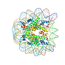 | | LIGAND BINDING ALTERS THE STRUCTURE AND DYNAMICS OF NUCLEOSOMAL DNA | | 分子名称: | Histone H2A.1, Histone H2B.1, Histone H3.2, ... | | 著者 | Suto, R.K, Edayathumangalam, R.S, White, C.L, Melander, C, Gottesfeld, J.M, Dervan, P.B, Luger, K. | | 登録日 | 2002-06-18 | | 公開日 | 2003-02-18 | | 最終更新日 | 2024-02-14 | | 実験手法 | X-RAY DIFFRACTION (2.45 Å) | | 主引用文献 | Crystal Structures of Nucleosome Core Particles in Complex with Minor Groove DNA-binding Ligands
J.Mol.Biol., 326, 2003
|
|
1M19
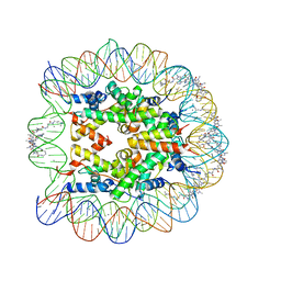 | | LIGAND BINDING ALTERS THE STRUCTURE AND DYNAMICS OF NUCLEOSOMAL DNA | | 分子名称: | 3-AMINO-(DIMETHYLPROPYLAMINE), 4-AMINO-(1-METHYLIMIDAZOLE)-2-CARBOXYLIC ACID, 4-AMINO-(1-METHYLPYRROLE)-2-CARBOXYLIC ACID, ... | | 著者 | Suto, R.K, Edayathumangalam, R.S, White, C.L, Melander, C, Gottesfeld, J.M, Dervan, P.B, Luger, K. | | 登録日 | 2002-06-18 | | 公開日 | 2003-02-18 | | 最終更新日 | 2023-11-15 | | 実験手法 | X-RAY DIFFRACTION (2.3 Å) | | 主引用文献 | Crystal Structures of Nucleosome Core Particles in Complex with Minor Groove DNA-binding Ligands
J.Mol.Biol., 326, 2003
|
|
1VFQ
 | |
1SGT
 | |
1SP2
 | |
1SP1
 | |
1W6S
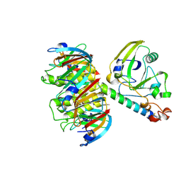
 | | The high resolution structure of methanol dehydrogenase from methylobacterium extorquens | | 分子名称: | CALCIUM ION, GLYCEROL, METHANOL DEHYDROGENASE SUBUNIT 1, ... | | 著者 | Williams, P.A, Coates, L, Mohammed, F, Gill, R, Erskine, P.T, Wood, S.P, Anthony, C, Cooper, J.B. | | 登録日 | 2004-08-23 | | 公開日 | 2004-12-21 | | 最終更新日 | 2019-05-22 | | 実験手法 | X-RAY DIFFRACTION (1.2 Å) | | 主引用文献 | The Atomic Resolution Structure of Methanol Dehydrogenase from Methylobacterium Extorquens
Acta Crystallogr.,Sect.D, 61, 2005
|
|
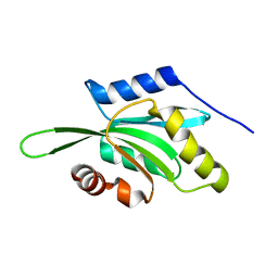
1W0H
 | | Crystallographic structure of the nuclease domain of 3'hExo, a DEDDh family member, bound to rAMP | | 分子名称: | 3'-5' EXONUCLEASE ERI1, ADENOSINE MONOPHOSPHATE, MAGNESIUM ION | | 著者 | Cheng, Y, Patel, D. | | 登録日 | 2004-06-04 | | 公開日 | 2004-09-30 | | 最終更新日 | 2011-07-13 | | 実験手法 | X-RAY DIFFRACTION (1.59 Å) | | 主引用文献 | Crystallographic Structure of the Nuclease Domain of 3'Hexo, a Deddh Family Member, Bound to Ramp
J.Mol.Biol., 343, 2004
|
|
1W0E
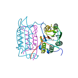
 | | Crystal structure of human cytochrome P450 3A4 | | 分子名称: | CYTOCHROME P450 3A4, PROTOPORPHYRIN IX CONTAINING FE | | 著者 | Williams, P.A, Cosme, J, Vinkovic, D.M, Ward, A, Angove, H.C, Day, P.J, Vonrhein, C, Tickle, I.J, Jhoti, H. | | 登録日 | 2004-06-03 | | 公開日 | 2004-07-22 | | 最終更新日 | 2024-05-08 | | 実験手法 | X-RAY DIFFRACTION (2.8 Å) | | 主引用文献 | Crystal Structures of Human Cytochrome P450 3A4 Bound to Metyrapone and Progesterone
Science, 305, 2004
|
|
1VFL
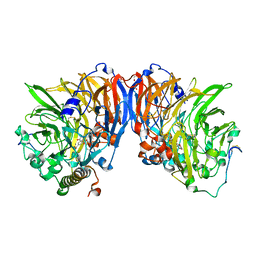
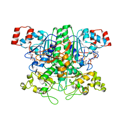
 | | Adenosine deaminase | | 分子名称: | Adenosine deaminase, ZINC ION | | 著者 | Kinoshita, T. | | 登録日 | 2004-04-16 | | 公開日 | 2005-08-16 | | 最終更新日 | 2023-12-27 | | 実験手法 | X-RAY DIFFRACTION (1.8 Å) | | 主引用文献 | Structural Basis of Compound Recognition by Adenosine Deaminase
Biochemistry, 44, 2005
|
|
1VGR
 | | Formyl-CoA transferase mutant Asp169 to Glu | | 分子名称: | COENZYME A, Formyl-coenzyme A transferase | | 著者 | Ricagno, S, Jonsson, S, Richards, N.G, Lindqvist, Y. | | 登録日 | 2004-04-28 | | 公開日 | 2004-08-03 | | 最終更新日 | 2023-10-25 | | 実験手法 | X-RAY DIFFRACTION (2.1 Å) | | 主引用文献 | Kinetic and mechanistic characterization of the formyl-CoA transferase from Oxalobacter formigenes
J.Biol.Chem., 279, 2004
|
|


