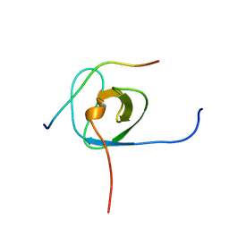
1SE0
 
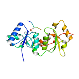
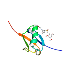
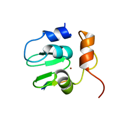 | | Crystal structure of DIAP1 BIR1 bound to a Grim peptide | | 分子名称: | Apoptosis 1 inhibitor, Cell death protein Grim, ZINC ION | | 著者 | Yan, N, Wu, J.W, Shi, Y. | | 登録日 | 2004-02-15 | | 公開日 | 2004-04-27 | | 最終更新日 | 2024-02-14 | | 実験手法 | X-RAY DIFFRACTION (1.75 Å) | | 主引用文献 | Molecular mechanisms of DrICE inhibition by DIAP1 and removal of inhibition by Reaper, Hid and Grim.
Nat.Struct.Mol.Biol., 11, 2004
|
|
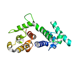
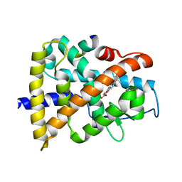
1S5I
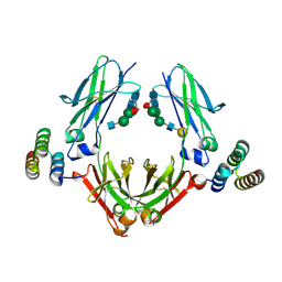
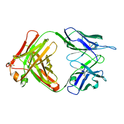 | | Fab (LNKB-2) of monoclonal antibody to Human Interleukin-2, crystal structure | | 分子名称: | Fab-fragment of monoclonal antibody | | 著者 | Pletnev, V.Z, Goryacheva, E.A, Tsygannik, I.N, Nesmeyanov, V.A, Pletnev, S.V, Pangborn, W, Duax, W. | | 登録日 | 2004-01-21 | | 公開日 | 2004-05-25 | | 最終更新日 | 2023-08-23 | | 実験手法 | X-RAY DIFFRACTION (2.7 Å) | | 主引用文献 | [A new crystal form of the Fab fragment of a monoclonal antibody to human interleukin-2: the three-dimensional structure at 2.7 A resolution].
Bioorg. Khim., 30
|
|
5U2E
 | |
3KJP
 | |
4Y2G
 | |
1SH6
 | |
5JH5
 | | Structural Basis for the Hierarchical Assembly of the Core of PRC1.1 | | 分子名称: | BCL-6 corepressor-like protein 1, Lysine-specific demethylase 2B, Polycomb group RING finger protein 1, ... | | 著者 | Wong, S.J, Taylor, A.B, Hart, P.J, Kim, C.A. | | 登録日 | 2016-04-20 | | 公開日 | 2016-09-14 | | 最終更新日 | 2016-10-19 | | 実験手法 | X-RAY DIFFRACTION (2.55 Å) | | 主引用文献 | KDM2B Recruitment of the Polycomb Group Complex, PRC1.1, Requires Cooperation between PCGF1 and BCORL1.
Structure, 24, 2016
|
|
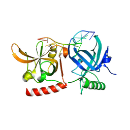
4XUS
 | | Crystal structure of the pre-catalytic ternary complex of DNA polymerase lambda with a templating A and an incoming dTTP | | 分子名称: | 2',3'-DIDEOXYCYTIDINE-5'-MONOPHOSPHATE, DNA (5'-D(*CP*AP*GP*TP*A)-3'), DNA (5'-D(*CP*GP*GP*CP*AP*GP*TP*AP*CP*TP*G)-3'), ... | | 著者 | Burak, M.J, Guja, K.E, Garcia-Diaz, M. | | 登録日 | 2015-01-26 | | 公開日 | 2016-02-03 | | 最終更新日 | 2024-02-28 | | 実験手法 | X-RAY DIFFRACTION (2.4 Å) | | 主引用文献 | Nucleotide binding interactions modulate dNTP selectivity and facilitate 8-oxo-dGTP incorporation by DNA polymerase lambda.
Nucleic Acids Res., 43, 2015
|
|
5TWV
 | | Cryo-EM structure of the pancreatic ATP-sensitive K+ channel SUR1/Kir6.2 in the presence of ATP and glibenclamide | | 分子名称: | ADENOSINE-5'-TRIPHOSPHATE, ATP-binding cassette sub-family C member 8, ATP-sensitive inward rectifier potassium channel 11 | | 著者 | Martin, G.M, Yoshioka, C, Chen, J.Z, Shyng, S.L. | | 登録日 | 2016-11-14 | | 公開日 | 2017-01-25 | | 最終更新日 | 2019-12-25 | | 実験手法 | ELECTRON MICROSCOPY (6.3 Å) | | 主引用文献 | Cryo-EM structure of the ATP-sensitive potassium channel illuminates mechanisms of assembly and gating.
Elife, 6, 2017
|
|
5J2B
 | |
3KEH
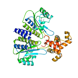
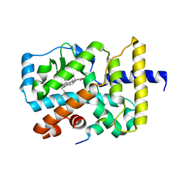
 | | Crystal Structure of N370S Glucocerebrosidase mutant at pH 7.4 | | 分子名称: | 2-acetamido-2-deoxy-alpha-D-glucopyranose, 2-acetamido-2-deoxy-beta-D-glucopyranose, 2-acetamido-2-deoxy-beta-D-glucopyranose-(1-4)-2-acetamido-2-deoxy-beta-D-glucopyranose, ... | | 著者 | Wei, R.R, Boucher, S, Pan, C.Q, Edmunds, T. | | 登録日 | 2009-10-26 | | 公開日 | 2010-10-27 | | 最終更新日 | 2024-04-03 | | 実験手法 | X-RAY DIFFRACTION (2.8 Å) | | 主引用文献 | Crystal Structure of Glucocerebrosidase Containing the N370S mutation: Implication on Chaperon Therapy
To be Published
|
|
5J2H
 | |
5J2R
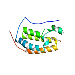
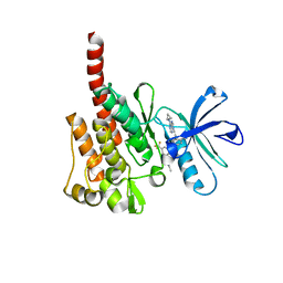
 | | Solution structure of Ras Binding Domain (RBD) of B-Raf | | 分子名称: | N-[2-methoxy-5-({[(E)-2-(2,4,6-trimethoxyphenyl)ethenyl]sulfonyl}methyl)phenyl]glycine, Serine/threonine-protein kinase B-raf | | 著者 | Dutta, K, Vasquez-Del Carpio, R, Aggarwal, A.K, Reddy, E.P. | | 登録日 | 2016-03-29 | | 公開日 | 2016-05-04 | | 最終更新日 | 2024-05-01 | | 実験手法 | SOLUTION NMR | | 主引用文献 | A Small Molecule RAS-Mimetic Disrupts RAS Association with Effector Proteins to Block Signaling.
Cell, 165, 2016
|
|
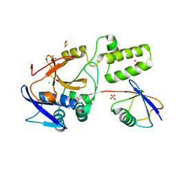
5JJA
 | | Crystal structure of a PP2A B56gamma/BubR1 complex | | 分子名称: | Mitotic checkpoint serine/threonine-protein kinase BUB1 beta, Serine/threonine-protein phosphatase 2A 56 kDa regulatory subunit gamma isoform | | 著者 | Wang, Z, Wang, J, Rao, Z, Xu, W. | | 登録日 | 2016-04-22 | | 公開日 | 2016-07-13 | | 最終更新日 | 2023-09-27 | | 実験手法 | X-RAY DIFFRACTION (2.35 Å) | | 主引用文献 | Crystal structure of a PP2A B56-BubR1 complex and its implications for PP2A substrate recruitment and localization.
Protein Cell, 7, 2016
|
|
4Y17
 | | SdiA in complex with 3-oxo-C8-homoserine lactone | | 分子名称: | 3-OXO-OCTANOIC ACID (2-OXO-TETRAHYDRO-FURAN-3-YL)-AMIDE, Transcriptional regulator of ftsQAZ gene cluster | | 著者 | Nguyen, N.X, Nguyen, Y, Sperandio, V, Jiang, Y. | | 登録日 | 2015-02-06 | | 公開日 | 2015-04-08 | | 最終更新日 | 2023-09-27 | | 実験手法 | X-RAY DIFFRACTION (2.84 Å) | | 主引用文献 | Structural and Mechanistic Roles of Novel Chemical Ligands on the SdiA Quorum-Sensing Transcription Regulator.
Mbio, 6, 2015
|
|
5U4Y
 | | IgG Fc bound to 3 helix of the B-domain from Protein A | | 分子名称: | 2-acetamido-2-deoxy-beta-D-glucopyranose-(1-2)-alpha-D-mannopyranose-(1-6)-[alpha-D-mannopyranose-(1-3)]alpha-D-mannopyranose-(1-4)-2-acetamido-2-deoxy-beta-D-glucopyranose-(1-4)-[alpha-L-fucopyranose-(1-6)]2-acetamido-2-deoxy-beta-D-glucopyranose, IgG1 fc, Immunoglobulin G-binding protein A, ... | | 著者 | Ultsch, M.H, Eigenbrot, C. | | 登録日 | 2016-12-06 | | 公開日 | 2017-05-17 | | 最終更新日 | 2023-10-04 | | 実験手法 | X-RAY DIFFRACTION (2.4994 Å) | | 主引用文献 | 3-2-1: Structural insights from stepwise shrinkage of a three-helix Fc-binding domain to a single helix.
Protein Eng. Des. Sel., 30, 2017
|
|
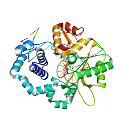
3KGQ
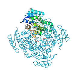
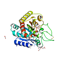 | | Carboxypeptidase A liganded to an organic small-molecule: conformational changes | | 分子名称: | ACETONE, CITRIC ACID, Carboxypeptidase A1, ... | | 著者 | Fernandez, D, Boix, E, Pallares, I, Aviles, F.X, Vendrell, J. | | 登録日 | 2009-10-29 | | 公開日 | 2010-10-27 | | 最終更新日 | 2023-11-01 | | 実験手法 | X-RAY DIFFRACTION (1.7 Å) | | 主引用文献 | Structural and Functional Analysis of the Complex between Citrate and the Zinc Peptidase Carboxypeptidase A
Enzyme Res, 2011, 2011
|
|
5U6G
 | |
5IXF
 | | Solution structure of the STAM2 SH3 with AMSH derived peptide complex | | 分子名称: | STAM-binding protein, Signal transducing adapter molecule 2 | | 著者 | Hologne, M, Cantrelle, F.X, Trivelli, X, Walker, O. | | 登録日 | 2016-03-23 | | 公開日 | 2016-12-07 | | 最終更新日 | 2024-06-19 | | 実験手法 | SOLUTION NMR | | 主引用文献 | NMR Reveals the Interplay among the AMSH SH3 Binding Motif, STAM2, and Lys63-Linked Diubiquitin.
J.Mol.Biol., 428, 2016
|
|
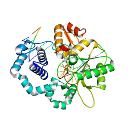
3KMR
 | | Crystal structure of RARalpha ligand binding domain in complex with an agonist ligand (Am580) and a coactivator fragment | | 分子名称: | 4-{[(5,5,8,8-tetramethyl-5,6,7,8-tetrahydronaphthalen-2-yl)carbonyl]amino}benzoic acid, Nuclear receptor coactivator 1, Retinoic acid receptor alpha | | 著者 | Bourguet, W, Teyssier, C. | | 登録日 | 2009-11-11 | | 公開日 | 2010-06-02 | | 最終更新日 | 2023-09-06 | | 実験手法 | X-RAY DIFFRACTION (1.8 Å) | | 主引用文献 | A unique secondary-structure switch controls constitutive gene repression by retinoic acid receptor.
Nat.Struct.Mol.Biol., 17, 2010
|
|
3KFA
 | |
5JQS
 | |
3KYT
 | |
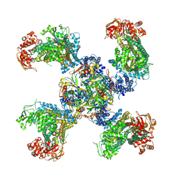
3KZF
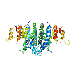 | | Structure of Giardia Carbamate Kinase | | 分子名称: | Carbamate kinase, GLYCEROL | | 著者 | Galkin, A, Herzberg, O. | | 登録日 | 2009-12-08 | | 公開日 | 2010-04-07 | | 最終更新日 | 2023-09-06 | | 実験手法 | X-RAY DIFFRACTION (3 Å) | | 主引用文献 | X-ray structure and characterization of carbamate kinase from the human parasite Giardia lamblia.
Acta Crystallogr.,Sect.F, 66, 2010
|
|
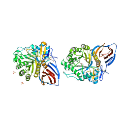
5JS6
 | | Crystal structure of 17beta-hydroxysteroid dehydrogenase 14 T205 variant in complex with NAD. | | 分子名称: | 17-beta-hydroxysteroid dehydrogenase 14, CHLORIDE ION, NICOTINAMIDE-ADENINE-DINUCLEOTIDE, ... | | 著者 | Bertoletti, N, Marchais-Oberwinkler, S, Heine, A, Klebe, G. | | 登録日 | 2016-05-07 | | 公開日 | 2016-07-13 | | 最終更新日 | 2024-01-10 | | 実験手法 | X-RAY DIFFRACTION (2.002 Å) | | 主引用文献 | New Insights into Human 17 beta-Hydroxysteroid Dehydrogenase Type 14: First Crystal Structures in Complex with a Steroidal Ligand and with a Potent Nonsteroidal Inhibitor.
J.Med.Chem., 59, 2016
|
|


